I. INTRODUCTION
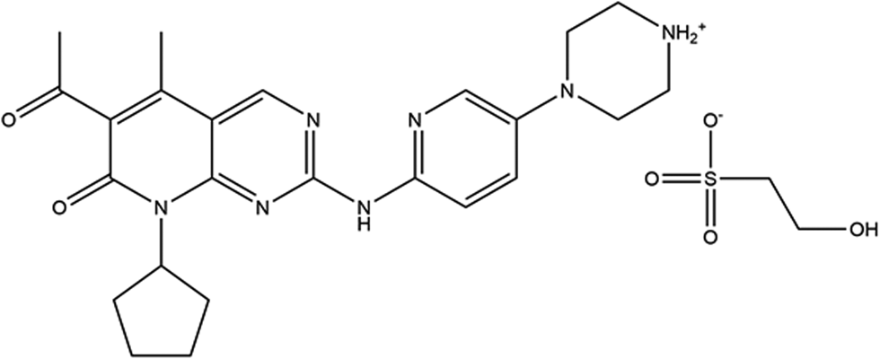
Palbociclib isethionate is the isethionate salt of palbociclib, an orally available cyclin-dependent kinase (CDK) inhibitor with potential anti-neoplastic activity, and is used for the treatment of HR-positive and HER2-negative breast cancer. Palbociclib selectively inhibits CDK4 and CDK6, thereby inhibiting retinoblastoma (Rb) protein phosphorylation early in the G1 phase leading to cell cycle arrest. This suppresses DNA replication and decreases tumor cell proliferation. CDK4 and 6 are serine/threonine kinases that are upregulated in many tumor cell types and play a key role in the regulation of cell cycle progression (PubChem; Kim et al., Reference Kim, Chen, Cheng, Gindulyte, He, He, Li, Shoemaker, Thiessen, Yu, Zaslavsky, Zhang and Bolton2019). Palbociclib is typically combined with letrozole or fulvestrant for a combination of drug treatment. The systematic name for palbociclib isethionate (CAS Registry No. 827022-33-3) is 6-acetyl-8-cyclopentyl-5-methyl-2-[(5-piperazinium-1-ylpyridin-2-yl)amino]pyrido[2,3-d]pyrimidin-7-one 2-hydroxyethanesulfonate. A two-dimensional molecular diagram is shown in Figure 1.
A 2D diagram of the molecular structure of palbociclib isethionate.

X-ray powder diffraction patterns of palbociclib isethionate Forms A, B, and D are reported in US Patents 7,345,171 B2 (Beylin et al., Reference Beylin, Blackburn, Erdman and Toogood2008; Warner–Lambert, now Pfizer) and 7,863,278 B2 (Beylin et al., Reference Beylin, Blackburn, Erdman and Toogood2011; Warner–Lambert). Powder patterns of palbociclib isethionate Forms α, β, γ, δ, and ε are reported in International Patent Application WO 2016/090257 A1 (Chen et al., Reference Chen, Zhang, Yang, Zhang, Li and Wang2016; Crystal Pharmatech), as well as patterns for many other salts and polymorphs of the free base. Powder patterns for palbociclib free base Forms A and B are reported in International Patent Applications WO 2016/156070 A1 (Lengauer and Kluschanzoff, Reference Lengauer and Kluschanzoff2016; Sandoz) and WO 2017/145054 A1 (Mane et al., Reference Mane, Nehate, Godbole and Singh2017; Lupin), and for Forms IX, X, and XII in International Patent Application WO 2017/067506 A1 (Chen et al., Reference Chen, Zhang, Yang, Yang and Zhang2017; Crystal Pharmatech). A single crystal structure of the free base is mentioned in passing in a European Medicines Agency report (2016), but no coordinates are provided. Powder data for crystalline forms of palbociclib monohydrochloride are reported in International Patent Application WO 2016/066420 A1 (Stefinovic, Reference Stefinovic2016; Sandoz). Powder data for additional salts of palbociclib are reported in International Patent Applications WO 2017/072543 A1 (Lukács et al., Reference Lukács, Márványos, Berecz, Heder, Milen, Peregi, Gudor, Volk and Tóthné Lauritz2017; Egis Gyógyszergyár) and WO 2016/127963 (Zvatora et al., Reference Zvatora, Dammer, Miksatko and Krejcik2016; Zentiva). No palbociclib structures are currently contained in the Cambridge Structural Database (Groom et al., Reference Groom, Bruno, Lightfoot and Ward2016) or the Powder Diffraction File (Gates-Rector and Blanton, Reference Gates-Rector and Blanton2019).
This work was carried out as part of a project (Kaduk et al., Reference Kaduk, Crowder, Zhong, Fawcett and Suchomel2014) to determine the crystal structures of large-volume commercial pharmaceuticals and include high-quality powder diffraction data for them in the Powder Diffraction File.
II. EXPERIMENTAL
Palbociclib isethionate was a commercial reference standard, purchased from Sigma (Lot #28914) and was used as-received. The white powder was packed into a 1.5 mm diameter Kapton capillary and rotated during the measurement at ~50 Hz. The powder pattern was measured at 295 K at beam line 11-BM (Antao et al., Reference Antao, Hassan, Wang, Lee and Toby2008; Lee et al., Reference Lee, Shu, Ramanathan, Preissner, Wang, Beno, Von Dreele, Ribaud, Kurtz, Antao, Jiao and Toby2008; Wang et al., Reference Wang, Toby, Lee, Ribaud, Antao, Kurtz, Ramanathan, Von Dreele and Beno2008) of the Advanced Photon Source at Argonne National Laboratory using a wavelength of 0.457912 Å from 0.5 to 50° 2θ with a step size of 0.001° and a counting time of 0.1 s step−1. The high-resolution powder diffraction data were collected using twelve silicon crystal analyzers that allow for high angular resolution, high precision, and accurate peak positions. Silicon (NIST SRM 640c) and alumina (SRM 676a) standards (ratio Al2O3:Si = 2:1 by weight) were used to calibrate the instrument and refine the monochromatic wavelength used in the experiment. The standard uncertainty on the wavelength is probably of the order of 5 ppm.
The pattern was indexed on a primitive triclinic unit cell with a = 8.71494, b = 9.32200, c = 17.73783 Å, α = 80.026, β = 82.359, γ = 76.159°, V = 1371.7 Å3, and Z = 2 using N-TREOR in EXPO2014 (Altomare et al., Reference Altomare, Cuocci, Giacovazzo, Moliterni, Rizzi, Corriero and Falcicchio2013). The space group was assumed to be P-1, which was confirmed by successful solution and refinement of the structure. A reduced cell search in the Cambridge Structural Database (Groom et al., Reference Groom, Bruno, Lightfoot and Ward2016) yielded no hits. The structure was solved by direct methods using EXPO2014. The atom types of O5 and C16 had to be changed to the correct types.
Rietveld refinement was carried out using GSAS-II (Toby and Von Dreele, Reference Toby and Von Dreele2013). Only the 1.3–30.0° portion of the pattern was included in the refinement (d min = 0.885 Å). Analysis of potential hydrogen bonding interactions in Mercury (Macrae et al., Reference Macrae, Sovago, Cottrell, Galek, McCabe, Pidcock, Platings, Shields, Stevens, Towler and Wood2020) indicated that N18 was protonated, so H73 was added to it in an approximate position. All non-H-bond distances and angles were subjected to restraints based on a Mercury/Mogul Geometry Check (Bruno et al., Reference Bruno, Cole, Kessler, Luo, Motherwell, Purkis, Smith, Taylor, Cooper, Harris and Orpen2004; Sykes et al., Reference Sykes, McCabe, Allen, Battle, Bruno and Wood2011) of the cation and anion. The Mogul average and standard deviation for each quantity were used as the restraint parameters. The restraints contributed 4.8% to the final χ 2. The hydrogen atoms were included in calculated positions, which were recalculated during the refinement using Materials Studio (Dassault, 2020). The Uiso were grouped by chemical similarity; the Uiso for the H atoms were fixed at 1.3× the Uiso of the heavy atoms to which they are attached. The peak profiles were described using the generalized microstrain model. The background was modeled using a 6-term shifted Chebyshev polynomial, along with peaks at 5.85° and 22.94° to model the scattering from the Kapton capillary and any amorphous component of the sample.
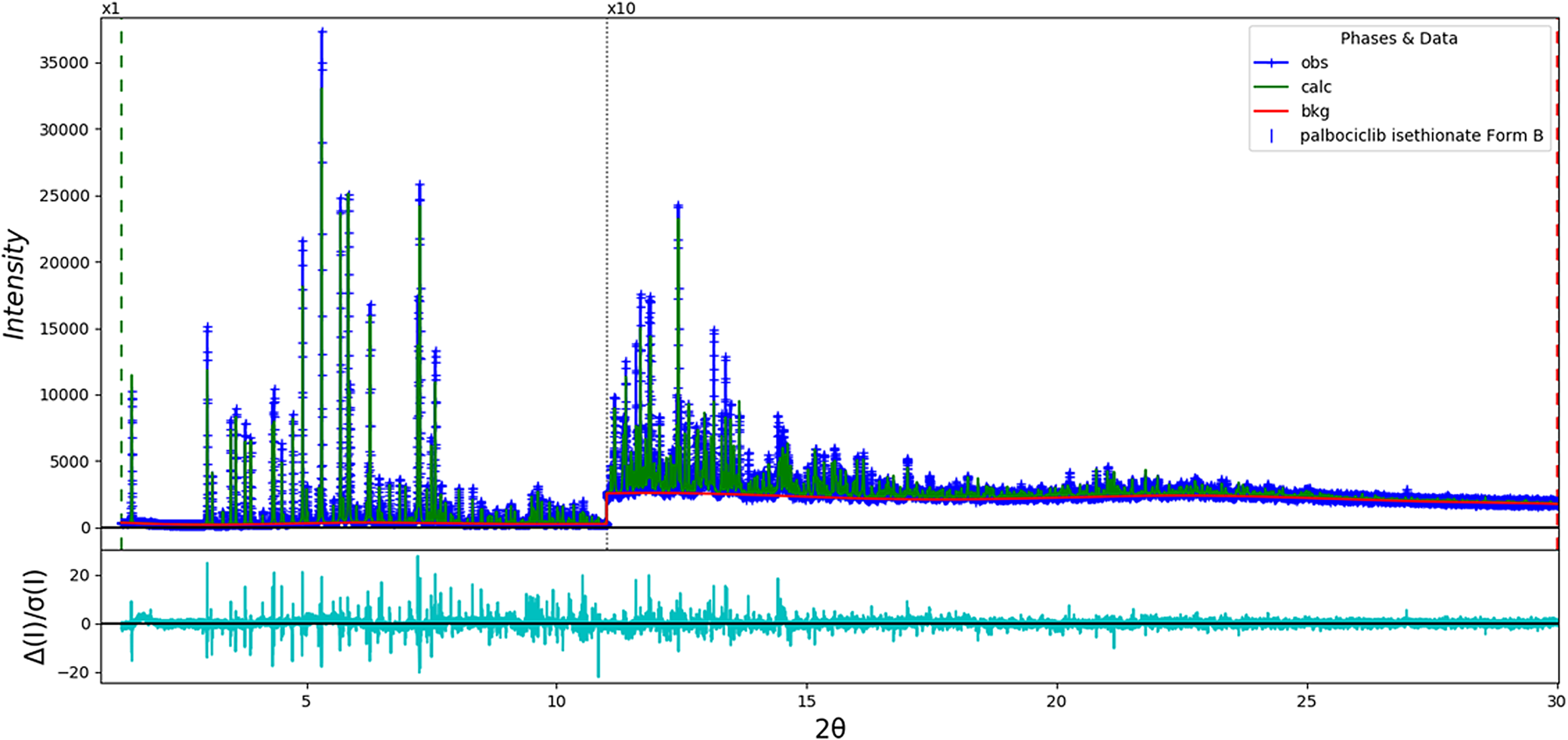
The final refinement (begun from the result of the density functional theory (DFT) calculation) of 148 variables using 28 746 observations and 104 restraints yielded the residuals Rwp = 0.1073 and GOF = 2.28. The largest peak (0.49 Å from O15) and hole (0.54 Å from O39) in the difference Fourier map were 0.90 and −0.49(11) eÅ−3, respectively. The largest errors in the fit (Figure 2) are in the shapes of the strong low-angle peaks and may represent subtle changes in the specimen during measurement or a more-complex microstructure than can be described by the generalized microstrain model.
The Rietveld plot for the refinement of palbociclib isethionate Form B. The blue crosses represent the observed data points, and the green line is the calculated pattern. The cyan curve is the normalized error plot. The vertical scale has been multiplied by a factor of 10× for 2θ > 11.0°.

A DFT geometry optimization (fixed experimental unit cell) was carried out using VASP (Kresse and Furthmüller, Reference Kresse and Furthmüller1996) through the MedeA graphical interface (Materials Design, 2016). The calculations were carried out on sixteen 2.4 GHz processors (each with 4 Gb RAM) of a 64-processor HP Proliant DL580 Generation 7 Linux cluster at North Central College. The calculations used the GGA-PBE functional, a plane wave cutoff energy of 400.0 eV, and a k-point spacing of 0.5 Å−1 leading to a 2 × 2 × 1 mesh. A single-point energy calculation, along with a Mulliken population analysis, was carried out using CRYSTAL14 (Dovesi et al., Reference Dovesi, Orlando, Erba, Zicovich-Wilson, Civalleri, Casassa, Maschio, Ferrabone, De Le Pierre, D'Arco, Noel, Causa, Rerat and Kirtman2014). The basis sets for the H, C, N, and O atoms were those of Gatti et al. (Reference Gatti, Saunders and Roetti1994), and the basis set for S was that of Peintinger et al. (Reference Peintinger, Vilela Oliveira and Bredow2013). The calculations were run on eight 2.1 GHz Xeon cores (each with 6 Gb RAM) of a 304-core Dell Linux cluster at IIT, used eight k-points and the B3LYP functional.
III. RESULTS AND DISCUSSION
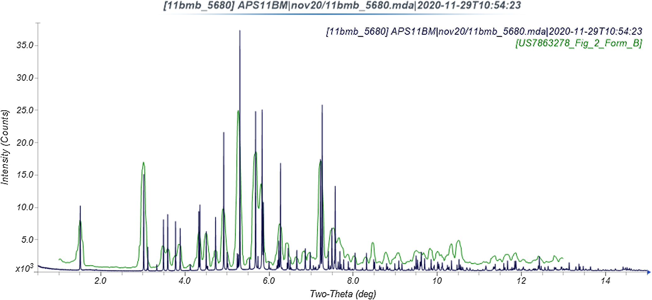
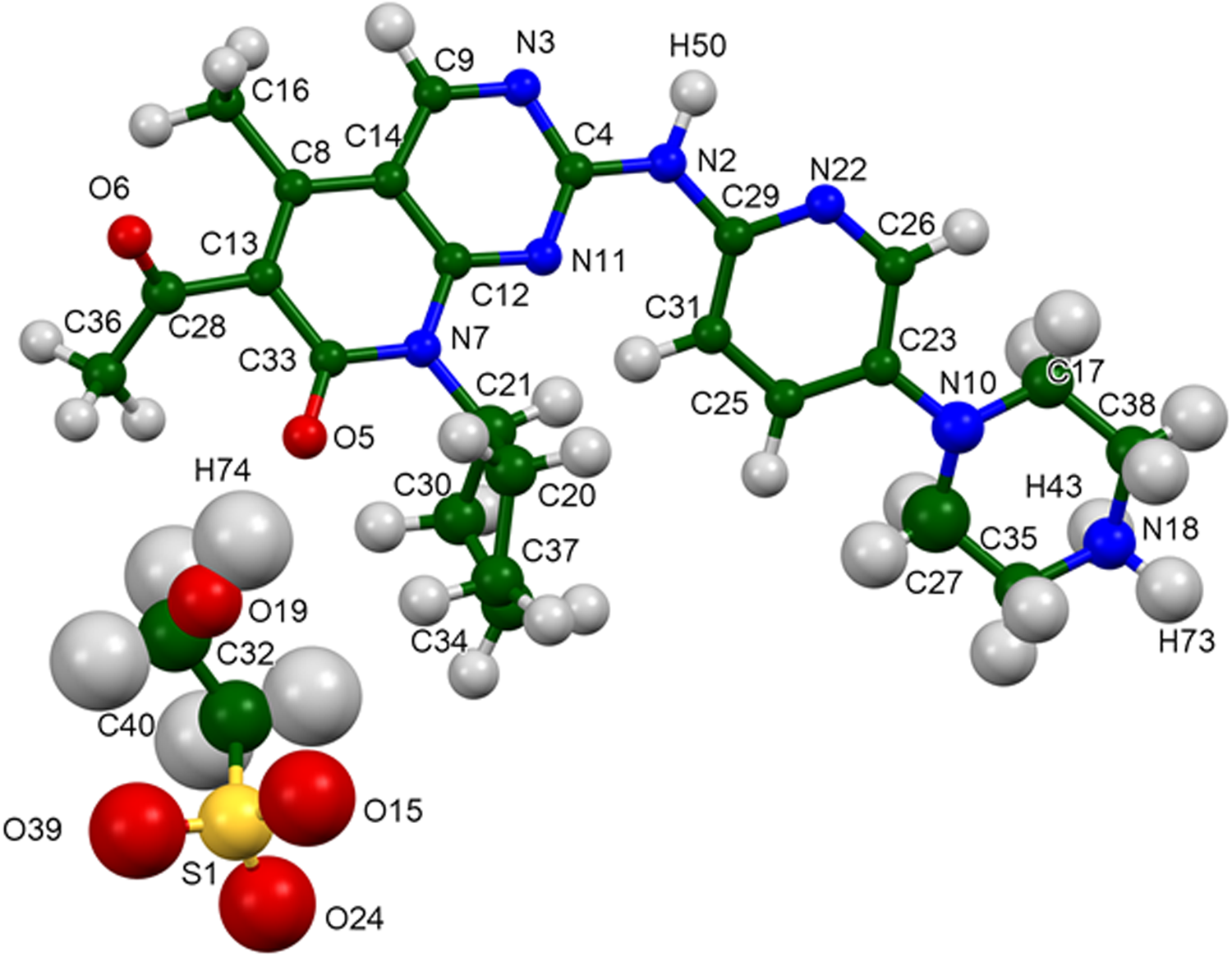
This synchrotron powder pattern is similar enough to that reported for Warner–Lambert's Form B (Figure 3) to conclude that they represent the same material. Form B is the most stable form (Beylin et al., Reference Beylin, Blackburn, Erdman and Toogood2008, Reference Beylin, Blackburn, Erdman and Toogood2011), so encountering it in a commercial sample is sensible. The refined atom coordinates (both from the Rietveld refinement and the DFT optimization) are reported in the CIFs which have been deposited with ICDD. The asymmetric unit (with atom numbering) is illustrated in Figure 4, and the crystal structure is presented in Figure 5.
Comparison of the synchrotron pattern of palbociclib isethionate to the pattern of Form B reported by Beylin et al. (Reference Beylin, Blackburn, Erdman and Toogood2011). The published pattern was digitized using UN-SCAN-IT (Silk Scientific, 2013) and scaled to the synchrotron wavelength of 0.457912 Å using MDI JADE Pro (MDI, 2019).

The asymmetric unit of palbociclib isethionate Form B, with the atom numbering. The atoms are represented by 50% probability spheroids.

The crystal structure of palbociclib isethionate Form B, viewed down the a-axis.

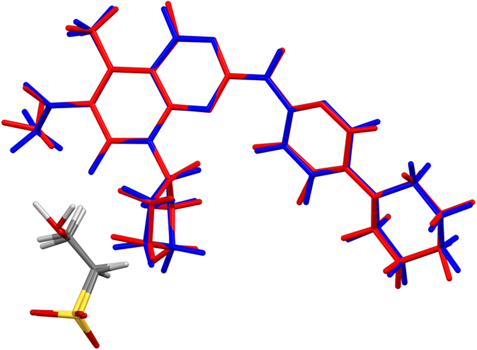
The root-mean-square Cartesian displacement of the non-hydrogen atoms in the Rietveld-refined and CRYSTAL14-optimized structures of the cations is 0.097 Å (Figure 6). The similar displacement for the anions is 0.075 Å. The good agreement is evidence that the experimental structure is correct (van de Streek and Neumann, Reference van de Streek and Neumann2014). The rest of the discussion will emphasize the CRYSTAL14 structure. The crystal structure is dominated by cation⋯anion and cation⋯cation hydrogen bonds, which result in layers roughly parallel to the (104) plane. The outer surfaces of the layers are formed by the anions and hydrocarbon portions of the cations.
Comparison of the Rietveld-refined (red) and CRYSTAL14-optimized (blue) structures of palbociclib isethionate form B.

Almost all of the bond distances, bond angles, and torsion angles fall within the normal ranges indicated by a Mercury/Mogul geometry check (Macrae et al., Reference Macrae, Sovago, Cottrell, Galek, McCabe, Pidcock, Platings, Shields, Stevens, Towler and Wood2020). The C12–N7–C33 angle of 120.7° (average = 122.0(4), Z-score = 3.2) is flagged as unusual. The uncertainty on the average is exceptionally low, resulting in an inflated Z-score. The C32–C40–S1–O15 torsion angle in the anion is unusual, lying on the tail of a broader gauche distribution.
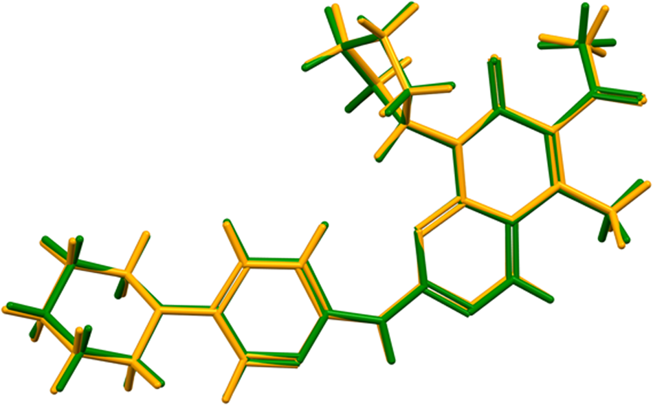
Quantum chemical geometry optimization of the palbociclib cation (DFT/B3LYP/6-31G*/water) using Spartan ‘18 (Wavefunction, 2020) indicated that the observed solid-state conformation is 6.9 kcal mol−1 higher in energy than the local minimum-energy conformation. The differences lie mainly in the conformation of the cyclopentane ring and the orientation of the pyridine ring. The local minimum conformation differs from the global minimum-energy conformation (Figure 7); the difference is mainly in the orientation of the methyl ketone group, and the rms Cartesian displacement is 0.472 Å. The differences become much smaller (0.105 Å) when the “flexibility” option of Mercury/Calculate/Molecule Overlay is invoked (Figure 8). This shows that intermolecular interactions are important in determining the observed solid-state conformation.
Comparison of the local minimum palbociclib conformation (green) to the minimum-energy conformation (orange).

Comparison of the local minimum palbociclib conformation (green) to the minimum-energy conformation (orange), after the “flexibility” option in mercury/calculate/molecule overlay is invoked.

Analysis of the contributions to the total crystal energy using the Forcite module of Materials Studio (Dassault, 2020) suggests that angle and torsion distortion terms dominate the intramolecular deformation energy. The intermolecular energy is dominated by electrostatic attractions, which, in this force-field-based analysis, include cation coordination and hydrogen bonds. The hydrogen bonds are better analyzed using the results of the DFT calculation.
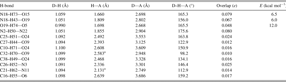
As expected, both H73 and H43 on the protonated nitrogen atom N18 participate in strong hydrogen bonds to the anion (Table I). H73 bonds to the sulfonate group, while H43 bonds to the hydroxyl group of the isethionate anion. The energies of these N–H⋯O hydrogen bonds were calculated using the correlation of Wheatley and Kaduk (Reference Wheatley and Kaduk2019). The hydroxyl group of the anion acts as a donor to the ketone oxygen atom O5 in the cation. The energy of this O–H⋯O hydrogen bond was calculated using the correlation of Rammohan and Kaduk (Reference Rammohan and Kaduk2018). There are also strong N2–H50⋯N22 hydrogen bonds, which occur in pairs linking the cations into dimers with rings having a graph set R2,2(8) (Etter, Reference Etter1990; Bernstein et al., Reference Bernstein, Davis, Shimoni and Chang1995; Shields et al., Reference Shields, Raithby, Allen and Motherwell2000). There are several C–H⋯O hydrogen bonds to O24 and O39 of the sulfonate group of the anion (one intramolecular), as well as one to the hydroxyl oxygen O19. Inter- and intramolecular C–H⋯O, C–H⋯N, and C–H⋯C hydrogen bonds contribute to the lattice energy.
Hydrogen bonds (CRYSTAL14) in form B of palbociclib isethionate.

a Intramolecular.
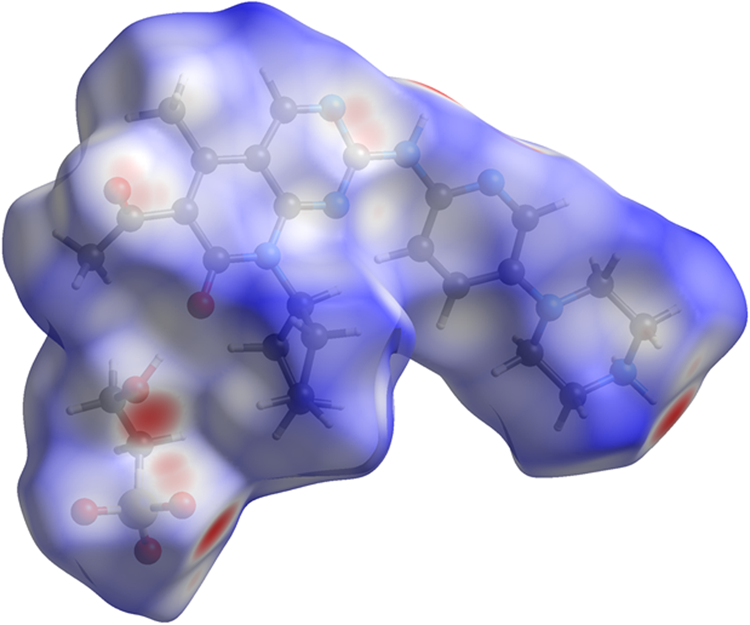
The volume enclosed by the Hirshfeld surface (Figure 9; Hirshfeld, Reference Hirshfeld1977; Turner et al., Reference Turner, McKinnon, Wolff, Grimwood, Spackman, Jayatilaka and Spackman2017) is 676.15 Å3, 98.61% of one-half the unit cell volume. The packing density is thus fairly typical. All of the significant close contacts (red in Figure 9) involve the hydrogen bonds. The volume/non-hydrogen atom is 16.7 Å3, lower than the usual 17.8(14) Å3 of our recent pharmaceutical structures. The smaller volume probably reflects the strong hydrogen bonds.
The Hirshfeld surface of palbociclib isethionate Form B. Intermolecular contacts longer than the sums of the van der Waals radii are colored blue, and contacts shorter than the sums of the radii are colored red. Contacts equal to the sums of radii are white.

The Bravais–Friedel–Donnay-Harker (Bravais, Reference Bravais1866; Friedel, Reference Friedel1907; Donnay and Harker, Reference Donnay and Harker1937) method suggests that we might expect platy morphology for palbociclib isethionate, with {001} as the principal faces. A second-order spherical harmonic model for preferred orientation was incorporated into the refinement. The texture index was 1.011(0), indicating that preferred orientation was slight in this rotated capillary specimen.
IV. DEPOSITED DATA
The powder pattern of palbociclib isethionate has been submitted to ICDD for inclusion in the Powder Diffraction File (PDF®). The Crystallographic Information Framework (CIF) files containing the results of the Rietveld refinement (including the raw data) and the DFT geometry optimization were deposited with the ICDD. The data can be requested at info@icdd.com.
ACKNOWLEDGEMENTS
We thank Lynn Ribaud and Saul Lapidus for their assistance in the data collection, and Andrey Rogachev for the use of computing resources at IIT.
FUNDING
Use of the Advanced Photon Source at Argonne National Laboratory was supported by the U.S. Department of Energy, Office of Science, Office of Basic Energy Sciences, under Contract No. DE-AC02-06CH11357. This work was partially supported by the International Centre for Diffraction Data.
CONFLICTS OF INTEREST
The authors have no conflicts of interest to declare.