Introduction
Studies on familial alcohol dependence (AD) have typically focused on the risk for, and manifestation of, AD in individuals. These studies show that a positive family history (FH) of AD (FH+) is associated with a higher risk for alcohol-use disorders (Hasin et al. Reference Hasin, Van Rossem, McCloud and Endicott1997; Nurnberger et al. Reference Nurnberger, Wiegand, Bucholz, O'Connor, Meyer, Reich, Rice, Schuckit, King, Petti, Bierut, Hinrichs, Kuperman, Hesselbrock and Porjesz2004), and, once developed, with a recurrent course and more impairments (Penick et al. Reference Penick, Powell, Bingham, Liskow, Miller and Read1987; Araujo & Monteiro, Reference Araujo and Monteiro1995; Milne et al. Reference Milne, Caspi, Harrington, Poulton, Rutter and Moffitt2009), remaining even after abstinence (Moriyama et al. Reference Moriyama, Muramatsu, Kato, Mimura and Kashima2006). In both AD patients and non-alcoholic individuals, a FH of AD may also contribute to a higher probability of psychiatric disorders other than AD (Araujo & Monteiro, Reference Araujo and Monteiro1995; Schuckit et al. Reference Schuckit, Hesselbrock, Tipp, Nurnberger, Anthenelli and Crowe1995; Dawson & Grant, Reference Dawson and Grant1998; Nurnberger et al. Reference Nurnberger, Wiegand, Bucholz, O'Connor, Meyer, Reich, Rice, Schuckit, King, Petti, Bierut, Hinrichs, Kuperman, Hesselbrock and Porjesz2004; Knappe et al. Reference Knappe, Lieb, Beesdo, Fehm, Low, Gloster and Wittchen2009; Sjoerds et al. Reference Sjoerds, Van Tol, Van den Brink, Van der Wee, Van Buchem, Aleman, Penninx and Veltman2012), such as mood and/or anxiety disorders. This implies a role of familial AD in the development of mood and anxiety disorders. However, little is known about the influence of familial AD on the neurophysiology of manifested mood and anxiety disorders.
Cognitive impairments often seen in alcoholic as well as non-alcoholic individuals with a FH of AD are disinhibition and poor behavioural control (Peterson et al. Reference Peterson, Finn and Pihl1992; Poon et al. Reference Poon, Ellis, Fitzgerald and Zucker2000). Brain areas involved in these cognitive control functions, including dorsal prefrontal areas and the cingulate gyrus, show abnormal activity in FH+ individuals during various neuropsychological tasks (Schweinsburg et al. Reference Schweinsburg, Paulus, Barlett, Killeen, Caldwell, Pulido, Brown and Tapert2004; Spadoni et al. Reference Spadoni, Norman, Schweinsburg and Tapert2008; Silveri et al. Reference Silveri, Rogowska, McCaffrey and Yurgelun-Todd2011) and decreased functional connectivity with other brain regions (Wetherill et al. Reference Wetherill, Bava, Thompson, Boucquey, Pulido, Yang and Tapert2011). Notably, these areas are also implicated in neurobiological models of emotion processing as part of a dorsal system, involved in top-down control and regulation of affective states and subsequent behaviours (Phillips et al. Reference Phillips, Drevets, Rauch and Lane2003a; Drevets et al. Reference Drevets, Price and Furey2008). Additionally, some studies have shown that the presence of familial AD is associated with abnormal emotion processing (Glahn et al. Reference Glahn, Lovallo and Fox2007; Hill et al. Reference Hill, Kostelnik, Holmes, Goradia, McDermott, Diwadkar and Keshavan2007; Bjork et al. Reference Bjork, Knutson and Hommer2008; Acheson et al. Reference Acheson, Robinson, Glahn, Lovallo and Fox2009). Para-limbic areas such as the insula, amygdala, and ventral prefrontal cortex, which are involved in emotional appraisal and identification of emotional significance of a stimulus (Phillips et al. Reference Phillips, Drevets, Rauch and Lane2003a), show structural and functional abnormalities in FH+ individuals (Hill et al. Reference Hill, De Bellis, Keshavan, Lowers, Shen, Hall and Pitts2001, Reference Hill, Wang, Kostelnik, Carter, Holmes, McDermott, Zezza, Stiffler and Keshavan2009; Bjork et al. Reference Bjork, Knutson and Hommer2008; Heitzeg et al. Reference Heitzeg, Nigg, Yau, Zubieta and Zucker2008). Importantly, impairments in these areas are also associated with the development of mood problems and negative affect (Tessner & Hill, Reference Tessner and Hill2010). Together with decreased cognitive control functions these limbic abnormalities may underlie an unfavourable symptom profile in patients with mood/anxiety disorders who have a FH of AD compared with those without such a FH (FH–).
Recently, evidence for an effect of a FH of AD on the neural substrate of mood/anxiety disorders was provided, i.e. depressed patients with a FH of AD demonstrated a significantly better anti-depressant response to the N-methyl-d-aspartate (NMDA)-receptor antagonist ketamine than patients without a FH of AD (Phelps et al. Reference Phelps, Brutsche, Moral, Luckenbaugh, Manji and Zarate2009). These findings indicate that depressed (and probably also anxious) patients may have a different neurobiological profile depending on the presence of a FH of AD, including differences in neurotransmitter and receptor profile, potentially leading to divergent performance during cognitive or emotional tasks. This experimental evidence suggests heterogeneity between depression/anxiety patients based on the presence or absence of a FH of AD and is consistent with the high variability in imaging findings in these populations (Fitzgerald et al. Reference Fitzgerald, Laird, Maller and Daskalakis2008), especially concerning cognitive functions. Therefore, it is desirable to further study the influence of familial AD on the neurophysiological profile in patients with mood/anxiety disorders to clarify the pathway through which a FH of AD predisposes to the development of mood and anxiety disorders.
The current study is the first to examine whether a FH of AD influences the functional neurobiology of mood/anxiety disorders, and whether this influence operates through pathways involved in cognitive or emotion processing, or both. In a sample of non-alcoholic adult patients with mood/anxiety disorders, functional magnetic resonance imaging (fMRI) was used to investigate regional brain activity during a cognitive planning task and an emotional appraisal task. We compared patients with and without a first-degree FH of AD. We hypothesized that the presence of a FH of AD affects cognitive control functions and modulates responsiveness to emotional stimuli, indicated by differences between the FH− and FH+ groups in performance levels and activation in brain areas associated with these functions. To facilitate the interpretation of our findings, differences in performance levels and brain activation between the FH− and the FH+ groups were post hoc compared with those of healthy controls (HCs) without a FH of AD.
Method
Sample
Participants were drawn from the observational multi-center Netherlands Study of Depression and Anxiety (NESDA). Within NESDA (n = 2981), a large neuroimaging study was embedded (n = 301), where 233 out-patients with a current (<6 months) Diagnostic and Statistical Manual of Mental Disorders, fourth edition, text revision (DSM-IV-TR) diagnosis of major depressive disorder (MDD) and/or anxiety disorder (panic disorder, social anxiety disorder, generalized anxiety disorder), and 68 HCs without any lifetime DSM-IV-TR diagnosis were studied using fMRI. Detailed descriptions of the NESDA study design and the NESDA neuroimaging study have been published elsewhere (Penninx et al. Reference Penninx, Beekman, Smit, Zitman, Nolen, Spinhoven, Cuijpers, De Jong, Van Marwijk, Assendelft, Van Der Meer, Verhaak, Wensing, De Graaf, Hoogendijk, Ormel and Van Dyck2008; Van Tol et al. Reference Van Tol, Van der Wee, Van den Heuvel, Nielen, Demenescu, Aleman, Renken, Van Buchem, Zitman and Veltman2010). Inclusion and exclusion criteria of the NESDA neuroimaging sample are described in the Supplementary material (Text S1).
For this report, we selected participants with a current diagnosis of MDD and/or anxiety disorder based on the Composite International Diagnostic Interview (Robins et al. Reference Robins, Wing, Wittchen, Helzer, Babor, Burke, Farmer, Jablenski, Pickens, Regier, Sartorius and Towle1988) who reported to have (a) at least one first-degree relative (parents/siblings) with a history of AD (FH+) or (b) no relatives at all with a history of AD or other substance-use disorder (FH–). We excluded (a) participants with imaging data of poor quality; (b) participants who lacked data on either of the two task paradigms, or who showed poor task involvement by having less than an overall performance of 75% for the planning task and/or over 40 missing responses for the emotional appraisal task; and (c) participants who had a current (<1 year) and/or lifetime (>1 year) diagnosis of alcohol abuse or dependence. Of the 233 patients, 77 met criteria for the FH− group and 31 for the FH+ group. Of the 68 HCs, 31 served as a comparison group without a FH of AD. More information on inclusion/exclusion steps can be found in the flowchart in Supplementary Fig. S3.
The research protocol was approved by the medical ethical review boards of the participating universities and all participants provided written informed consent.
Clinical assessment
FH was obtained using the family-tree method (Fyer & Weissman, Reference Fyer and Weissman1999), an interview assessing the presence of psychiatric disorders, including AD, among relatives of the participant with acceptable inter-rater reliability (Andreasen et al. Reference Andreasen, Endicott, Spitzer and Winokur1977). The Alcohol Use Disorder Identification Test (AUDIT; Babor et al. Reference Babor, Kranzler and Lauerman1989) for alcohol use and dependence, and the Fagerström questionnaire (Heatherton et al. Reference Heatherton, Kozlowski, Frecker and Fagerstrom1991) for the assessment of smoking behaviour and nicotine dependence were used to provide a more detailed description of alcohol and nicotine use and related problems. On the scanning day, depression severity and mood state were assessed using Dutch versions of the Inventory of Depressive Symptomatology (IDS; Rush et al. Reference Rush, Gullion, Basco, Jarrett and Trivedi1996) and the Montgomery–Åsberg Depression Rating Scale (MADRS; Montgomery & Asberg, Reference Montgomery and Asberg1979), whereas anxiety severity was measured with the Beck Anxiety Inventory (BAI; Beck et al. Reference Beck, Epstein, Brown and Steer1988).
Task paradigms
To assess executive functions, we used a parametric visuospatial planning task (Tower of London; ToL; Van den Heuvel et al. Reference Van den Heuvel, Groenewegen, Barkhof, Lazeron, Van Dyck and Veltman2003). This task involves recruitment of the dorsolateral prefrontal cortex (DLPFC) and anterior cingulate cortex (ACC), brain areas involved in cognitive control/planning and regulation of emotion processing (Phillips et al. Reference Phillips, Drevets, Rauch and Lane2003a). The ToL task has previously been associated with functional impairments in FH+ compared with FH− individuals (Dolan et al. Reference Dolan, Bechara and Nathan2008). An emotional word memory (EWM) task (Daselaar et al. Reference Daselaar, Veltman, Rombouts, Raaijmakers and Jonker2003) was used to study emotional responsiveness during encoding of mood-congruent (negative), mood-incongruent (positive) and neutral words. The EWM task has been associated with activation of (para)limbic structures involved in primary appraisal of emotional significance (Phillips et al. Reference Phillips, Drevets, Rauch and Lane2003a). A detailed description of the two tasks can be found in the Supplementary material (Text S2).
Briefly, during the ToL task, participants were shown a starting configuration of beads and were asked to compute the minimal number of steps (ranging from 1 to 5) to reach a target configuration (Supplementary Fig. S4). The EWM task consisted of an encoding phase, during which participants had to indicate whether the shown words had a positive, neutral or negative valence, and a recognition phase in which participants had to indicate whether they had seen the words during the encoding phase among novel distracter words (Supplementary Fig. S5). For the current analysis we focused on performance and brain activity during words that were successfully encoded (hence, subsequently remembered during the recognition phase) in each condition (positive/negative/neutral). During baseline trials, participants were requested to perform button presses as indicated (‘≪left’, ‘≪middle≫‘, ‘right≫‘).
Image acquisition and processing
Imaging data were acquired using Philips 3-T MR-systems (Best, The Netherlands) located at the Academic Medical Center Amsterdam (AMC), the Leiden University Medical Center (LUMC) and the University Medical Center Groningen (UMCG). A SENSE-8 (UMCG, LUMC) and a SENSE-6 (AMC) channel head coil were used for radio frequency transmission and reception. For each subject, echo-planar images were obtained using a T2*-weighted gradient echo sequence [repetition time (TR) = 2300 ms; echo time (TE) = 30 ms (UMCG: 28 ms); matrix size: 96 × 96 (UMCG: 64 × 64); 35 axial slices (UMCG: 39); interleaved acquisition; 2.29 × 2.29 mm in-plane resolution (UMCG: 3 × 3 mm); 3 mm slice thickness]. Anatomical imaging was performed between the encoding and recognition phases of the EWM task, and included a sagittal three-dimensional gradient-echo T1-weighted sequence (TR = 9 ms; TE = 3.5 ms; matrix 256 × 256; voxel size: 1 × 1×1 mm; 170 slices) for co-registration with the functional data.
Images were pre-processed with SPM5 [Wellcome Trust Centre for Neuroimaging, London (http://www.fil.ion.ucl.ac.uk/spm/software/)] implemented in Matlab 7.3.0 (The Mathworks Inc., USA). Before pre-processing, images were manually reoriented to the anterior commissure. Temporal and spatial preprocessing of the data included slicetime correction, image realignment, co-registration between the anatomical and mean echo planar imaging (EPI) images, warping to the standard Montreal Neurological Institute (MNI) space as defined by the SPM5 T1 template, re-sampling into a 3 × 3×3 mm grid, and spatial smoothing using a Gaussian kernel (8 mm full-width at half-maximum). Data of participants with movement greater than 3 mm in more than one direction were excluded. To remove low-frequency temporal noise, a high-pass filter was applied, with a cut-off of 128 s, to the fMRI time-series.
Statistical analyses
Sample characteristics and task performance
Sample characteristics and performance data were analysed using SPSS (SPSS Inc., USA). Normally distributed characteristics and clinical data were analysed using independent-samples t tests. If data did not meet assumptions required for parametric analysis, log transformations were made; if following log transformation a variable was still not normally distributed, the non-parametric Mann–Whitney U test for two independent samples was used. Categorical data were analysed using χ2 tests.
Performance data on both fMRI tasks (accuracy and response times) were examined by means of a repeated-measures analysis of covariance. Dependent factors for the ToL task were the proportion correct scores and mean response times per trial type (1–5 steps). For the EWM task the dependent factors were the proportion of subsequently remembered words and mean response time during successful encoding per valence category (positive/negative/neutral). For both tasks the between-subject factor was group (FH–/FH+), whereas age, education and current diagnosis (MDD only, anxiety only, or co-morbid MDD/anxiety) were added as covariates. Significance was set at p < 0.05.
Imaging data
Analysis of imaging data was performed in the context of the general linear model (Friston et al. Reference Friston, Holmes, Poline, Grasby, Williams, Frackowiak and Turner1995) in SPM8. Onsets of each stimulus type were modelled with a delta function convolved with a synthetic haemodynamic response function and modulated using response times. For the ToL task, contrast images containing parameter estimates for task load (steps 1–5) were calculated per participant on a voxel-by-voxel basis. These were entered into second-level analyses for group comparisons using a whole-brain two-sample t test. For the EWM task, successfully encoded items (i.e. items during the encoding phase that were correctly recognized during the retrieval phase) were contrasted with baseline items at first level, and entered in a 2 (group: FH–/FH+) × 3 (valence: positive/negative/neutral) full factorial second-level model to test for the main effect of task. Group interactions were assessed by valence in three separate two-sample t tests.
All analyses were performed with age and scanning location as covariates, in line with previous imaging studies on the ToL and EWM within the NESDA cohort (Van Tol et al. Reference Van Tol, Van der Wee, Demenescu, Nielen, Aleman, Renken, Van Buchem, Zitman and Veltman2011, Reference Van Tol, Demenescu, Van der Wee, Kortekaas, Nielen, Den Boer, Renken, van Buchem, Zitman, Aleman and Veltman2012). To capitalize on our sample size we included participants using selective serotonin reuptake inhibitor (SSRI) medication, since previous findings from our group demonstrated that SSRI use did not affect imaging results of these tasks (Van Tol et al. Reference Van Tol, Van der Wee, Demenescu, Nielen, Aleman, Renken, Van Buchem, Zitman and Veltman2011, Reference Van Tol, Demenescu, Van der Wee, Kortekaas, Nielen, Den Boer, Renken, van Buchem, Zitman, Aleman and Veltman2012). However, we performed post hoc analyses with SSRI use as an additional covariate. In order to study possible effects of state-dependent severity of depression and anxiety, we additionally added BAI and IDS scores as covariates to the main analyses.
Main task effects over groups are reported in Supplementary Table S7 at a threshold of p < 0.001 uncorrected (p uncorr). To restrict the search for interaction effects to voxels which were identified in the main task effects, t test group comparisons were masked with the orthogonal main task effect over all groups at p uncorr <0.001. Group × task interaction effects were visually inspected at a threshold of p uncorr <0.001 and cluster size >3 voxels for effects in a priori regions of interest (ROIs) as defined by the Automated Anatomical Labeling Atlas (Maldjian et al. Reference Maldjian, Laurienti, Kraft and Burdette2003), implemented in the WFU Pickatlas toolbox (Wake Forest University School of Medicine). For the ToL task the ROIs comprised the DLPFC and ACC. For the EWM task the ROIs were defined as the hippocampus, amygdala, insula, inferior frontal gyrus and medial prefrontal gyrus. Following previous findings of smaller grey matter volumes in patients with an alcoholic FH in non-alcoholic adults (Sjoerds et al. Reference Sjoerds, Van Tol, Van den Brink, Van der Wee, Van Buchem, Aleman, Penninx and Veltman2012), we additionally designated the (right) parahippocampal gyrus as an area of interest for both fMRI tasks, applying the small volume correction implemented in SPM with a sphere twice the size of the smoothing kernel centred over the peak coordinates of the previously reported finding (MNI: x = 20, y = –37, z = –11).
Post hoc comparison with HCs
In order to test and interpret how effects of FH in patients with mood/anxiety disorders are related to normal regional brain function, we post hoc compared the observed differences between the FH+ and FH− groups with findings from a group of HCs without a FH of AD. We therefore added the HC group as a third group to post hoc analysis of variance (ANOVA) for the parametric contrast of the ToL task and per valence for the EWM task. Comparable secondary analyses were done for the comparison of baseline characteristics and performance data, adding HCs as a third group in the repeated-measures ANOVAs.
Results
Sample characteristics of FH+ and FH− patients
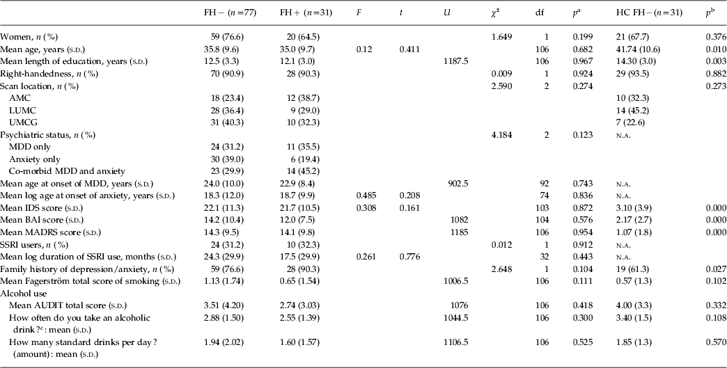
Sample characteristics are listed in Table 1. The two patient groups did not differ significantly (p > 0.05) on sociodemographic variables, SSRI use, depression and anxiety scores, alcohol use and smoking. Distribution of current MDD only, current anxiety only and co-morbid MDD and anxiety was the same for both groups, as well as age of onset for the affective disorders. The groups only differed on the presence of alcoholic FH, and not on the presence of familial depression/anxiety.
Sample characteristics

FH − , No relatives at all with a history of alcohol dependence or other substance-use disorder; FH + , positive family history of alcohol dependence; df, degrees of freedom; HC, healthy controls; s.d., standard deviation; AMC, Academic Medical Center; LUMC, Leiden University Medical Center; UMCG, University Medical Center Groningen; n.a., not applicable; MDD, major depressive disorder; log, logarithm transformation; IDS, Inventory of Depressive Symptomatology; BAI, Beck Anxiety Inventory; MADRS, Montgomery–Åsberg Depression Rating Scale; SSRI, selective serotonin reuptake inhibitor; AUDIT, Alcohol Use Disorder Identification Test.
a Significance between FH− and FH+ patient groups.
b Significance on the three-group analysis of variance between the FH− and FH+ patient groups and the HC group.
c 1 = Never; 2 = monthly or less; 3 = 2–4 times a month; 4 = 2–3 times a week; 5 = 4 or more times a week.
Performance and fMRI results of FH+ and FH− patients
ToL task
Performance data
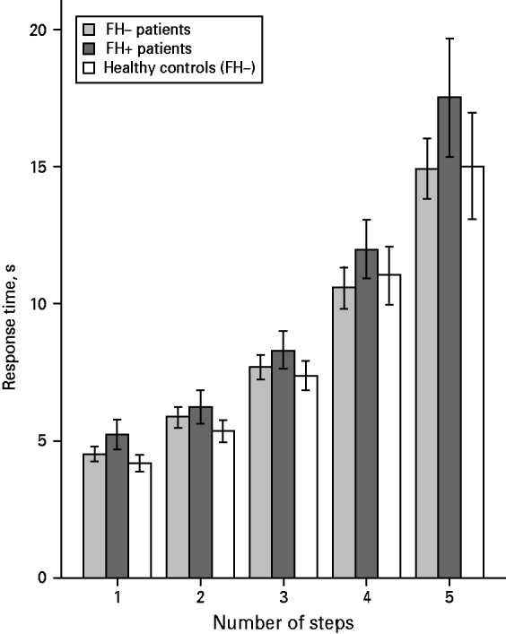
Overall, no effect of FH of AD was observed on planning accuracy (F 2.4,246.99 = 0.46, p = 0.67). However, a significant main group effect on response times was found (F 1,103 = 6.73, p = 0.01), and the group × task load interaction was significant due to slower responding by the FH+ group during the more difficult trials (4 and 5 steps; Fig. 1) (F 1.59,163.45 = 3.62, p = 0.04). For raw performance data, see Supplementary Table S6.
Response times during the visuospatial planning task (Tower of London) by the three groups: family history-negative (FH–) patients, family history-positive (FH+) patients and family history-negative healthy controls. Values on the y axis are mean response times with 95% confidence intervals represented by error bars.
Imaging data
FH+ patients showed significantly more activation in the right superior [Brodmann's area (BA) 6; Z = 3.21] and right middle (BA 46; Z = 3.54) frontal gyrus, compared with FH− patients (Fig. 2 a). At a slightly more liberal threshold (p uncorr <0.005), the ACC (BA 24; Z = 2.81) showed increased activation in FH+ patients, an area of a priori interest. Results were unaffected when variations of depression/anxiety severity and SSRI use were taken into account. No significant clusters were visible in the FH– > FH+ contrast, even at a threshold of p < 0.005 whole-brain uncorrected. The main task effect is displayed in Supplementary Table S7.
(a) Superior and middle frontal gyrus activation in family history-positive patients (FH+) compared with family history-negative patients (FH–) on the parametric contrast of the Tower of London task showing plots with the three groups activating on the peak voxel in the (I) superior frontal gyrus and (II) middle frontal gyrus. (b) Right insula activation in FH+ compared with FH− during positive encoding on the Emotional Word Memory task showing a plot (III) with the three groups activating on the peak voxel in the right insula. Both brain figures are presented at p < 0.005, masked by main task effect. Coloured bars in the left panels represent Z scores. Values on the y axis of the plots are parameter estimates with 95% confidence intervals represented by error bars. Coordinates given on the y axes are in Montreal Neurological Institute (MNI) space.
EWM task
Performance data
The two groups did not differ in classification behaviour (F 1.17,114.71 = 0.046, p = 0.96) (see Supplementary Table S6). A main effect of valence (positive/negative/neutral) on response times was observed (F 2,208 = 3.83, p = 0.02). However, no main effect of FH + /FH− group and no interaction of valence × group was seen on performance accuracy (F 1,104 = 0.59, p = 0.44 and F 1.74,181.33 = 0.52, p = 0.57, respectively) or mean response times (F 1,104 = 0.04, p = 0.84 and F 2,208 = 0.26, p = 0.77, respectively).
Imaging data
During positive word encoding the FH+ group activated the right insula (BA 13; Z = 4.22) significantly more than the FH− group (Fig. 2 b). The opposite contrast, testing for increased brain activity in FH− compared with FH+ patients, did not show significant results. Activation during negative and neutral word encoding did not reveal group differences. Adding SSRI use and depression/anxiety severity to the main analysis did not influence the results. Main task effects are displayed in Supplementary Table S7.
Comparison with HCs
The HC group was significantly older and higher educated than the two patient groups (FH− and FH+). HCs scored, as expected, lower on depression and anxiety symptom severity questionnaires. Additionally, the HC group contained relatively few participants with a FH of depression/anxiety.
The patient groups did not differ from HCs in performance accuracy for both tasks. However, HCs showed trendwise shorter response times compared with the FH+ group on the ToL (F 3.861,38.989 = 2.003, p = 0.094; Fig. 1).
During the ToL task, brain activation in the superior and middle frontal gyrus (FH + >FH–) in the FH+ group was significantly higher compared with the HC group, an effect that was particularly robust in the middle frontal gyrus. During the EWM task, activation in the right insula in the FH+ group was comparable with that of HCs during positive word encoding. Plots of the three groups in the described areas are shown in Fig. 2 (right panels).
Discussion
In the present study, we investigated the effects of a FH of AD on neural correlates of cognitive functions and emotional perception in patients with mood/anxiety disorders. Although extensive studies are available on the association between FH of AD and manifestation of AD, the current study suggests that a FH of AD also affects the underlying neurophysiology of non-alcoholic patients with mood/anxiety disorders. To our knowledge, this is the first study examining the neurophysiological correlates of a FH of AD in patients with mood/anxiety disorders. Results indicate that the presence of a FH of AD has a negative influence on cognitive functions during a visuospatial planning task and a normalizing effect during positive word encoding in these patients. These results could not be explained by differences in depression and anxiety severity or medication use and therefore confirm our hypothesis that a FH of AD independently affects cognitive functions and modulates emotion processing in patients with mood/anxiety disorders.
In patients with a FH of AD, increased response times and greater regional dorsolateral and trendwise dorsomedial brain activity during planning coupled with equal performance accuracy suggest the need to recruit additional resources to attain the same level of accuracy (i.e. reduced neuronal efficiency), as compared with patients without such a history and HCs. Reduced neuronal efficiency of frontal areas during cognitively challenging tasks was reported earlier in FH+ youth (Silveri et al. Reference Silveri, Rogowska, McCaffrey and Yurgelun-Todd2011), heavy cannabis users (Kanayama et al. Reference Kanayama, Rogowska, Pope, Gruber and Yurgelun-Todd2004) and after sleep deprivation (Drummond et al. Reference Drummond, Gillin and Brown2001, Reference Drummond, Meloy, Yanagi, Orff and Brown2005). As the DLPFC and ACC are part of a network involved in top-down emotional regulation (Phillips et al. Reference Phillips, Drevets, Rauch and Lane2003a; Drevets et al. Reference Drevets, Price and Furey2008), impairments in these regions could lead to a decreased capacity for emotion regulation, further contributing to an unfavourable course of mood/anxiety disorders.
During positive word encoding we observed that a FH of AD was associated with increased recruitment of the right posterior insula compared with FH–, in the absence of behavioural differences. This activation in FH+ patients, however, was comparable with that in the HC group, indicating that a hypoactivation, as was seen in FH− patients, was absent in the presence of a FH of AD. Hypoactivation during positive word encoding was recently reported in the hippocampus in depressed and anxious patients, independent of illness severity (Van Tol et al. Reference Van Tol, Demenescu, Van der Wee, Kortekaas, Nielen, Den Boer, Renken, van Buchem, Zitman, Aleman and Veltman2012). We did not find FH group differences in the hippocampus, but rather in the more posterior part of the right insula, which is, in connection with the anterior insula, involved in bottom-up detection of and autonomic reactivity towards salient stimuli (Craig, Reference Craig2003; Menon & Uddin, Reference Menon and Uddin2010). Hypoactivation in the insula (as seen in FH− patients) may be related to a blunted response to positive stimuli, which would be in concordance with the mood-incongruence bias hypothesis. Mood-incongruent bias, i.e. attentional and memory bias away from positive information (Burt et al. Reference Burt, Zembar and Niederehe1995; Van Wingen et al. Reference Van Wingen, Rijpkema, Franke, Van Eijndhoven, Tendolkar, Verkes, Buitelaar and Fernandez2010; Van Tol et al. Reference Van Tol, Demenescu, Van der Wee, Kortekaas, Nielen, Den Boer, Renken, van Buchem, Zitman, Aleman and Veltman2012) is associated with the decreased ability to enjoy positive situations, possibly reflective of anhedonia (Bower, Reference Bower1981). On the other hand, mood-congruent bias, i.e. attentional and memory bias towards negative information (Bradley et al. Reference Bradley, Mogg and Millar1996; Elliott et al. Reference Elliott, Rubinsztein, Sahakian and Dolan2002; Phillips et al. Reference Phillips, Drevets, Rauch and Lane2003b), contributes to the development and persistence of negative affective symptoms (Elliott et al. Reference Elliott, Rubinsztein, Sahakian and Dolan2002). We did not observe a decreased insula response to positive stimuli in FH+ patients, suggesting a different pathway through which depression and anxiety symptoms are maintained in patients with depressive/anxiety disorders with a FH of AD, for example through decreased cognitive functions, as shown above with respect to the ToL task, contributing to a decreased ability to regulate negative affect. These differences in the neurophysiology of depressed/anxious patients with and without a FH of AD could also be related to differences in receptor profiles, as suggested by treatment studies (Phelps et al. Reference Phelps, Brutsche, Moral, Luckenbaugh, Manji and Zarate2009).
The current analyses were controlled for severity of current depression/anxiety and the observed results therefore cannot be attributed to differences in state depression/anxiety between the FH+ and FH− groups. This implies that the observed effects of a FH of AD may have been present prior to the onset of the depression/anxiety either through environmental, genetic or epigenetic pathways. Including a fourth group of HCs with a FH of AD could have allowed us to further address this issue; however, within the NESDA neuroimaging sample this subgroup was too small (n = 6) for meaningful analyses. Notably, a previous fMRI study from the NESDA neuroimaging sample (Van Tol et al. Reference Van Tol, Van der Wee, Demenescu, Nielen, Aleman, Renken, Van Buchem, Zitman and Veltman2011) found that patients with more severe depression showed increased prefrontal activation during visuospatial planning, a similar blood oxygen level-dependent (BOLD) effect as found within our FH+ group. Comparing these results with ours thus suggests that patients with depression/anxiety constitute a heterogeneous group, which can partly be explained by the presence or absence of family histories such as AD. However, direct comparisons between these two studies are not straightforward, because in the present study we focused on the effect of FH of AD on associated psychopathology, and not on current diagnosis per se. Therefore, we pooled a selected sample of depressed/anxious patients, since FH of AD predisposes to both depression and anxiety disorders. Moreover, splitting FH groups based on current diagnosis would result in groups too small for valid analysis. Finally, such a strategy would only account for the current status of psychopathology, whereas a substantial subgroup of the patients with current ‘pure’ depression or anxiety had met diagnostic criteria for the other psychopathology type at some point during their lifetime. Separating patient groups based on current psychopathology would therefore still give relatively heterogeneous groups.
According to the mean scores on severity questionnaires like the MADRS and IDS, severity of current psychopathology was mild to moderate only, so that a potential dose–response association may have been missed.
Results were unaffected by SSRI use, confirming earlier reports within the NESDA neuroimaging study that also failed to observe effects of SSRIs on fMRI parameters (Van Tol et al. Reference Van Tol, Van der Wee, Demenescu, Nielen, Aleman, Renken, Van Buchem, Zitman and Veltman2011, Reference Van Tol, Demenescu, Van der Wee, Kortekaas, Nielen, Den Boer, Renken, van Buchem, Zitman, Aleman and Veltman2012). It should, however, be noted that the current findings are based on cross-sectional analyses; thus the possibility of within-subject changes in BOLD responses as an effect of SSRI use cannot be fully excluded.
Another potential limitation is the indirect assessment of familial AD, since NESDA did not interview first-degree relatives. Using the family-tree method, relying on participants' self-reported data of FH is likely to lead to under-reporting of familial AD. However, this would, if anything, have resulted in an underestimation of the true effects of familial AD.
Importantly, none of the participants in the current study had a lifetime diagnosis of AD and the groups did not differ in alcohol use, or the presence of alcohol-use symptoms according to the AUDIT, ruling out the possibility that observed effects are due to the neurotoxic effects of alcohol use.
Together, the results of this study demonstrate that a FH of AD influences the neurophysiological profile of mood/anxiety disorders through adverse effects on cognitive functions, possibly decreasing the ability to regulate emotional states and increasing the risk of sustained negative symptoms seen in mood/anxiety disorders. On the other side, the presence of a FH of AD may have a protective effect on decreased positive emotional appraisal in patients with mood/anxiety disorders. The current neurophysiological findings may be associated with previous findings that a FH of AD is associated with less dysphoric mood induction in HCs after ketamine administration (Petrakis et al. Reference Petrakis, Limoncelli, Gueorguieva, Jatlow, Boutros, Trevisan, Gelernter and Krystal2004) and with better treatment effects in treatment-resistant patients with a major depression (Phelps et al. Reference Phelps, Brutsche, Moral, Luckenbaugh, Manji and Zarate2009). Therefore, the results of the present study may contribute to a better characterization of the heterogeneity within the group of patients with a mood/anxiety disorder (Wardenaar et al. Reference Wardenaar, Giltay, Van Veen, Zitman and Penninx2012), to a better understanding of heterogeneous reactions of patients with mood/anxiety disorders to pharmacological interventions (Phelps et al. Reference Phelps, Brutsche, Moral, Luckenbaugh, Manji and Zarate2009), to an effective profiling of patients, and finally to the development of personalized medicine in the treatment of mood and anxiety disorders (Bartova et al. Reference Bartova, Berger and Pezawas2010; Uher, Reference Uher2011).
Supplementary material
For supplementary material accompanying this paper visit http://dx.doi.org/10.1017/S003329171200222X.
Acknowledgements
The infrastructure for the NESDA study (www.nesda.nl) is funded through the Geestkracht program of the Netherlands Organisation for Health Research and Development (ZonMw, grant no. 10-000-1002) and is supported by participating universities and mental health care organizations (VU University Medical Center, GGZ inGeest, Arkin, Leiden University Medical Center, GGZ Rivierduinen, University Medical Center Groningen, Lentis, GGZ Friesland, GGZ Drenthe, Scientific Institute for Quality of Healthcare (IQ healthcare), Netherlands Institute for Health Services Research (NIVEL) and Netherlands Institute of Mental Health and Addiction (Trimbos Institute). Data analyses were supported by grant no. 31160004 from the Netherlands Organization for Health Research and Development. The Netherlands Organization for Health Research and Development had no further role in study design, in the collection, analysis and interpretation of data, in the writing of the report, and in the decision to submit the paper for publication.
Declaration of Interest
A.T.F.B. has received grants for research from Eli Lilly, Astra Zeneca, Janssen and Shire and as a speaker from Lundbeck and Eli Lilly.