Introduction
With the continuous progress of industrialization and increasing demands of new materials [Reference Ma, Zhu, Wang, Yang, Song and Huang1, Reference Ma, Li, Wu, Song, Li, Han, Wang and Huang2, Reference Ma, Zhu, Feng, Song, Huang, Liu, Zhang, Fan, Hou and Guo3, Reference Zhu, Cui, Zhang, Chen, Dong, Liu, Shao, Ding, Wu and Guo4, Reference Yang, Yang, Wang, Dong, Liu, Wujcik, Shao, Wu, Ding and Guo5, Reference Lin, Lin, Wang, Chen, Wang, Dong, Gao, Shao, Ding, Liu, Wu and Guo6, Reference Gu, Xu, Dong, Xie, Shao, Fan, Liu, Wu, Wei and Guo7, Reference Shi, Xu, Deng, Dong, Murugadoss, Liu, Shao, Wu and Guo8, Reference Xu, Shi, Zhao, Deng, Dong, Liu, Murugadoss, Mai and Guo9, Reference Shi, Jia, Wang, Deng, Xu, Wu, Dong and Guo10, Reference Zhao, Bai, Misra, Dong, Guan, Li, Zhang, Tan, Gao, Ding, Du and Guo11, Reference Gong, Liu, Wang, Xie, Dong, Dong, Liu, Shao, Lu, Murugadoss, Ding and Guo12, Reference Chen, Yin, Dong, Gao, Qian, Wang, Dong, Shao, Liu, Han, Ding, Guo and Wang13] and energy [Reference Luo, Pei, Wang, Qian, Miao, Pan, Chen and Feng14, Reference Liu, Shi, Yan, Zhuo, Wu, Wang, Liu and Guo15, Reference Yang, Shi, Jiang, Liu, Yan, Liu and Guo16, Reference Sheng, Yang, Wang, Liu, Liu, Yan and Guo17, Reference Luo, Pan, Pei, Jin, Miao, Yang, Qian, Chen and Feng18, Reference Shen, Liu, Cao, Zhou, Liu, Tang, Guo, Huang and Chen19, Reference Zhao, Guan, Zhang, Zhao and Bai20, Reference Liu, Yang, Sun, Lai, Zhao, Peng and Liu21, Reference Liu, Li, Zhou, Chen, Liu and Liu22, Reference Liang, Qi, Ma, Xiao, Wang, Liu, Zhang, Guo, Liu, Xie, Ding and Lu23, Reference Ma, Wang, Qi, Shi, Wei, Xiao, Huang, Liu, Yu, Teng, Liu, Murugadoss, Zhang, Wang and Guo24, Reference Ren, Hou, Chen, Liu, He, Wang, Shao, Dong, Wu, Wang, Lin, Luo and Guo25, Reference Lin, Lin, Chen, Yu, Li, Gao, Dong, Shao, Wu, Ding and Guo26, Reference Le, Wang, Wang, Wang, Shao, Murugadoss, Wu, Liu, Liu, Gao and Guo27, Reference Ma, Hou, Zhang, Zhang, Liu, Wu and Guo28, Reference Li, Zhu, Fu, Liang, Chen, Luo, Dong, Shao, Lin, Wei and Guo29, Reference Idrees, Batool, Kong, Zhuang, Liu, Shao, Lu, Feng, Wujcik, Gao, Ding, Wei and Guo30], serious pollution of environment is caused by the release of many chemicals such as heavy metals [Reference Yuan, Yu, Wen, Li, Guo, Wang and Wang31, Reference Li, Yang, Liu, Zhang, Xie, Wang, Liu and Guo32, Reference Huang, Zeng, Guo, Lan, Zhang and Cao33, Reference Zhao, An, Lin, Feng, Murugadoss, Ding, Liu, Shao, Mai, Wang, Gu, Angaiah and Guo34, Reference Qian, Yuan, Wang, Liu, Zhang, Shi, Guo and Wang35], organics [Reference Guo, Yang, Yang, Luo, Wang, hao, Yang, Lai, Zhao, Gao, Shao, Wu, Ding, fu, Mai, Dong and Lin36, Reference Xu, Lv, Zeng and Cao37, Reference Sun, Yang, Pu, Dou, Wang, Wang, Hao, Chen, Shao, Dong, Wu, Ding and Guo38], gases [Reference Shi, Wu, Gu, Liang, Xu, Liu, Zhang, Hou, Zhang and Guo39, Reference Wang, Jing, Tian, Chen, Dong, Wang, Yan, Liu, Ding, Xie and Guo40], and oils that are harmful to animals and plants [Reference Niu, Yang, Wang, Ren, Guo and Gao41, Reference Lin, Zhang and Lan42, Reference Lin, Fan, Zhang, Yang and Zhang43, Reference Shi, Wu, Wu, Liu, Xu, Deng, Gu, Zhang, Umar, Ma and Guo44, Reference Wang, Lan, He, Xie, Zhao, Hou, Guo, Murugadoss, Liu, Shao, Gao, Ding, Wei and Guo45]. Environmental estrogen or endocrine disruptor (EDC) is an important pollutant [Reference Andreescu and Sadik46]. After entering the human body, it has two main effects on human beings. Firstly, it competes with the body’s own estrogen to bind serum protein, affecting the normal functions of its own estrogen. Secondly, its binding to nuclear receptors affects the hormonal homeostasis [Reference Andreescu and Sadik46]. Bisphenol A (BPA), namely, 2,2-bis-(4-hydroxyphenyl)propane, as the endocrine disruptor, has been widely used as an industrial raw material for synthesizing polymers such as food packaging and medical materials. However, the large-scale use of BPA brings great harms to human life by interfering with the human endocrine system, affecting fertility as well as immune functions, and increasing the risk of cancer [Reference Yu, Gou, Zhou, Bao and Gu47]. The traditional methods for detecting BPA, including high-performance liquid chromatography [Reference Chen, Chen and Zhu48], gas chromatography–mass spectrometry [Reference Sun, Zhang, Xing and Wang49], and fluorescence method [Reference Du, Zhang, Wang, Liu, Zhang and Wang50], have their disadvantages such as inaccurate measurement and difficult control. By contrast, electrochemical methods have received extensive attention because of their high sensitivity, simple instrumentation, high responsiveness, potential for miniaturization, and in situ analysis.
Electrochemical sensors [Reference Lu, Li, Tao, Song, Wang, Li and Gao51, Reference Shao, Wang, Wu, Liu, Aksay and Lin52, Reference Liu, Qu, Guo, Chen, Liu and Dong53] possess the characteristics of simple operation, low cost, fast analysis speed, high sensitivity, and online analysis. They are of great interest in environmental testing, food industry, biomedical research, and fermentation industrial production [Reference Luo, Morrin, Killard and Smyth54, Reference Wu, He, Tan, Wang and Zhang55, Reference Ge, Yan, Lu, Zhang, Yu, Yu, Song and Yu56, Reference Ameen, Akhtar and Shin57]. To better detect BPA via the electrochemical method, various nanomaterials and nanocomposites have been used to modify the bare electrode. Among lots of nanomaterials, graphene attracts much attention because of its unique planar and lamellar crystal structure nowadays. Graphene presents to be a high specific surface area and the ordered and long-order π bond structure delivers the excellent thermal conductivity [Reference Shao, Wang, Wu, Liu, Aksay and Lin52, Reference Zhang, Li, Zhang, Wang, Tang, Liu, Shao, Ding, Umar and Guo58], electrical conductivity [Reference Shao, Wang, Wu, Liu, Aksay and Lin52, Reference Schwierz59], and biocompatibility [60, Reference Geim and Novoselov61], being suitable for various applications, e.g., energy storage, catalysis, sensing, and biology [Reference Hou, Tai, Zhao, Zhai, Hou, Fan, Dang, Wang and Liu62, Reference Zhang, Zhang, Jiao, Yang, Li, Zhang and Gao63, Reference Xia, Chen, Li and Tao64, Reference Jiao, Zhang, Liu, Liang, Li, Zhou and Zhang65, Reference Cheng, Li, Li, Zhou and Fang66, Reference Zhang, Liang, Wang, Zhou, Li, Zhang, Feng, Lu, Wang and Guo67, Reference Sun, Li, Xu, Bai, Yao and Shi68, Reference Chen, Zhang, Liu and Li69, Reference Li, Jing, Xu, Tian, Dong, Shao, Wang, Wang, Zheng, Yang and Guo70, Reference He, Chen, Liu, Zhang, Wu, Lu, OuYang, Jiang, Wu, Zhang, Li, Fan, Liu and Guo71, Reference Huang, Yin, Qian, Zhao, Liu, Liu, Fan, Hou and Guo72, Reference Kirubasankar, Murugadoss, Lin, Ding, Dong, Liu, Zhang, Li, Wang, Guo and Angaiah73]. Furthermore, graphene holds a great promise to be an ideal electrical sensor support material [Reference Li, Geng, Guo, Rong, Gong, Wu, Zhang, Li, Xu, Cheng, Sun and Liu74, Reference Yoon, Jun, Yang, Zhou, Yang and Cheng75, Reference Zhang and Cui76, Reference Kulkarni, Reddy, Zhong and Fan77, Reference Choi, Choi, Kim, Choe, Chung, Shin, Kim, Youn, Kim, Lee, Choi, Kim, Choi and Yu78] such as graphene-based H2O2 sensor [Reference Sun, Li, Xu, Bai, Yao and Shi68], DNA sensor [Reference Chen, Zhang, Liu and Li69], dopamine sensor [Reference Li, Jing, Xu, Tian, Dong, Shao, Wang, Wang, Zheng, Yang and Guo70], and glucose sensor [Reference Li, Geng, Guo, Rong, Gong, Wu, Zhang, Li, Xu, Cheng, Sun and Liu74]. Metal nanoparticles (NPs) with their large specific surface areas and some unique chemical and physical properties also exhibit excellent catalytic respondence [Reference Jiang, Wang, Li, Sun, Wu, Yan, Xing, Qi, Li, Liu, Xie, Wang, Ding and Guo79, Reference Li, Zhang, Jiang, Zhao, Liu, Zhang, Fan, Guo and Huang80, Reference Zhang, Liu, Yang, Shi, Zhang, Shan, Mi, Liu, Shen and Guo81, Reference Wang, Zhao, Li, Yu, Sun, Ge, Guo, Xie, Jiang, Wujcik, Huang, Wang and Guo82]. Cu NPs prepared based on chemical reduction of copper(II) nitrate trihydrate possessed the 10−8 M of detection limits for BPA, outstanding stability and repeatability [Reference Guo, Sun, Wang, He and Zhu83]. The three-dimensional Cu-MOF exhibited a great prospect for sensitive and rapid detection of BPA because of the p–p stacking interactions between BPA and Cu-MOF [Reference Wang, Lu, Wu and Chen84]. Unfortunately, the metal NPs with their high specific surface areas and surface energy tend to aggregate together, resulting in decreasing the catalytic respondence. Therefore, it is necessary to anchor metal NPs to some supporters to decrease the agglomeration and enhance their stability. Two-dimensional nanomaterials and porous materials show great advantages. In addition, Fe3O4 NPs have been attractive because of their low level of toxicity, superparamagnetism, and biocompatibility. The Fe3O4 decorated with chitosan to reduce the agglomeration tendency displayed the sensitive electrochemical determination of BPA with the limit of detection of 8.0 × 10⁻⁹ mol/dm3 [Reference Yu, Gou, Zhou, Bao and Gu47].
The electrochemical enzyme biosensors have both molecular and selective recognition catalytic functions of enzymes, while rapidly measuring the concentration of target substances. They feature a sensitive detection, easy operation, and rapid chemical reaction [Reference Chen, Chen and Zhu48, Reference Zhu, Luo and Wang85, Reference Portaccio, Di Tuoro, Arduini, Moscone, Cammarota, Mita and Lepore86, Reference Dempsey, Diamond and Collier87, Reference Zheng, Wang, Li, Sun, Gao and Zhou88, Reference Shan, Yang, Han, Zhang, Ivaska and Niu89]. Therefore, in this work, to prevent the agglomerate tendency of nanomaterials and exploit the synergistic effect, the Cu and Fe3O4 NPs were decorated to the surface of 3D-GN by a modified chemical reduction method. A laccase biosensor was prepared by coating the 3D-GN/Cu/Fe3O4 with laccase to measure the BPA content in a liquid. Functional graphene composites can provide active sites for enzymatic reactions with a high conductivity. Laccase has been used as a molecular recognition unit, with a low specificity of the substrate, mild reaction conditions, and simple operation. The graphene composite–modified electrode was synthesized through the modified chemical reduction protocol. The current response of graphene composites was analyzed by cyclic voltammetry (CV), and the detection limit of enzyme biosensor was measured by differential pulse voltammetries. The biosensor used to detect the BPA content features a low detection limit, wide linear range, and high selectivity, and can be used for detecting the content in tap water samples.
Results and discussion
Characterization of electrode materials
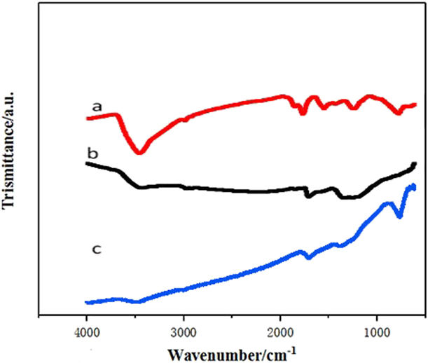
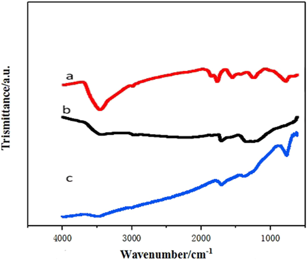
Firstly, 3D-GO/GCE, 3D-GN/Cu/GCE, and 3D-GN/Cu/Fe3O4/GCE were characterized by Fourier transform infrared (FI-IR) to check the chemical structure (see Fig. 1). For the 3D-GO spectrum [Fig. 1(a)], the absorption peak at 1017 cm−1 denotes the C–O–C stretching, the absorption peak at 3423 cm−1 represents the O–H stretching, and the stretching vibration absorption peak at C=O is 1622 cm−1. The spectrum of 3D-GN/Cu [Fig. 1(b)] and 3D-GN/Cu/Fe3O4 [Fig. 1(c)] shows that the absorptive oxygen-containing functional groups are reduced. This indicates that 3D-GO is reduced to 3D-GN. The absorption peak of 569 cm−1 in 3D-GN/Cu/Fe3O4 corresponds to the stretching vibration of Fe–O, indicating the presence of Fe3O4.
Infrared spectra of composites: (a) 3D-GO, (b) 3D-GN/Fe3O4, and (c) 3D-GN/Cu/Fe3O4.
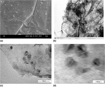
The 3D-GN/Cu/Fe3O4 composites were characterized by transmission electron microscopy (TEM) and scanning electron microscope (SEM) (Fig. 2) to observe the microstructure and morphology. It can be clearly seen from the SEM image [Fig. 2(a)] that the graphene exhibits the stable fold and wrinkles and the Cu and Fe3O4 NPs are uniformly decorated on the surface of graphene. The TEM image [Fig. 2(b)] further displays the layer structure, and 3D graphene presents a three-dimensional skeleton structure. The edge sheets are thin and transparent. This indicates that the 3D graphene is single or few layers. These properties provide a good environment for further loading NPs. Figure 2(b) also demonstrates that the NPs supported without agglomeration on the graphene sheets are evenly distributed and their particle size distribution is about 15 nm. It can be seen that the layered structure of the composites have wrinkles from the HRTEM [Figs. 2(c) and 2(d)], which prove the three-dimensional structure of graphene and uniform dispersion of the NPs on the surface. The diameter of the Cu NPs is between 10 and 30 nm, and the added Fe3O4 NPs have no obvious agglomeration phenomenon, which proves that the dispersibility is good.
(a) SEM, (b) TEM, and (c) and (d) HRTEM images of 3D-GN/Cu/Fe3O4.

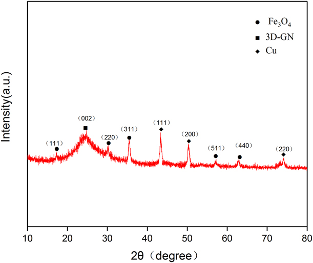
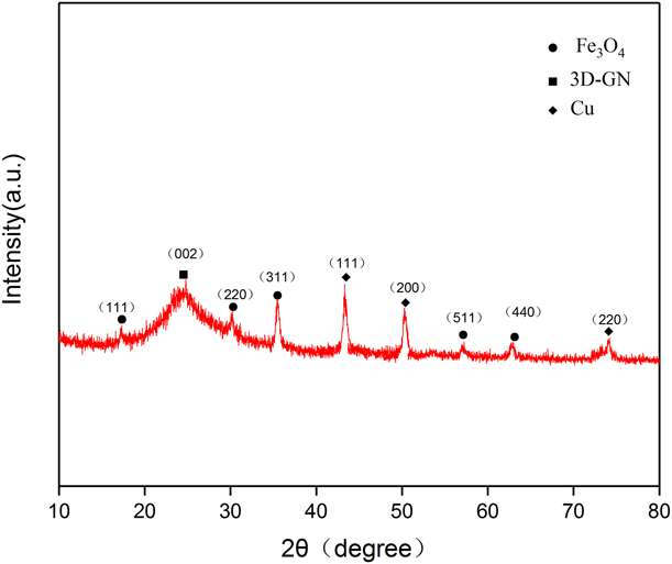
The component of the sample was investigated by XRD. As shown in Fig. 3, the broad peak observed from 20 to 28° was attributed to the diffraction of graphene. The three peaks at 43.5, 50.5, and 74.5° in the range of 40–80° can be assigned to the diffraction from the (111), (200), and (220) planes of Cu(0) with a cubic phase, respectively. The peaks at 2θ = 18.9°, 30.2°, 35.5°, 57.2°, and 62.9° can be, respectively, indexed to (111), (200), (311), (400), (511), and (400) diffractions of Fe3O4. From Fig. 3, it can be seen that the component was successfully prepared.
XRD spectra of 3D-GN/Cu/Fe3O4 composites.
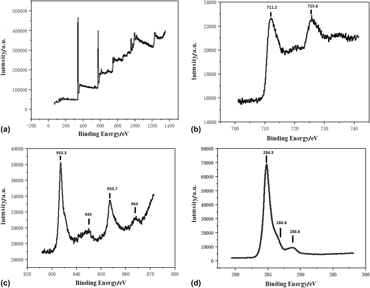
The 3D-GN/Cu/Fe3O4/GCE is further characterized by X-ray photoelectron spectroscopy (XPS) to check the element and valence state. Figure 4(a) is the full spectrum of the XPS of 3D-GN/Cu/Fe3O4 composites, and Figs. 4(b)–4(d) are the high-resolution spectra of Fe 2p, Cu 2p, and C 1s, respectively. Besides C and O elements, Fe 2p can be seen from the full spectrum [Fig. 4(a)]. The peaks of Cu 2p appeared at 710 and 935 eV. The high-resolution spectrum of Fe 2p [Fig. 4(b)] reveals the two peaks at 711.2 and 725.6 eV, corresponding to Fe 2p 3/2 and Fe 2p 1/2, respectively. The peak about 720 eV (corresponding to γ-Fe2O3) is not observed, indicating that only Fe3O4 is present in the electrode and is not oxidized [Reference Yuan, Yu, Wen, Li, Guo, Wang and Wang31]. The two peaks at 933.3 and 953.7 eV correspond to Cu 2p 3/2 and Cu 2p 1/2, respectively, for the spectrum of Cu 2p [Fig. 4(c)], indicating the presence of Cu. In addition, the two weak peaks at about 945 and 964 eV (corresponding to Cu2+) indicate the residual Cu2+ in the 3D-GN/Cu/Fe3O4/GCE. There is a strong C–C signal at 284.3 eV, a weak C–O signal at 286.8 eV, and an O–C=O signal at 288.6 eV, implying a partial reduction of GO. So, the main component of prepared sample is 3D-GN/Cu/Fe3O4.
(a) XPS spectrum of 3D-GN/Cu/Fe3O4 composites, (b) Fe 2p spectrum of 3D-GN/Cu/Fe3O4/composites, (c) Cu 2p spectrum of 3D-GN/Cu/Fe3O4 composites, and (d) C 1s spectrum of 3D-GN/Cu/Fe3O4 composites.

Electrochemical behavior of the modified electrode
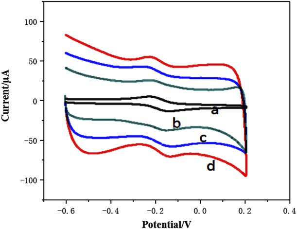
The CV test for GCE, 3D-GN/GCE, 3D-GN/Cu/GCE, and 3D-GN/Cu/Fe3O4/GCE was performed in the range of −0.6–0.6 V, and the sweep speed of 1 mV/s was executed in a 0.10 mol/L KNO3 solution containing 5.0 × 10⁻³ mol/L K3Fe(CN)6 (see Fig. 5). From Fig. 4, the CV curves for all samples show a clear redox peak, and the redox peaks all are symmetrical. The peak current ratio is about 1:1, and the voltage difference is less than 200 mV, confirming that the electron transport between the carrier and the electrode is stable. The current response on the electrode is normal, and the reaction is reversible.
CV of different modified electrodes: (a) bare electrodes, (b) 3D-GN/GCE, (c) 3D-GN/Cu/GCE, and (d) 3D-GN/Cu//Fe3O4/GCE.
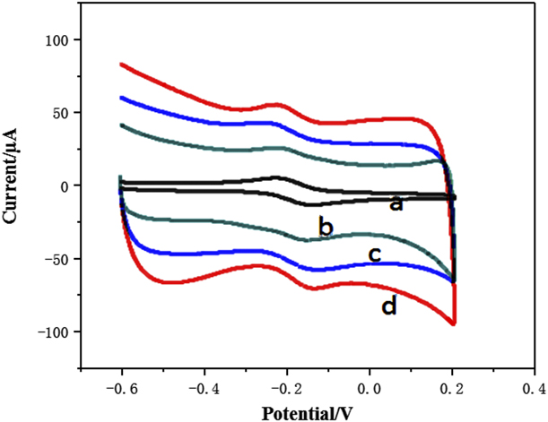
By comparing the electrochemical response of bare electrode in the solution [Fig. 5(a)] and that of the 3D-GN material coated on the surface of the bare GCE [Fig. 4(b)], the later has a reversible oxidation peak at −0.22 V with a peak current of 0.359 μA. Moreover, the 3D-GN/Cu/GCE [Fig. 5(c)] shows a reversible reduction peak at −0.17 V with a peak current of 0.489 μA. The loading of Cu improves the catalytic activity and the conductivity of the 3D-GN/GCE. When the 3D-GN/Cu/Fe3O4/GCE is prepared by mixing Fe3O4 NPs in the 3D-GN/Cu material, the peak current of the oxidation current of the electrode is −0.22 V, and the peak current increases to 0.684 μA, indicating that the redox process of the 3D-GN/Cu/Fe3O4/GCE has a higher activity [Fig. 5(d)]. Both Fe3O4 NPs and 3D-GN/Cu exhibit a high electrochemical activity, and these two materials are simultaneously introduced into the electrode surface to exert a synergistic effect. Besides, the 3D-GN material facilitates the dispersion of NPs. As a good electronic media, Cu and Fe3O4 increase the electron transfer rate and can effectively enhance the conductivity of the electrode. In addition, the high specific surface area of 3D-GN effectively expands the electrochemical effective surface area of the electrode, which makes more electrolyte contact the electrode surface to enhance the response signal, so that the 3D-GN/Cu/Fe3O4/GCE shows a higher electrochemical activity.
Dynamic performance
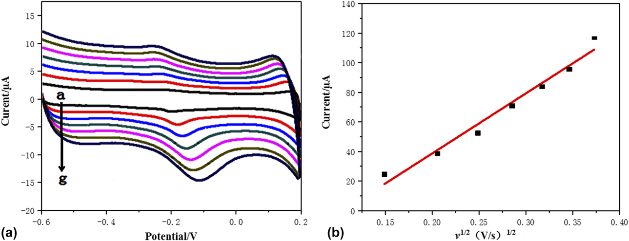
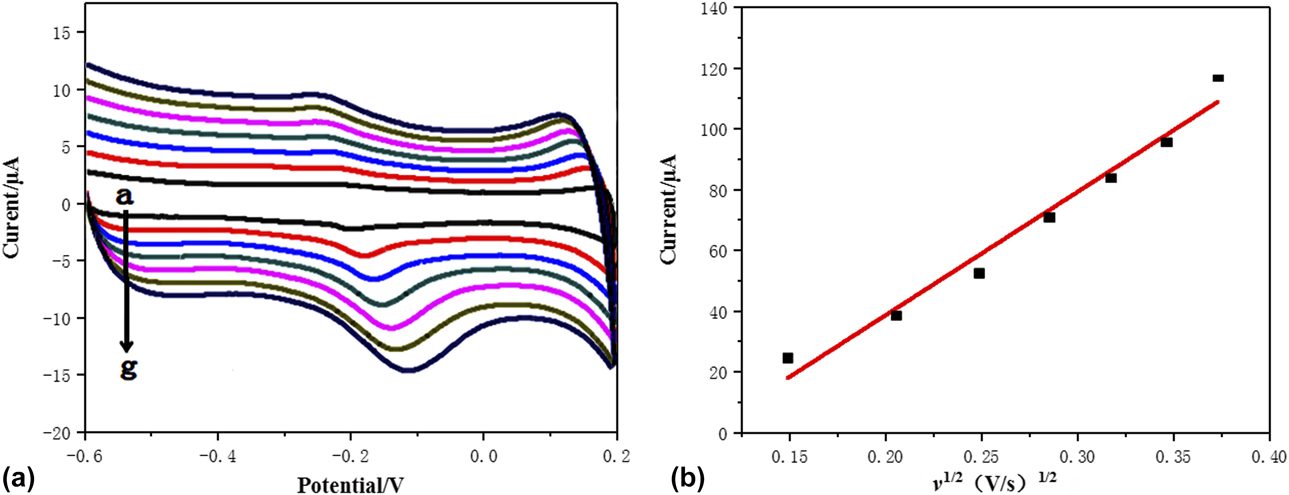
The laccase solution was loaded onto the 3D-GN/Cu/Fe3O4/GCE by the dipping method. The relationship between the potential scanning rate (V) and the electrochemical behavior of BPA was investigated by CV. The Lac-3D-GN/Cu/Fe3O4/GCE biosensor was tested in pH 4.0 phosphate-buffered saline (PBS) and with a potential range of −0.6–0.2 V. Figure 6 shows that the square root of the sweep speed was linear to the peak current. The linear equation follows: I = 4.36 + 82.1v 1/2 (V/s)1/2, r = 0.9973. A larger sweep speed indicates a higher peak current. The reaction of BPA on the modified electrode is a surface control process. As the sweep rate increases, it is also confirmed that the enzyme activity remains remarkable.
(a) CV of Lac-3D-GN/Cu/Fe3O4 biosensor at sweep speeds of 20, 40, 60, 80, 100, 120, and 140 mV/s (from a to g), and (b) the relationship between Lac-3D-GN/Cu/Fe3O4 biosensor sweep speed and peak current.
Effect of solution pH
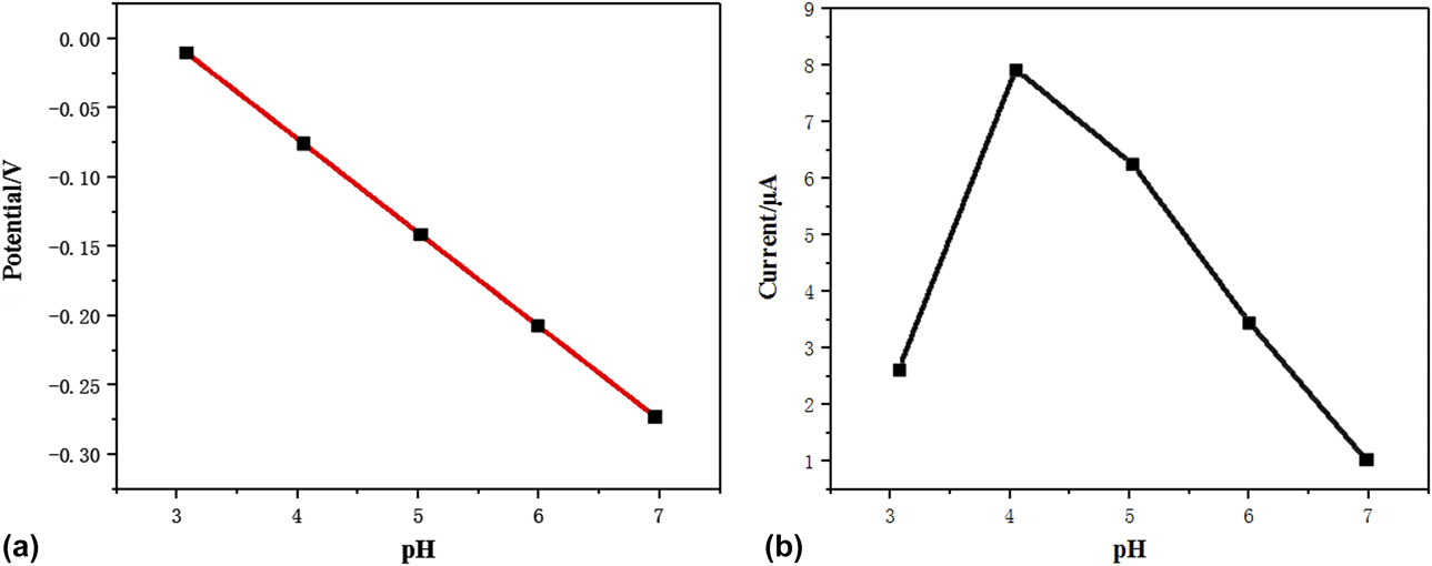
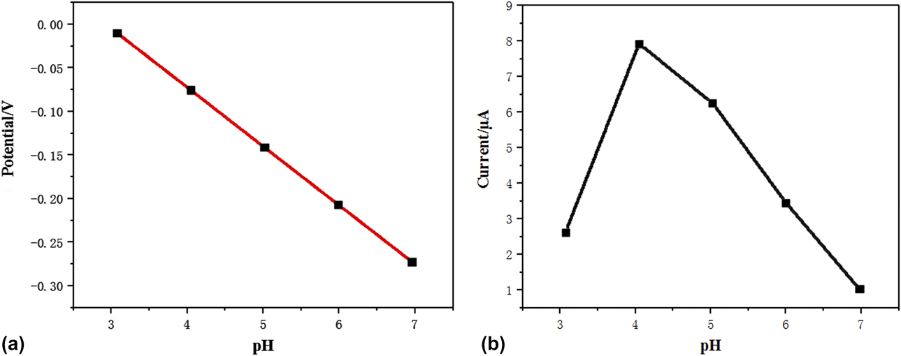
CV was used to study the electrical behavior of the solution pH of BPA in the PBS buffer solution (Fig. 7). The oxidation peak current first increases and then decreases in the range of pH 3–7. At pH 4.0, the maximum oxidation peak current occurs in the PBS solution. As the pH increases, the oxidation peak current gradually decreases until the enzyme electrode has no catalytic activity at pH 7.0. This can be ascribed to the hydroxyl and cyanide interfered with the oxygen molecules at the active site, which result in the loss of the electrocatalytic activity of laccase. It is proved that the catalytic activity of the enzyme is the best when the pH of the PBS buffer solution is 4.0. In the range of pH 3–7, the oxidation peak potential of the electrode gradually shifts negatively with increasing the pH because the protons participate in the electrode reaction and has a linear relationship with pH. The linear equation is as follows: E = 0.2018–0.0686 pH, r = −0.9964, indicating that BPA underwent four electron transfers and had four proton transfers.
Relationship between potential and pH (a) and current value and pH (b) of Lac-3D-GN/Cu/Fe3O4/GCE biosensor in a 0.2 M citric acid buffer solution of pH 3–7.
Differential pulse voltammetry for determining the BPA content
Linear range and detection limit
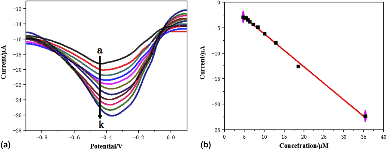
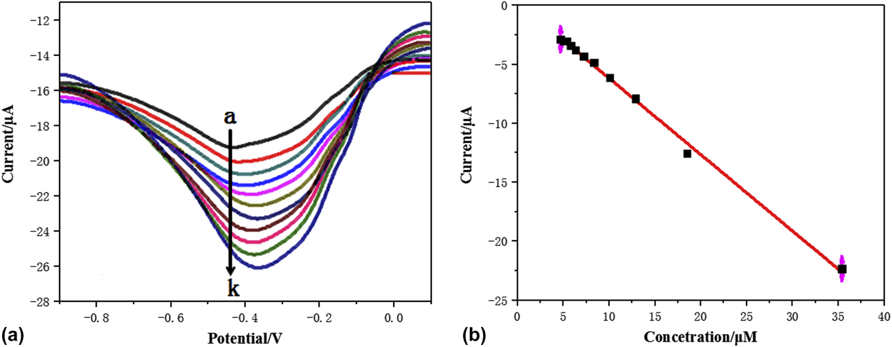
Differential pulse voltammetry was applied to determine the BPA content with different concentration gradients at pH = 4.0. Figure 8 shows that the relationship between the peak current of BPA and its concentration is linear in the range of 7.2–18 μmol/L. The linear equation is as follows: I = −1.0944 × 10⁻⁶ − 0.356 × 10⁻⁶c, the correlation coefficient r = 0.996, and the detection limit is 1.7 μM (S/N = 3). Compared with the biosensor prepared by Dempsey et al. [Reference Choi, Choi, Kim, Choe, Chung, Shin, Kim, Youn, Kim, Lee, Choi, Kim, Choi and Yu78], this biosensor has a considerable detection limit and a wide linear range. This biosensor is equivalent to the biosensor prepared by Portaccio et al. [Reference Portaccio, Di Tuoro, Arduini, Moscone, Cammarota, Mita and Lepore86] because of the excellent carrier effect of 3D-GN, resulting in obtaining NPs with small particle size and uniform dispersion.
(a) Differential pulse curve of Lac-3D-GN/Cu/Fe3O4/GCE biosensor at different concentrations of BPA of pH 4.0 (a–k: 3.3, 3.6, 4, 4.5, 5.1, 6, 7.2, 9, 12, 18, and, 36 μmol/L) and (b) the relationship between Lac-3D-GN/Cu/Fe3O4//GCE biosensor concentration and peak current.
Reproducibility of biosensor
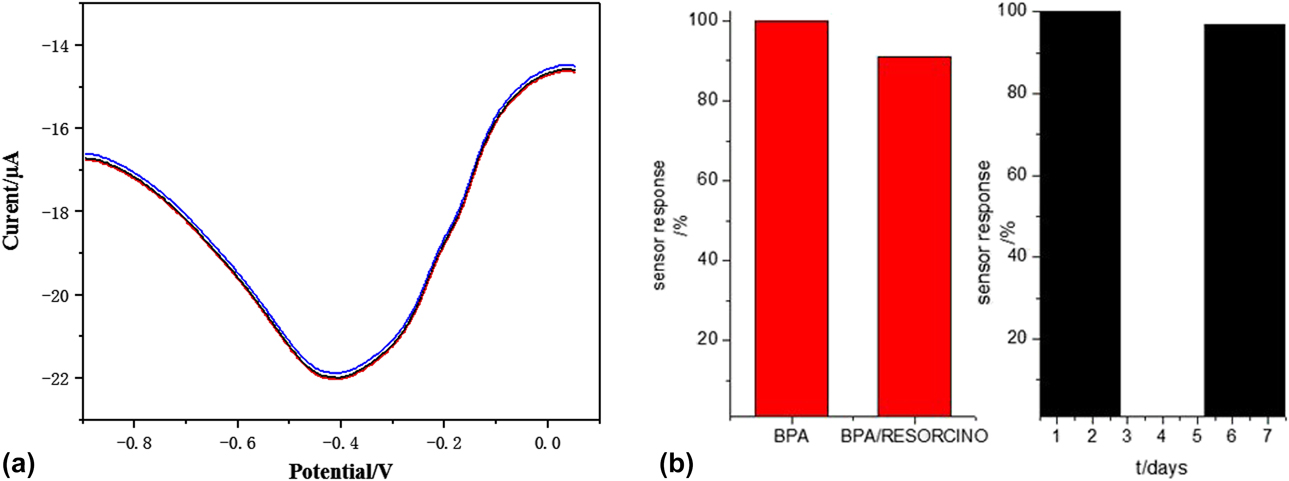
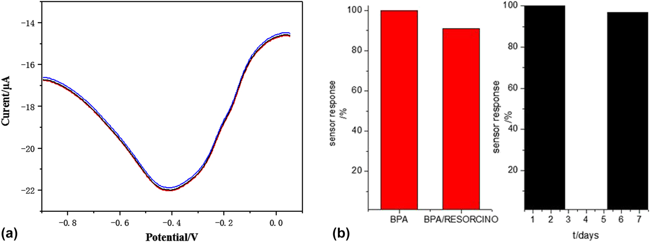
The reproducibility of biosensor test was performed by differential pulse voltammetry with a BPA solution of 7.2 μmol/L. Three Lac-3D-GN/Cu/Fe3O4/GCE biosensors were prepared under the identical conditions, and the same test was carried out in the same solution system [Fig. 9(a)]. The three curves were found to be close and almost identical, and the peak value was unchanged. The measured standard deviation of the peak current was 6.3%, indicating the high reproducibility of the biosensors.
(a) Reproducibility of Lac-3D-GN/Cu/Fe3O4/GCE biosensor and (b) anti-interference and storage stability of Lac-3D-GN/Cu/Fe3O4/GCE biosensor.
Anti-interference and stability
Differential pulse voltammetry was used to test the change in the current response before and after adding the interfering substances. 0.5 g m-diphenol was added to a BPA solution with a concentration of 7.2 μmol/L at pH 4.0. The current response to BPA was 100% before adding the interfering substances. The responsivity of the current was 91.1% after adding resorcinol [Fig. 9(b)]. The biosensor was found to have a strong anti-interference ability and selectivity to BPA. The laccase biosensor was placed at 4 °C for one week at a pH of 4.0 and the change in the current response was tested with a BPA concentration of 7.2 μmol/L via differential pulse voltammetry. The current response after one week was 98.9%. Laccase had a good biological activity on the biosensors. The laccase biosensor has an acceptable storage stability.
Water sample test
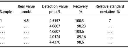
The water sample was taken from laboratory running water, and the pH of tap water was adjusted to 4.0. It was continuously measured five times, and the relative standard deviation (RSD) of the measurement results was 7%. The measured value and the true value were calculated from the linear relationship between the current and the concentration in Fig. 7. The recovery test of sample water was carried out, and the recovery rate was obtained (Table I). The recovery rate was between 96 and 102%. Therefore, the biosensor was found to have an excellent testability for BPA in the actual samples and could be used to detect the concentration of BPA in nature.
Determination of BPA content in tap water samples.

Conclusions
The prepared 3D-GN/Cu/Fe3O4 composites present a remarkable dispersibility, large specific surface area, and adequate active sites. The physical and chemical properties of the electrode are obviously improved. The excellent conductivity and uniform dispersion and synergistic effect of Fe3O4 NPs, Cu, and 3D-GN facilitate the electrochemical activity. The resulted composite materials can be widely used as an excellent carrier to enhance the performance of electrochemical biosensors. The Lac-3D-GN/Cu/Fe3O4/GCE had an excellent laccase activity. At pH 4.0, the detection of BPA has a low detection limit, wide linear range, and excellent selectivity. The reproducibility, selectivity, and stability of the biosensor are turned out to be extraordinary. The detection of real water samples is only 7% RSD, indicating the adaptability to determine the BPA content in nature. These materials combining the conductive copper and graphene and the magnetic magnetite have potential applications for other fields such as electromagnetic interference (EMI) shielding [Reference Jiang, Murugadoss, Wang, Lin, Ding, Wang, Shao, Wang, Liu, Lu, Wei, Subramania and Guo90, Reference Wang, Murugadoss, Kong, He, Mai, Shao, Chen, Guo, Liu, Angaiah and Guo91, Reference Wu, Xu, Wang, Wang, Liu, Liu, Shao, Liu, Gao and Guo92] and strain sensors [Reference Jiang, Wang, Li, Sun, Wu, Yan, Xing, Qi, Li, Liu, Xie, Wang, Ding and Guo79, Reference Gu, Zhang, Ma, Sun, Liu, Dai, Zhang, Wei, Ding and Guo93, Reference Liu, Li, Zhang, Yin, Liu, He, Dai, Shan, Guo, Liu, Shen, Wang, Wang, Wang, Wei and Guo94, Reference Li, Liu, Zhang, Zhang, Liu, He, Mi, Zhang, Liu, Shen and Guo95].
Experimental section
Materials
Analytically pure BPA was obtained from Tianjin Kemi Chemical Reagent Co., Ltd. (Tianjin, China), natural flake graphite was purchased from Qingdao Jinrilai Graphite Co., Ltd. (Qingdao, China), and laccase was supplied by Cool Biotechnology Co., Ltd. (Fuyang, China). Potassium permanganate (KMnO4), copper sulfate (CuSO4) powder, absolute ethanol, sodium nitrate, sodium citrate, natrium aceticum, hydrogen peroxide, and hydrazine hydrate were obtained from Kemi Ou Chemical Reagent Co., (Tianjin, China). The other reagents used in the experiment were analytically pure, and the solution was formulated using ultrapure water.
The synthesis of 3D-GO, 3D-GN, and Fe3O4
Three-dimensional graphene oxide (3D-GO) and graphene (3D-GN) were prepared by methods described previously [Reference Li, Jing, Xu, Tian, Dong, Shao, Wang, Wang, Zheng, Yang and Guo70]. Briefly, 5 g natural flake graphite, 3 g sodium nitrate, and 120 mL concentrated H2SO4 were gradually added into the absolute dry flask and stirred to disperse completely the mixture in ice-water bath (10–15 °C). 20 g KMnO4 were slowly added into the above mixture to stir for 2 h at 15 °C. The mixture was continued to stir for half an hour at 35 °C, 2 min at 98 °C, and then cooled to room temperature. The hydrogen peroxide was added dropwise into the mixture to remove excess KMnO4. The dilute hydrochloric acid and warm deionized water were used to wash until the pH value of solution was close to neutral. The obtained product was dispersed in ultrapure water and ultrasonic treated for 1 h, and then freeze-dried to obtain 3D-GO. The reducing agent hydrazine hydrate was added in to 100 mL GO (0.05 g/L) water solution and stirred for 24 h at 60 °C. The mixture was flushed and filtrated by ethanol and ultrapure water for several times. After being freeze-dried, the sample was collected, which was named as 3D-GN.
The hydrothermal method was used to prepare Fe3O4 NPs (Fe3O4 NPs). The 2.2 g FeCl3·6H2O was completely dissolved in 80 mL ethylene glycol. Then, natrium aceticum and sodium citrate mixture was added to the above solution. The colloidal solution was transferred in to hydrothermal reactor and heat-treated at 200 °C for 20 h to obtain Fe3O4 NPs.
The synthesis of electrode materials
For preparing 3D graphene/copper (3D-GN/Cu) composites, 0.040 g of 3D-GO was mixed with ultrapure water and placed in an ultrasonic cleaner to ultrasonically dissolve the clear and nonparticulate matter in the solution. Furthermore, 0.012 g of copper sulfate powder was weighed and put into the suspension prepared in the first step. The reducing agent sodium borohydride solution was slowly added dropwise as the solution changed from dark brown to black, and then heated in a water bath at 80 °C for 4 h. The prepared 3D-GN/Cu was washed with water until the pH was neutral and kept for further use. Then, the treated Fe3O4 NPs were added to the prepared 0.012 g of 3D-GN/Cu solution by simple physical mixing based on the ultrasonic treatment to obtain a 3D-GN/Cu/Fe3O4 composite material, which was freeze-dried and collected for further use.
The electrode was treated before decoration as follow. The glassy carbon electrode (GCE, Φ = 3 mm) was continuously polished with α-Al2O3 powder (particle size is 10, 0.3, and 0.05, respectively) until mirror surface was formed. The polished electrode was rinsed with water and HNO3–H2O solution [HNO3:H2O = 1:1 (volume ratio)], and ultrasonic cleaned for 3 min. Then, the electrode was dipped in 0.5 mol/L H2SO4 and cyclic scanned until the current maintained stable at 100 mV/s sweep speed and at −0.6 to ∼0.2 V potential range. The electrode was then rinsed with ultrapure water and the surface of the electrode was blow-dried with nitrogen for further use.
The aforementioned 3D-GN, 3D-GN/Cu, and 3D-GN/Cu/Fe3O4 composite materials were separately dispersed in the solution, dropped on the surface of the above electrode (GCE), and dried naturally. The modified electrode was represented as 3D-GN/GCE, 3D-GN/Cu/GCE, and 3D-GN/Cu/Fe3O4/GCE, respectively. The laccase electrode Lac-3D-GN/Cu/Fe3O4/GCE was prepared by dripping the laccase buffer solution on the 3D-GN/Cu/Fe3O4/GCE.
Characterization of electrode materials
The dispersibility, surface morphology, and spatial structure of the electrode materials were observed by transmission electron microscopy (TEM) (Hitachi S-7650, Tokyo, Japan), and scanning electron microscope (SEM) (Hitachi S-4300, Tokyo, Japan). The surface composition and valence state were analyzed by Thermo Scientific K-Alpha X-ray photoelectron spectroscopy (XPS) (Thermo Fisher, Waltham, Massachusetts). The surface chemistry of the electrode materials was examined by Fourier transform infrared (FT-IR) (PE Spectrum One B FTIR, PerkinElmer, Hopkinton, Massachusetts) based on KBr tablet from 500 to 4000 cm−1.
Electrochemical tests
A CHI 660C electrochemical workstation (CH Instruments, Shanghai, China) was employed via a three-electrode system: modified GCE, namely, electrode materials as the working electrode, Pt electrode as the counter electrode, and saturated Ag/AgCl electrode as the reference electrode. Electrochemical tests were conducted to obtain CV curve. For GCE, 3D-GN/GCE, 3D-GN/Cu/GCE, and 3D-GN/Cu/Fe3O4/GCE, the 0.10 mol/L KNO3 solution containing 5.0 × 10⁻³ mol/L K3Fe(CN)6 was used as electrolyte solution for the CV test at the range of −0.6–0.6 V and at the sweep speed of 1 mV/s. For Lac-3D-GN/Cu/Fe3O4 biosensor, PBS and 0.10 mol/L KNO3 solution contain BPA at different sweep speeds. PBS was obtained with 0.1 mol/L Na2HPO4 and 0.1 mol/L NaH2PO4 in different proportions.
Acknowledgments
The authors are grateful for the Project supported by the Graduate innovation research projects for Qiqihar University (Item No.: YJSCX2018-030X).