Introduction
The sphenoid sinus is the most posterior paranasal sinus, and has a complex anatomy and variations. The degree of sinus pneumatisation and its relation to vital surrounding structures, such as the optic nerve, internal carotid artery, pituitary gland, maxillary nerve, cavernous sinus and vidian nerve, are highly variable.Reference Tomovic, Esmaeili, Chan, Shukla, Choudhry and Liu1
The sphenoid sinus starts development as a constriction of the sphenoethmoidal recess at the third fetal month. Major sinus pneumatisation begins in the third year of life.Reference Aoki, Dillon, Barkovich and Norman2 The pneumatisation progresses gradually in all directions, and the sinus reaches its adult configuration and three-dimensional measurements at the age of 16 years.Reference Barghouth, Prior, Lepori, Duvoisin, Schnyder and Gudinchet3
Chronic rhinosinusitis is a relatively common disease in children. It has a great impact on quality of life, and is associated with major financial and healthcare resource burdens.Reference Gilani and Shin4, Reference Heath, Hartzell, Putt and Kennedy5 It is mainly diagnosed clinically, based on the presence of longstanding sinonasal symptoms (nasal purulent discharge, headache, cough or nasal obstruction).Reference Silviu-Dan6 Anterior rhinoscopy performed with an otoscope may show congested nasal mucosa and the presence of discharge. In older and co-operative children, fibre-optic nasoendoscopy is helpful. A sinus computed tomography (CT) scan is necessary when a complication is suspected, in presence of polyps, and in children for whom medical treatment has failed and endoscopic sinus surgery is being considered.Reference Heath, Hartzell, Putt and Kennedy5, Reference Silviu-Dan6
Advancement in navigation systems and power instrumentation has allowed access to the sphenoid sinus and the adjacent vital region endoscopically, with minimal morbidity and complications. Accurate pre-operative knowledge and evaluation of anatomical variations is important for accessing the sinus and avoiding potential serious complications.Reference Al-Qudah7 The CT scan gives detailed information about sphenoid sinus pneumatisation and adjacent neurovascular structure variations.
Although sphenoid configuration and the prevalence of anatomical variation in the sphenoid sinus area have been thoroughly studied in adults, there are scant data on the paediatric age group in the literature. This study aimed to report the prevalence of different anatomical variations of the sphenoid sinus and its related structures among paediatric patients with or without chronic rhinosinusitis, and to assess the role of age and sinusitis on the frequency of these variations.
Materials and methods
After obtaining institutional review board approval, consecutive high-resolution CT scans of 50 paediatric patients with chronic rhinosinusitis were retrospectively reviewed and compared to scans of 50 paediatric patients without chronic rhinosinusitis. All images were studied by the senior authors (HO and MA). When there were discrepancies, further evaluations of the scans were carried out by the senior authors together and a mutual consensus was reached.
A diagnosis of chronic rhinosinusitis was made based on a duration of more than 12 weeks of symptoms and signs, which included: mucopurulent nasal discharge; nasal obstruction and congestion; facial pain, pressure or fullness; and a decreased sense of smell. Anterior rhinoscopy and fibre-optic endoscopy, performed in older and co-operative children, show purulent discharge, congestion, or oedema in the middle meatus or anterior ethmoid region, and sinus CT scans show opacification in the paranasal sinuses.
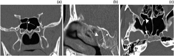
The type of sphenoid sinus pneumatisation, position of the vidian canal, lateral aeration of the sphenoid sinus, presence of an Onodi cell, presence of a single sphenoid septum or multiple septa, and internal carotid artery bulging in the sphenoid sinuses were thoroughly examined and analysed (Figure 1).
High-resolution computed tomography scans of the sinuses: (a) coronal view showing a right-sided Onodi cell (arrow); (b) sagittal view showing sphenoid sinusitis (short arrow) and a conchal sphenoid configuration (long arrow); and (c) axial view showing internal carotid artery bulging (long arrow) and lateral pneumatisation of the sphenoid sinus (short arrow).

The patients were divided into three age groups, namely 4–8 years, 9–12 years and 13–16 years, to assess the effect of growth on the aforementioned variations.
The sphenoid sinus was classified into three types based on its relation to the sella turcica: (1) conchal type, when there is thick bone separating the posterior wall of the sphenoid from the sella; (2) pre-sellar type, when there is a thin layer of bone between the posterior wall of the sinus and the sella; and (3) sellar type, when the sella turcica bulges to a variable degree in the sinus, and the posterior wall of the sphenoid ends beyond the anterior wall of the sella.Reference Hamberger, Hammer and Norlen8
The vidian canal was classified into three types, according to Lee et al:Reference Lee, Kao, Hsu and Lin9 type 1, when the vidian canal is completely within the sphenoid sinus; type 2, when the canal is on the floor of the sphenoid sinus or partially protruding into the sphenoid sinus; and type 3, when it is completely embedded in the sphenoid corpus.
Patients with craniofacial anomalies, facial trauma, nasal or facial neoplasms, immunodeficiency, cystic fibrosis, or those who had undergone previous sinus surgery were excluded.
The chi-square test and the unpaired t-test were used for the statistical analysis; a p-value of less than 0.05 was accepted as statistically significant.
Results
A total of 100 paediatric patients with high-resolution sinus CT scans were included. There were 54 boys and 46 girls, with ages ranging from 4 to 16 years (mean of 10.9 years). Fifty patients had chronic rhinosinusitis and 50 had no sinusitis; the 2 groups were matched in age and gender.
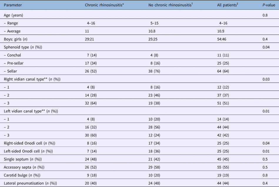
Table 1 demonstrates the prevalence of the studied variations based on the presence or absence of chronic rhinosinusitis. There were statistically significant differences between paediatric patients with or without sinusitis in terms of: sphenoid sinus pneumatisation type, vidian canal type and Onodi cell presence.
Sphenoid sinus pneumatisation types and surrounding structure variations in patients with or without sinusitis

*n = 50; †n = 50; ‡n = 100. **Vidian canal type 1 = vidian canal is completely within the sphenoid sinus; type 2 = canal is on the floor of the sphenoid sinus or partially protruding into the sphenoid sinus; and type 3 = canal is completely embedded in the sphenoid corpus.
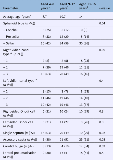
Table 2 shows the distribution of the studied variables based on age. The sellar type of sphenoid sinus was the commonest in all groups and was seen in 64 cases, while the conchal type was the least common and was observed in 11 cases (11 per cent). There were significant differences between the three age groups in terms of: sphenoid sinus pneumatisation type, single sinus septum and multiple septa presence, and internal carotid bulging. Our results indicate that as children advance in age, they are more likely to have a pneumatised sinus, accessory septa and internal carotid artery bulging in the sphenoid sinus.
Sphenoid sinus pneumatisation types and surrounding structure variations based on age

*n = 24; †n = 41; ‡n = 35. **Vidian canal type 1 = vidian canal is completely within the sphenoid sinus; type 2 = canal is on the floor of the sphenoid sinus or partially protruding into the sphenoid sinus; and type 3 = canal is completely embedded in the sphenoid corpus.
No significant differences were observed between the sides of the sphenoid sinus (right or left) for all examined variations, even when patients were divided based on age or presence of sinusitis.
Discussion
The sphenoid sinus is an anatomically complex structure. It varies in size and there are great variations in nearby landmarks, and it is situated close to many vital organs. A comprehensive understanding of the extent of sinus pneumatisation and its relation to adjacent neurovascular structures is essential for safe endoscopic surgery.Reference Al-Qudah7
Sphenoid surgery is important for treating diseases inside the sinus walls; it is also a corridor for accessing the skull base. An endoscopic trans-sphenoidal approach has become the preferred route to the sella turcica. It provides excellent minimally invasive exposure to the pituitary gland region, and prevents serious complications associated with external approaches.Reference Kim, Kim, Kim and Cho10
In this study, we reported on the prevalence of sphenoid sinus configurations and described the frequency of variations in surrounding structures in paediatric patients using high-resolution CT. We also examined differences in these variables based on age and the presence of chronic rhinosinusitis.
Although the anatomy and degree of sphenoid sinus pneumatisation in adults have been extensively studied, few articles in the literature have investigated variations in the paediatric age group.Reference Kim, Kim, Kim and Cho10, Reference Al-Qudah11 The degree of sphenoid pneumatisation can affect surgical outcome; a highly pneumatised sinus may thin the sinus bony walls and put the surrounding vital structures at an increased risk of iatrogenic injury,Reference Sirikci, Bayazit, Bayram, Mumbuc, Gungor and Kanlikama12 whereas poor pneumatisation can narrow the exposed surgical field and necessitate extensive bone drilling.
Szolar et al.Reference Szolar, Preidler, Ranner, Braun, Kern and Wolf13 reviewed magnetic resonance imaging (MRI) scans of the sphenoid sinuses for 401 paediatric patients. They found that 85 per cent of the studied cases had complete sinus pneumatisation at the age of 7 years and all had complete pneumatisation at the age of 10 years. They concluded that pneumatisation starts at the age of 15 months and progresses rapidly, and is completed by the age of 10 years. They recommended the clinical investigation of any children with absent sphenoid pneumatisation at the age of nine years.
Barghouth et al.Reference Barghouth, Prior, Lepori, Duvoisin, Schnyder and Gudinchet3 conducted a retrospective study to establish the age-related three-dimensional size of the sphenoid sinus by evaluating 95 MRI head scans of children aged less than 17 years. Children who had undergone MRI for sinus diseases or surgery were excluded. The sphenoid sinus septum was not seen in 60 per cent of the cases.Reference Heath, Hartzell, Putt and Kennedy5 Magnetic resonance imaging is the best radiological modality for detecting paranasal sinus soft tissue pathology, whereas CT is better at evaluating bony wall integrity and septation, drainage pathway, and anatomical variations around the sphenoid sinus. Recent advances in CT technology allow for more rapid imaging, with substantially lower radiation exposure, potentially mitigating concerns related to radiation exposure.Reference Callahan, MacDougall, Bixby, Voss, Robertson and Cravero14
As the child grows up, the sphenoid sinus volume increases.Reference Szolar, Preidler, Ranner, Braun, Kern and Wolf13, Reference Vaezi, Cardenas, Pinheiro-Neto, Paluzzi, Branstetter and Gardner15, Reference Fujioka and Young16 We found a significant association between age and sphenoid sinus pneumatisation type. Our results confirmed Szolar and colleagues’Reference Szolar, Preidler, Ranner, Braun, Kern and Wolf13 finding: after the age of nine years, the majority of children have a good sized sphenoid sinus, and endoscopic surgery for sphenoid or pituitary disorders can be performed with similar outcomes as those in adults.
We found that paediatric patients with chronic rhinosinusitis tend to have a less pneumatised sphenoid sinus. Kim et al.Reference Kim, Friedman, Sulek, Duncan and McCluggage17 studied paranasal sinus development in children with: chronic or recurrent sinusitis without cystic fibrosis (n = 19); cystic fibrosis (n = 15); or no previous sinus issue (n = 16). The pneumatisation of the sphenoid sinus was assessed by CT and subjectively graded as: well pneumatised, moderately pneumatised or minimally pneumatised. They found that cystic fibrosis and chronic rhinosinusitis patients had a less pneumatised sinus than those with no previous sinus issue. They explained their finding by hypothesising that the sphenoid sinus undergoes a conversion from red to yellow fatty bone marrow in the first few years of life, with the earliest pneumatisation usually occurring at or shortly after the age of two years. The occurrence of sinusitis during this crucial period may interfere with the normal development of the sphenoid sinus.Reference Aoki, Dillon, Barkovich and Norman2
The vidian canal is located inferomedial to the foramen rotundum and the palatovaginal canal. It has a close relationship to critical neurovascular structures in the posterior wall of the pterygopalatine fossa and to the surrounding sphenoid sinus structures.Reference Wiebracht and Zimmer18 Vescan et al.Reference Vescan, Snyderman, Carrau, Mintz, Gardner and Branstetter19 studied the relationship between the vidian canal and the internal carotid artery. They found that the anterior genu of the petrous internal carotid artery was always located superior-medial to the vidian canal. We found a significant difference in vidian canal type in the paediatric patients based on the presence or absence of chronic rhinosinusitis.
Aksoy et al.Reference Aksoy, Yenigun, Goktas and Ozturan20 examined the sinus CT scans of 347 adult patients to evaluate the prevalence of accessory sphenoid septation and its relationship with neighbouring anatomical structures. Accessory septa were present in 50 per cent of patients. A significant association between the presence of accessory septa and the vidian canal configuration was found. The authors concluded that the presence of an accessory sphenoidal septum indicates an increased risk of surgical complications because of its attachment to vital structures such as the internal carotid artery and optic nerve. We found that the prevalence rates of accessory septa and internal carotid bulging inside the sphenoid sinus increase with age, but do not differ between patients with or without sinusitis. It is possible that as the sphenoid sinus increases in size, more accessory septa develop in the late stage of the sphenoid sinus maturation.
• Sphenoid configuration and anatomical variations in the sphenoid sinus area have been thoroughly studied in adults but not in children
• The sellar type of sphenoid sinus was most common, while the conchal type was the least common
• There were significant differences between paediatric patients with or without sinusitis in: pneumatisation type, vidian canal type and Onodi cell presence
• Paediatric patients with chronic rhinosinusitis had a less pneumatised sphenoid sinus
• Age and sinusitis have a significant impact on sphenoid pneumatisation type and surrounding structure variation
The Onodi cell (sphenoethmoidal air cell) represents a pneumatisation of the most posterior ethmoid cell superiorly and laterally to the sphenoid sinus. It has a close relation to the optic nerve. Pre-operative identification of these cells by CT viewed in different planes is highly important to avoid complications in sphenoid sinus surgery. The incidence of an Onodi cell reported in the literature is variable, and ranges from 7 per cent to 65 per cent. Differences in age, race, methods of cell identification (endoscopic or CT) and the CT plane used to determine the cells’ presence may explain this variability.Reference Sareen, Agarwal, Kaul and Sethi21 In a recent study, Chmielik and ChmielikReference Chmielik and Chmielik22 detected an Onodi cell in 40 per cent of 196 paediatric paranasal sinus CT scans. They concluded that the prevalence of an Onodi cell was higher than previously reported, and they recommended evaluating CT scans in all three planes to avoid missing or over-detecting an Onodi cell. They found axial and sagittal planes to be preferable for Onodi cell identification.Reference Chmielik and Chmielik22 Overall, we found an Onodi cell in 25 per cent of patients. In addition, we did not find that the frequency of the Onodi cell's presence differed in terms of age; however, it was significantly less common in children with sinusitis, which may support Kim and colleagues’ hypothesis.Reference Kim, Friedman, Sulek, Duncan and McCluggage17
Conclusion
Sphenoid sinus configuration varies significantly among paediatric age groups, which warrants the comprehensive interpretation of CT scans prior to surgery. Recognition of these variations can be useful for mapping this region, and enables a safer and more efficient endoscopic surgical procedure. We found that age and the presence of sinusitis have a significant impact on the type of sphenoid pneumatisation and the frequency of anatomical variations surrounding the sphenoid sinus. However, prospective studies and large sample sizes are needed to confirm these findings.
Competing interests
None declared