Introduction
In sea urchins, both adult and larval forms produce a calcareous endoskeleton. The larval skeleton consists of two branched spicules, while the adult skeleton is more complex and consists of a test, spines and teeth. In both the larvae and the adult, mineralized hard parts are composed of magnesium-bearing calcite along with an occluded organic matrix; development of both structures is thought to occur by secretion of mineral into a syncytial cavity by skeletogenic cells (see reviews by Wilt, Reference Wilt1999, Reference Wilt2002; and Wilt et al., Reference Wilt, Killian and Livingston2003). Some of the spicule matrix proteins from larvae that have been characterized have been found to also reside in adult mineralized structures (Harkey et al., Reference Harkey, Klueg, Sheppard and Raff1995; Killian & Wilt, Reference Killian and Wilt1996; Ameye et al., Reference Ameye, Hermann, Killian, Wilt and Dubois1999; Wilt, Reference Wilt1999). Recent proteome and genome analyses have revealed additional spicule matrix proteins and other biomineralization-related proteins (Livingston et al., Reference Livingston, Killian, Wilt, Cameron, Landrum, Ermolaeva, Sapojnikov, Maglott, Buchanan and Ettensohn2006; Mann et al., Reference Mann, Poustka and Mann2008, Reference Mann, Wilt and Poustka2010).
The formation of the larval skeleton by primary mesenchyme cells (PMCs) in sea urchins has been studied intensively (see reviews by Decker & Lennarz, Reference Decker and Lennarz1988; Ettensohn & Ingersoll, Reference Ettensohn, Ingersoll, Rossomando and Alexander1992; Wilt, Reference Wilt1999, Reference Wilt2002). The PMCs ingress into the blastocoel just prior to gastrulation, migrate along the wall of the blastocoel and subsequently fuse with one another to form a ring of cells connected by filopodial cables. This process leads to the formation of a membranous cavity where calcium and spicule matrix proteins accumulate. Skeletogenesis begins in two prominent clusters of cells (the ventrolateral clusters) where triradiate spicules are produced. These nascent spicules are subsequently thickened, branched and elongated to form the pluteus larval skeleton. The shape of the developing spicule is determined by the position of the primary mesenchyme cell bodies and interconnecting syncytial filopodia.
Calcium and spicule matrix glycoproteins appear to be routed to the spicule cavity by a secretory pathway (Decker et al., Reference Decker, Morrill and Lennarz1987; Wilt, Reference Wilt1999). In this study, supporting evidence is provided by a transcellular pathway by which material is taken up at the PMC cell surface and deposited into the internal matrix of the spicules.
Materials and methods
Lytechinus pictus or Strongylocentrotus purpuratus sea urchins were purchased from Marinus (Long Beach, CA, USA) and maintained in Instant Ocean seawater (Aquarium Systems, Mentor, OH, USA) in refrigerated marine aquaria. Gametes were collected by injecting 0.5 M KCl into the body cavity and eggs were fertilized with a dilute sperm solution. Embryos were then diluted to <1% (v/v) with Instant Ocean seawater and reared at 16°C with constant, gentle stirring. At the desired developmental stage, embryos were collected on a 35 μm mesh and transferred to artificial seawater (ASW; 423 mM NaCl, 9 mM KCl, 9.3 mM CaCl2, 22.9 mM MgCl2, 25.5 mM MgSO4, 2.1 mM NaHCO3, pH 8).
Embryos were briefly treated with calcium–magnesium-free seawater (CMFSW; 462 mM NaCl, 10.7 mM KCl, 7 mM Na2SO4, 5.0 mM NaHCO3, 1 mM EDTA, pH 8.0) which disrupts cellular junctions, allowing substances to diffuse into the blastocoel as described by Itza & Mozingo (Reference Itza and Mozingo2005). Embryos were monitored microscopically and when the apical surfaces of the cells began to round up, embryos were pelleted by hand centrifugation, washed once with ASW, and then incubated in ASW that contained 10–25 μg/ml fluorescein isothiocyanate-conjugated wheat germ agglutinin (WGA–FITC; Sigma-Aldrich, St. Louis, MO, USA) for 30 min at 16°C. After washing in ASW, embryos were examined with epifluorescence microscopy either immediately after fluorescent labelling or 4–72 h after fluorescent labelling. Experiments were repeated at least three times with qualitatively similar results. For double-labelling experiments, mesenchyme blastula stage embryos were first labelled with WGA–FITC as described above. After 24 h, embryos were treated briefly with CMFSW, monitored microscopically and when the apical surfaces of cells began to round up, embryos were pelleted by hand centrifugation, washed once with ASW, and then incubated in ASW that contained 10–25 μg/ml tetramethylrhodamine isothiocyanate-conjugated wheat germ agglutinin (WGA–TRITC; Sigma-Aldrich, St. Louis, MO, USA) for 30 min at 16°C. Embryos were then washed and examined with epifluorescence microscopy as described above. To remove membranous material associated with spicules (Cho et al., Reference Cho, Partin and Lennarz1996), 2.5–5% NaOCl was added to double-labelled embryos until spicules were free of cellular debris. Cleaned spicules were then washed in distilled H2O and examined with epifluorescence microscopy.
Results and Discussion
Early gastrula-stage embryos were treated with a brief pulse of divalent cation-depleted seawater to permeabilize the embryonic epithelium (Latham et al., Reference Latham, Martinez, Cazares, Hamburger, Tully and Oppenheimer1998; Itza & Mozingo, Reference Itza and Mozingo2005). The embryos were then incubated with WGA–FITC for 30 min, washed and examined either immediately after labelling, or 4–24 h after labelling. Immediately after labelling, bright fluorescence was observed on the surface of PMCs and, to a lesser extent, on the apical surface of embryos (Fig 1 a, b). Intracellular labelling of the PMCs was not apparent at this time. This result is consistent with previous studies that have demonstrated that WGA binds specifically to PMCs (DeSimone & Spiegel, Reference DeSimone and Spiegel1986a, Reference DeSimone and Spiegel1986b). In control embryos, which were not pulsed with CMFSW, no fluorescence was observed on the surface of the PMCs (not shown). When embryos were examined 4 h after labelling, there was a significant redistribution of fluorescence. Fluorescent labelling of the PMC plasma membrane had largely disappeared and intracellular PMC vesicles were now brightly fluorescent (Fig. 1 c, d).
WGA–FITC accumulates in intracellular granules. Differential interference contrast (DIC) (A, C) and fluorescent (B, D) images of early gastrula-stage embryos. Embryos were permeabilized with CMFSW and examined immediately after labelling with WGA–FITC (A, B) or after a 4 h incubation period (C, D) indicating that WGA–FITC preferentially binds to primary mesenchyme cells (PMCs). Immediately after labelling, fluorescence is associated primarily with the surface of PMCs (inset, B). After 4 h, fluorescent label is visible in intracellular granules (inset, D). Bars = 20 μm.

To further examine redistribution of WGA–FITC, embryos labelled with WGA–FITC at the early gastrula stage were allowed to develop for an additional 24 h at 16°C and then re-examined. In late gastrula-stage embryos, PMCs had migrated, fused and commenced spiculogenesis in a normal fashion and fluorescence was still associated with the PMCs (Fig. 2). However, higher magnification views indicated that fluorescence had almost completely disappeared from the surface of the PMCs and was instead associated with prominent intracellular vesicles (Fig. 2, inset). The fluorescent vesicles observed in this study were reminiscent of calcium-containing vesicles that have been implicated in the delivery of calcium to the spicule (Decker et al., Reference Decker, Morrill and Lennarz1987; Beniash et al., Reference Beniash, Addadi and Weiner1999; Wilt et al., Reference Wilt, Killian, Hamilton and Croker2008).
Fluorescence accumulates in prominent intracellular granules after 24 h of incubation. Fluorescent image of an embryo incubated for 24 h after labelling. Primary mesenchyme cells (PMCs) migrated, fused and commenced spiculogenesis in a normal fashion. Inset shows intense fluorescence in intracellular PMC granules. Bar = 20 μm.

To further explore the redistribution of WGA–FITC, embryos were examined approximately 48 h after labelling. At this stage, there was still some granular fluorescence associated with the PMCs, however most of the fluorescence now appeared to be emanating from the spicule (Fig. 3 a, b). To verify that fluorescence was associated with the internal structure of the spicule rather than peripherally associated with the surface, embryos were placed in NaOCl to remove membranous material associated with surface of spicule (Cho et al., Reference Cho, Partin and Lennarz1996). Cleaned spicules were still brightly fluorescent, indicating that the fluorescent label had been incorporated into the internal matrix of the spicule (Fig. 3 c, d).
Incorporation of fluorescent label into the spicule in late stage embryos. Transmitted light (A) and fluorescent (B) images of embryos examined approximately 48 h after labelling. Fluorescence appears to be associated with the internal matrix of the spicule. Transmitted light (C) and fluorescent (D) image of a NaOCl-cleaned spicule. Bar = 10 μm in (A/B). Bar = 20 μm in (C, D).

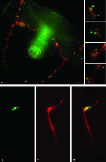
Double-labelling experiments indicated that fluorescently labelled lectin probes were sequentially taken up by PMCs and incorporated into distinct regions of the internal spicule matrix. Mesenchyme blastula stage embryos were labelled with WGA–FITC, washed and allowed to develop. The same embryos were labelled with WGA–TRITC at the late gastrula stage and then examined 24 h later. Low magnification views of embryos double-labelled in this way demonstrated green fluorescence (WGA–FITC) associated with the larval gut and red (WGA–TRITC) fluorescence associated with the larval skeleton. Higher magnification views of the PMCs demonstrate that both fluorescent probes were incorporated into intracellular vesicles and that there was very little co-localization of the two fluorescent probes (Fig. 4 a, insets). Instead, the probes appear to reside in different intracellular vesicles, which indicated that they might be at different stages of an intracellular trafficking pathway. If this situation is the case, it is predicted that the two probes might become incorporated into different parts of the spicule. The accumulation of fluorescent probe into the spicule in double-labelled samples was examined by cleaning spicules from pluteus stage embryos. Cleaned spicules from double-labelled embryos showed a distinctive fluorescent labelling pattern. Green fluorescence was associated with a small area of the spicule that is proximal to the initial site of calcite deposition. Red fluorescence is seen throughout a larger area that both surrounds and extends beyond the green fluorescent region (Fig. 4 B–D). As with the intracellular fluorescent vesicles, there was very little co-localization of these two fluorescent probes (Fig. 4 D).
Double-labelling experiments indicated that fluorescent lectins were sequentially taken up by primary mesenchyme cells (PMCs) and incorporated into distinct regions of the spicule. Fluorescent image (A) of a double-labelled embryo. The two fluorescent labels reside in different intracellular granules (insets). NaOCl-cleaned spicule from double-labelled samples at the pluteus stage of development (B–D). Accumulation of (B) WGA–FITC (green) and (C) WGA–TRITC (red) into the spicules. (D) is an overlay of (B, C). Bar = 20 μm in (A). Bar = 10 μm in (D).

This study provides evidence for a transcellular pathway in which material is taken up from the cell surface, incorporated into intracellular vesicles and then deposited into the developing spicule. Using calcein, a vital fluorescent label for calcium, Wilt et al. (Reference Wilt, Killian, Hamilton and Croker2008) have visualized calcium-containing vesicles in PMCs and the subsequent deposition of fluorescence in the spicule. The fluorescent vesicles observed in this study and the vesicles observed by Wilt et al. (Reference Wilt, Killian, Hamilton and Croker2008) have a similar appearance and, in both cases, fluorescence is ultimately deposited into the spicule. Further, the double-labelling experiments described in this study reveal a distinctive pattern of fluorescent lectin incorporation into spicules that mirrors the growth pattern of spicules whereby spicules increase in girth and length (Ettensohn & Malinda, Reference Ettensohn and Malinda1993; Wilt, Reference Wilt2002). Thus, the cellular pathway observed in this study may parallel an endogenous pathway for routing calcium to the spicule from seawater and the methods described here may prove useful for future studies aimed at characterizing this process.
Acknowledgements
This study was supported in part by a Research Challenge grant from the Ohio Board of Regents and a Faculty Mini-Grant from CSU Channel Islands to NMM.