Introduction
Epistaxis is a common ENT emergency, often requiring hospital admission. Its prevalence in the population is around 10 per cent. However, less than 10 per cent of these patients seek medical treatment. Only 1–2 per cent of epistaxis patients seeking medical care will require surgical treatment.Reference Shaheen1
Most cases of epistaxis are managed conservatively with chemical cautery or packing. However, when this fails, ligation of vessels is required. Previously, ligation of the internal maxillary artery through a trans-antral approach was commonly used. However, this was often associated with a number of complications, including facial swelling, facial numbness and oro-antral fistula.Reference McDonald and Pearson2 More recently, ligation of the sphenopalatine artery has been adopted, which is associated with fewer complications and a shorter hospital stay compared with ligation of the internal maxillary artery.Reference Feusi, Holzmann and Steurer3 Ligation of the sphenopalatine artery was first described in 1976 by Prades.Reference Prades4 Endoscopic sphenopalatine artery ligation is now a recognised and frequently used treatment for control of intractable posterior epistaxis that has not been controlled by conventional methods.Reference McGarry5, Reference White6 However, locating the sphenopalatine foramen in a bleeding patient can be difficult; thus, good knowledge of the local anatomy is essential.
The sphenopalatine artery is the main vessel supplying the mucosa of the nasal septum, fontanelle, superior, middle and inferior turbinates, and the lateral nasal wall.Reference Padgham and Vaughan-Jones7 The sphenopalatine artery arises in the pterygopalatine fossa from the maxillary artery and divides either proximal to or within the sphenopalatine foramen. The sphenopalatine artery supplies about 90 per cent of the nasal mucosa.Reference Lang8 The operation relies on identification of the bony sphenopalatine foramen. This foramen is bounded superiorly by the body of the sphenoid, anteriorly by the orbital process of the palatine bone, posteriorly by the sphenoidal process of the palatine bone and inferiorly by the perpendicular plate of the palatine bone.Reference McGarry5 There remains a debate in the literature about the exact position of the sphenopalatine foramen. Lee et al. showed that the foramen was located within the superior meatus, between the middle turbinate and the posterior horizontal end of the lamella of the superior turbinate, attaching to the lateral nasal wall in 90 per cent of cases. This was a cadaveric study in an Asiatic population; therefore, its applicability to the UK is unclear. In 10 per cent of cases, the foramen extended superiorly beyond the posterior horizontal portion of the lamella of the superior turbinate.Reference Lee, Kim, Kim, Son, Kim and cho9 Wareing studied 238 lateral nasal walls and suggested an osteological classification for the position of the sphenopalatine foramen.Reference Wareing and Padgham10 These two articles highlight the natural anatomical variation of the sphenopalatine foramen and raise the possibility of ethnic variation.
Objectives
The aim of this study was to examine the distance between key bony landmarks and the sphenopalatine foramen, in order to derive surgically useful coordinates which could be used in clinical practice.
Materials and methods
Ethics
Ethical approval was gained from the Glasgow Royal Infirmary local research ethics committee.
Surgical navigation system
The BrainLAB© surgical navigation system (BrainLab Limited, Cambridge, UK) was used to take anatomical measurements. The computed tomography (CT) scans of 28 patients were used. The CT scans had been done for other clinical reasons and were loaded in the BrainLAB surgical navigation system. No CTs were done solely for the purpose of this study. No patient had undergone previous sphenopalatine artery ligation.
The BrainLAB system is a surgical navigation tool allowing the surgeon to make detailed measurements in millimetres and degrees. The computer software allows the surgeon to display the three different views of each CT scan (sagittal, coronal and axial) on one screen together with a three-dimensional reconstruction. This system allows identification of the sphenopalatine foramen on each view; this fixed anatomical landmark can then be identified on the other views using a ‘drag and drop’ facility.
The two fixed bony landmarks used were chosen for clinical applicability. These were the anterior nasal spine and the plane of the piriform aperture. Three measurements were taken on the right and left of each CT: the distance from the sphenopalatine foramen to the nasal spine; the angle of elevation from the nasal floor to the sphenopalatine foramen; and the distance from the sphenopalatine foramen to the piriform aperture. The distance measurements were taken from the centre of the sphenopalatine foramen and were recorded in millimetres to the nearest 0.1 mm. The angle of elevation was measured to the nearest degree. All measurements taken were to scale, as checked with the CT software.
Measurements
A full set of measurements was taken on two occasions by three different observers of varying experience (one consultant, one specialist registrar and one senior house officer). The measurements were then used to compute the average distance and angle of elevation from the entrance of the nose to the sphenopalatine foramen. Inter- and intra-observer agreement was assessed by calculating the intra-class correlations between measurements.Reference Bland and Altman11 Between-group comparisons (comparing men versus women and left versus right) were done on the average measures using t-tests, with the results displayed as confidence intervals for the differences along with p values. All analyses were done using Minitab (version 14) software at a significance level of 5 per cent.
Comparison of BrainLAB and skull data
Prior to this study, research on this subject had mainly relied upon anatomical dissection. In order to determine the overall scale of measurements likely, we carried out a preliminary, benchmarking anatomical study.
Ten Caucasoid skulls obtained from the Glasgow University anatomy department were measured and studied. Two independent observers made 40 measurements on these skulls. Measurements of the distance from the nasal spine to the sphenopalatine foramen ranged from 52 to 63 mm, with a mean distance of 57.5 mm. Measurements of the distance from the piriform aperture to the sphenopalatine foramen ranged from 42 to 56 mm, with a mean distance of 48 mm. These figures were comparable to the data obtained from the BrainLAB study, and thus supported its use as an alternative to osteological studies.
Results and analysis
There were 28 patients, eight women and 20 men. The mean age was 49.9 years (range 20–83 years). A total of 1008 observations was made.
The mean distance from the nasal spine to the sphenopalatine foramen was 59.027 mm (n = 336, standard deviation (SD) = 3.978 mm, median = 59 mm, range = 50–69 mm). The mean distance from the piriform aperture to the sphenopalatine foramen was 48.481 mm (n = 336, SD = 3.512 mm, median = 48.2 mm, range = 40.5–57.5 mm). The mean angle of elevation from the nasal floor was 22.560° (n = 336, SD = 3.214°, median = 22°, range = 14–32°).
Table I shows the intra-observer correlations for each observer over the two assessments for each measure. Correlations were higher for the spine and piriform aperture measurements than for the angle measurements. Inter-observer agreement is shown in Table II. Again, inter-observer correlations were markedly higher for the spine and piriform aperture measurements than for the angle measurements.
Intra-observer correlations for each observer over the two assessments

R = right; L = left; spine = distance from anterior nasal spine to the sphenopalatine foramen; angle = angle of elevation from nasal floor to sphenopalatine foramen; lateral = distance from piriform aperture to sphenopalatine foramen
Inter-observer correlation over the two assessments

R = right; L = left; spine = distance from anterior nasal spine to the sphenopalatine foramen; angle = angle of elevation from nasal floor to sphenopalatine foramen; lateral = distance from piriform aperture to sphenopalatine foramen
Comparison of the average measures (over the two time points) for men and women are shown in Table III. Spine and piriform aperture measurements were significantly less in women.
Comparison of women vs men for each measurement

* For men vs women. †Calculated by t-test. CIs = confidence intervals; R = right; L = left; SD = standard deviation; spine = distance from anterior nasal spine to the sphenopalatine foramen; lateral = distance from piriform aperture to sphenopalatine foramen; angle = angle of elevation from nasal floor to sphenopalatine foramen
Table IV shows that there were no obvious differences between the left and right measurements.
Comparison for right vs left for each measurement

* For right (R) vs left (L). †Calculated by paired t-test. CIs = confidence intervals; SD = standard deviation; spine = distance from anterior nasal spine to the sphenopalatine foramen; lateral = distance from piriform aperture to sphenopalatine foramen; angle = angle of elevation from nasal floor to sphenopalatine foramen
Overall inter-observer correlation values were calculated using the average of the two measurements for each of the spine, angle and piriform aperture recordings.
The inter-observer correlations were as follows: 0.866 for sphenopalatine foramen to nasal spine measurements; 0.441 for angle of elevation measurements (between the nasal floor and the sphenopalatine foramen); and 0.828 for sphenopalatine foramen to piriform aperture measurements.
The intra-observer correlations were as follows: 0.822 for sphenopalatine foramen to nasal spine measurements; 0.499 for angle of elevation measurements; and 0.779 for sphenopalatine foramen to piriform aperture measurements.
Between-group comparisons were calculated based on the average of measurements taken at the two time points.
Discussion
A number of recent publications have described the use of endoscopic sphenopalatine artery ligation for the arrest of posterior epistaxis that has failed to settle with conservative management.Reference McGarry5, Reference White6 This procedure has replaced trans-antral ligation of the internal maxillary artery, as the sphenopalatine artery procedure has lower rates of morbidity, hospital stay, complications and failure.Reference Feusi, Holzmann and Steurer12,Reference Umapathy, Quadri and Skinner13 There is continued debate in the literature about the exact position of the sphenopalatine foramen, with a recent Korean paper by Lee et al. reporting its position as within the superior meatus in 90 per cent of cases, and Wareing suggesting three different areas in which the foramen can arise anatomically.Reference Lee, Kim, Kim, Son, Kim and cho9, Reference Wareing and Padgham10 We propose that the measurements reported here represent useful surgical coordinates which may assist endoscopic sphenopalatine artery ligation.
• Endoscopic sphenopalatine artery ligation is becoming the management of choice for refractory epistaxis
• Locating the sphenopalatine foramen can be surgically challenging
• The sphenopalatine foramen is clearly visible on computed tomography scans
• Coordinates derived from image guidance software may aid surgery of the sphenopalatine foramen
• This study used a mathematical model to provide clinically relevant and useful measurements for locating the sphenopalatine foramen during endoscopic sphenopalatine artery ligation
The distance from the sphenopalatine foramen to the nasal spine had the highest inter- and intra-observer correlation coefficient for each assessment. It would therefore be the most reliable and clinically applicable measurement. The angle of elevation of the sphenopalatine foramen from the nasal floor had the lowest inter- and intra-observer correlation, and thus should be viewed as a rough guide only.
How could this knowledge be used?
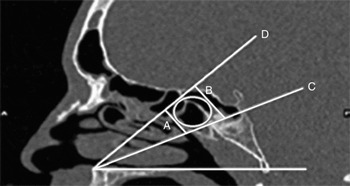
Intra-operatively, the surgeon could advance a ball-ended probe 5.9 cm from the nasal spine, or 4.8 cm from the ipsilateral piriform aperture, along the nasal floor, then elevate it to an angle of 22° and move the probe's tip to touch the lateral nasal wall. The ball of the probe would then indicate the centre of a circle marking the most likely location of the sphenopalatine foramen (Figure 1).
Sagittal computed tomography scan showing the measurements taken and the most likely location of the sphenopalatine foramen. A = minimum distance from anterior nasal spine to sphenopalatine foramen (=55 mm); B = maximum distance from anterior nasal spine to sphenopalatine foramen (=63 mm); C = minimum angle of elevation from nasal floor to sphenopalatine foramen (=18°); D = maximum angle of elevation from nasal floor to sphenopalatine foramen (=26°); circle = most likely location of sphenopalatine foramen

We stress that the use of such parameters is not a substitute for detailed knowledge of the relevant anatomy, which remains essential.
We have also shown that the BrainLAB surgical navigation system is a useful and reliable tool for obtaining anatomical measurements, and could potentially be used for training and validation.