In the 19th and early 20th centuries the brain was very much shrouded in mystery and as such was considered a ‘black-box’. The greatest advance to emerge during this period was the discovery of regional specialisation, namely, that different brain areas serve different functions. One of the earliest examples of this is documented in the work of German physiologists Fritsch and Hitzig who when electrically stimulating a patch of cortex in the brain of a dog observed movement in its right front paw. When the same patch of cortex was surgically removed from two other dogs, they noticed that the dogs not only could not use the paw, but also seemed unaware of it. These early rudimentary studies questioned the ‘black-box’ concept and precipitated a surge in the study of the brain over the ensuing half-century in which scientists poked, prodded, stimulated and removed portions of animal (and human) brain in an attempt to uncover its mysteries. Even with so much ‘brain-power’ being directed towards demystifying the workings of the brain, progress was slow and it is only more recently that significant advances have been made. In particular, over the past several decades technological advances in physics and computing have contributed to the development of sophisticated technology, which permits an unprecedented ability to dissect the ‘working brain’ and better understand its neurobiology.
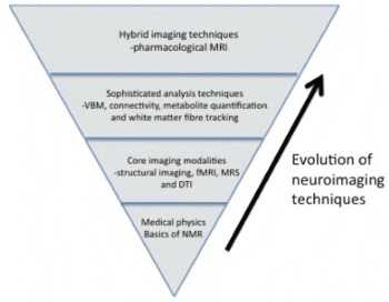
In this thematic issue of Acta Neuropsychiatrica, dedicated to imaging, the opportunity arises to chronicle some of the imaging techniques that have been influential in advancing our understanding of brain function in neuropsychiatry. The various techniques and modalities that have come together to form the field of neuroimaging can be conceptualised as a pyramid (Fig. 1). At the very top of the pyramid is the basic science that underpins the imaging technique of nuclear magnetic resonance (NMR) from which magnetic resonance imaging (MRI) has evolved. The next layer represents the various core modalities that have been developed as a direct result of NMR, and beneath this layer reside the analysis techniques that have been developed to unlock the potential of the various modalities. Finally, the bottom layer represents the latest hybrid-imaging modalities to emerge that incorporate external manipulations to normal brain function to investigate brain dynamics.
Schematic representation of the evolution of neuroimaging techniques.

It is self-evident that discoveries in the field of medical physics have spawned the development of the vast array of imaging techniques that we have at our disposal today. In particular, the field of NMR has been integral to developing techniques such as structural imaging, functional MRI and in vivo spectroscopy, which have allowed unprecedented investigations of brain structure, function and neurochemistry, respectively. Since its inception in the early 1970s, MRI has evolved into an indispensable modality in both the clinical and research domains as it provides us with a completely non-invasive tool to study the brain. Although in its early days MRI was used primarily for structural imaging, its role in research has quickly grown owing to its ability to provide functional and biochemical measures. Throughout the history of MRI, its development has been related to the increase of magnet strength and this increase in strength, despite early doubts, has resulted in more sophisticated imaging techniques Reference Sharma(1). Moreover, progress in the MRI field has been accelerated through iteration between an increasing knowledge base and methodological developments. There are essentially four core-imaging modalities (structural imaging, functional MRI, spectroscopy and diffusion tensor imaging) that are derivations of MRI and each of these has significantly impacted on our understanding of brain function.
The first of these modalities is structural imaging. Using computational imaging techniques brain tissue segmentation can be measured very accurately. The basic premise of structural imaging is to make inferences about the brain of separate groups on the basis of differences in tissue classifications. Structural imaging techniques are relatively straightforward and as computing power is relatively inexpensive and easily attainable, most of the computation can be automated, minimising the need for expert-user intervention Reference Lagopoulos(2). These techniques have enhanced our understanding of structure-behaviour relationships. Similarly, in the rapidly growing field of cognitive neuroscience, functional MRI has revolutionised the manner in which brain function is studied by providing a mechanism for examining the working of the brain Reference Sharma(3). Using blood oxygenated level-dependent (BOLD) imaging, it is possible to non-invasively obtain information relating to brain function. The BOLD signal indirectly measures neural activity by localising the metabolic activity that follows, on the basis of changes in blood flow associated with mental activity. Functional MRI has been a revolutionary development of the past decade, which has made it possible to non-invasively map areas of increased neuronal activity in the human brain without the use of an exogenous contrast agent Reference Sharma(4). The functional MRI methodology is particularly useful in neuropsychiatry for investigating issues such as aberrant circuitry involved in processing emotion and decision-making Reference Sharma(3) and this modality of research now makes up the majority of the psychiatry neuroimaging literature.
Another imaging modality that has gained prominence is that of magnetic resonance spectroscopy (MRS). MRS has traditionally been used as a diagnostic tool in the biochemical characterisation of pathophysiological processes Reference Saether, Das and Lagopoulos(5). However, in recent years, in line with increases in magnet-field strength, MRS has seen a resurgence in its use and uptake. While in the early days of development it was used predominantly to characterise brain tumours, abscesses and brain-related changes associated with stroke, it is now increasingly and successfully applied to neuropsychiatric disorders to investigate underlying neurochemistry. MRS allows the non-invasive determination and quantification of metabolites in specific brain regions, essentially enabling direct ‘chemical sampling’ of the brain's metabolites. However, unlike MRI, which uses the signal from protons to construct anatomical images, MRS determines metabolite concentrations and accordingly data is depicted as a spectrum of individual metabolite concentrations. Nowadays, MRS is being used to elucidate changes in neurotransmitter systems such as those underpinned by glutamate and GABA both of which are implicated in psychiatric disease. In additional, MRS can also be used to investigate brain energetics, response to treatment such as lithium as well as receptor binding efficiency.
The final core modality that warrants attention is that of diffusion tensor imaging (DTI). Unlike the structural imaging and fMRI which largely rely on the analysis of relationships to grey matter anatomy, investigation into the brain's white matter tracts is inherently complex and thus determining functional associations has been difficult and until recently, very limited. The main reason for this has been the fact that white matter is more opaque than grey matter when using routine imaging techniques and as such is less visible Reference Lagopoulos(6). However, a profound discovery made towards the end of the 1990s revealed that it was possible to identify the brain's underlying neuronal microstructure, normally ‘invisible’, using conventional structural imaging. By using conventional diffusion weighted MRI and measuring the rate at which water molecules diffuse within the brain, it was possible to determine the paths followed by water allowing for the first time the visualisation of the location, orientation and the anisotropy of the brain's white matter tracts. The ability of DTI in determining the white matter architecture is unparalleled by any other imaging modality and this modality is leading the way in studies of connectivity.
Clearly, the advent of the aforementioned core-imaging modalities has been a significant advance; however, the accompanying development of sophisticated algorithms and analysis techniques that allow the raw signal derived from these modalities to be meaningfully interrogated has been equally important. Within the structural modality, for instance, voxel-based morphometry (VBM) has been widely adopted because of its ability to make inferences on the basis of differences in tissue classifications Reference Lagopoulos(2). In the functional domain, the general linear model (GLM) has provided the foundation for all fMRI analyses with newer approaches, such as those described within the frameworks of connectivity Reference Das and Lagopoulos(7), shedding new light on the integration within and between specialised brain regions. These connectivity analytical approaches allow greater inferences on a network level rather than simply depicting brain function as static activation foci on brain maps. Within the MRS modality, considerable development efforts have focused on the processes by which MRS data are collected and post-processed allowing the actual in vivo metabolite concentrations for the sampled voxel of interest to be determined Reference Lagopoulos(8). The advantage of obtaining absolute measures is that it provides an unambiguous representation of the change in metabolite concentrations independent of any secondary influence of other metabolites. This is important especially in neuropsychiatry where there may be changes in several observed metabolites or even T1 and T2 relaxation effects. Finally, advanced fibre-tracking algorithms have been developed for the DTI modality Reference Lagopoulos(6), which makes it possible to reconstruct 3D trajectories of white matter tracts.
Looking to the future, understanding the neural substrates underlying modification of neurotransmitter systems is emerging as a significant research domain and accordingly is critical in the development of suitable drug therapies for neuropsychiatric disorders. To this end, hybrid-imaging modalities have been developed in an effort to disentangle complex brain-behaviour relationships. Psychophysiological MRI is leading the way in investigating the effects of psychoactive substances on human brain functioning Reference Rasmussen(9). The application of microangiographic techniques to pharmacology is also being used to show the regions of the brain that respond to different psychotropics. This novel and exciting application of existing imaging techniques holds great promise in providing quantitative biomarkers of psychotropic drug effects as well as offering new insights into which patients are likely to respond to psychopharmacotherapy and is an invaluable tool in the development of novel drugs.
Over the past several decades, neuroimaging has made a tremendous impact upon our understanding of the brain. While currently the majority of neuropsychiatric neuroimaging is research-orientated, it is important to note its enormous clinical potential and understand the rapid advances that have been made in a comparatively short period. Given this rapid progress it seems inevitable that a growing number of imaging modalities and techniques, some of which are mentioned in this issue of Acta Neuropsychiatrica, could soon be incorporated into routine clinical practice.