Persistent left superior caval vein draining into the left atrium is responsible for a right-to-left shunt that can be a source of oxygen desaturation and systemic embolism.Reference Troost, Gewillig and Budts 1 – Reference Kim, Park, Jung, Kim and Choi 4 Percutaneous closure using different devices is the preferred treatment in most centres;Reference Troost, Gewillig and Budts 1 – Reference Kim, Park, Jung, Kim and Choi 4 however, because of the rarity of this congenital abnormality, there are only a few case reports published in the literature describing the use of different devices while providing some procedural suggestions and clinical experiences.Reference Troost, Gewillig and Budts 1 – Reference Kim, Park, Jung, Kim and Choi 4 Surprisingly, data regarding long-term outcomes are also lacking in the literature.
We aim to report our single-centre experience and provide long-term follow-up data by using contrast-enhanced CT.
Methods
From our centre’s clinical database, patients with persistent left superior caval vein draining into the left atrium were selected, excluding patients with heterotaxy syndromes and patients with re-opening of the persistent left superior caval vein after bidirectional cavopulmonary connection.Reference Filippini, Ovaert, Nykanen and Freedom 5 A total of three patients were included in the present study. All of them underwent percutaneous treatment in our centre between 2005 and 2015. Pre- and post-procedural clinical documents as well as echocardiographic and catheterisation videos and images were retrospectively reviewed. The study was approved by the Institute’s Ethics Committee. Informed, written consent was obtained from all patients/parents, and all three patients underwent standard contrast-enhanced CT without electrocardiogram-synchronisation 1, 2, and 10 year(s) after the left superior caval vein occlusion, respectively.Reference Ou, Celermajer, Calcagni, Brunelle, Bonnet and Sidi 6 , Reference Marini, Defilippi, Bordese and Agnoletti 7 From the conventional angiographic or fluoroscopic images, measures of the left superior caval vein diameter, device waist diameter, and height were obtained in the anteroposterior view and in the lateral view when available. From the set of CT images, the same measures were taken from the multiplanar reconstructions using the standard double-oblique technique.Reference Schoenhagen, Numburi and Halliburton 8
Results
Patients
Patients’ characteristics are summarised in Table 1. In our study, one patient had persistent left superior caval vein draining into the left atrium (Patient 1); two patients had persistent left superior caval vein draining into the left atrium connected to the right superior caval vein through a bridging vein (Patients 2 and 3). Associated abnormalities were patent foramen ovale in Patient 1 and restrictive perimembranous ventricular septal defect in Patient 2.
Patients’ characteristics.

LA=left atrium; LSVC=left superior caval vein; yr=years
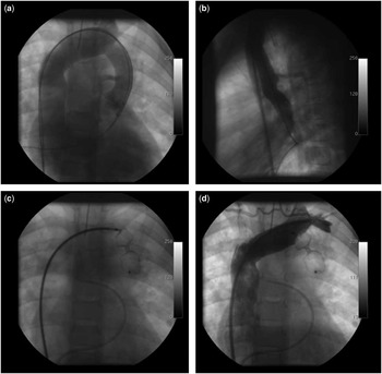
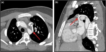
Age at diagnosis was 29 months, 5 years, and 34 years, respectively. In Patient 1, left superior caval vein draining into the left atrium was suspected because of low oxygen saturation (90%), whereas in Patient 3 the suspicion was cyanosis (93%) and multiple episodes of arterial embolism and thrombosis of lower limbs. In Patient 2, initially, partial anomalous pulmonary venous return was suspected because of progressive right ventricle dilation. Surprisingly, cardiac catheterisation showed a normal pulmonary venous return, but revealed the presence of a persistent left superior caval vein with a left-to-right shunt from the left atrium back to the innominate vein (Fig 1). In the absence of left-sided obstructive disease, we speculate this occurred because of abnormal left ventricle compliance. Evaluation of the levoatriocardinal vein was considered, but was eventually excluded in this patient because the vein was anterior to the left pulmonary artery (Fig 2).Reference Marini, Agnoletti and Pace Napoleone 9
Percutaneous occlusion of the left superior caval vein in Patient 2. Left superior caval vein angiography showing a retrograde shunt from the innominate vein to the right superior caval vein in the anteroposterior view ( a ). Please note the accessory hemiazygos vein in the lateral view ( b ). Amplatzer® Septal Occluder deployment through the right femoral vein ( c ). Innominate vein angiography excluding the right-to-left shunt; however, the patency of the accessory hemiazygos vein could not be assessed after left superior caval vein occlusion ( d ).

Contrast-enhanced CT in Patient 2; 10 years after the procedure, CT angiography revealed a persistent left-to-right shunt through a patent accessory hemiazygos vein (red arrow) in the axial view ( a ) and the sagittal/oblique view ( b ).

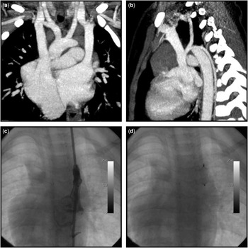
In Patients 1 and 3, clinical suspicion of left superior caval vein draining into the left atrium was confirmed by contrast-enhanced CT (Fig 3).
In Patient 1, contrast-enhanced CT was useful to confirm the clinical suspicion of left superior caval vein draining into the left atrium, excluding the presence of a bridging vein connecting the right superior caval vein and the left superior caval vein ( a ). Please note the accessory hemiazygos vein draining into the left superior caval vein in the sagittal/oblique view ( b ). Percutaneous occlusion of left superior caval vein was obtained by the left internal jugular vein approach. Post-procedural left superior caval vein angiography showed residual intradevice shunt ( c ). Please note the dog-bone shape of the Amplatzer® Vascular Plug type-1 ( d ).

Procedural data and early results
Procedural data and early results are summarised in Tables 2a and 2b. The procedure was performed through the left internal jugular vein in Patients 1 and 3 as previously reported,Reference Abadir, Bouzguenda, Boudjemline and Agnoletti 3 and through the right femoral vein in Patient 2. In Patients 1 and 3, the Amplatzer® Vascular Plug type-I (St. Jude Medical, St. Paul, Minnesota, United States of America) was preferred, whereas in Patient 2 the procedure was performed in 2005 when Amplatzer® Vascular Plug devices were unavailable in the market. In this case, the operator chose the off-label use of an Amplatzer® Septal Occluder. This was preceded by a balloon test occlusion as described by Dehghani et alReference Dehghani, Benson and Horlick 2 , which showed an increasing wedge capillary and increasing left atrial pressures from 12 and 11 mmHg to 17 and 16 mmHg, respectively.
Procedural data.

LIJV=left internal jugular vein; RFV=right femoral vein
Early results.

ASO=Amplatzer Septal Occluder; AVP=Amplatzer Vascular Plug; LSVC=left superior caval vein
In Patients 1 and 3, the device size/left superior caval vein diameter ratio was 1.7 and 1.2, respectively. For Patient 2, an Amplatzer® Septal Occluder measuring 11 mm was chosen, as the child had a left superior caval vein measuring 10 mm in diameter.
There was a mild intradevice residual shunt in all patients immediately after the procedure (Fig 3). The measured height/nominal height ratio of the plugs was 2.2 and 1.4, and the measured waist/nominal diameter ratio was 0.7 and 0.9 in Patients 1 and 3, respectively.
In Patient 2, the Amplatzer® Septal Occluder device was used as a rough cork; owing to its double-disc morphology and abnormal deployment, which caused it to resemble the Amplatzer® Vascular Plug type-4 (Fig 1), we judged that fine measurements of device deformation after deployment were of no interest in this case, and thus we have not reported them in the Results section or in any tables of this article.
Fluoroscopy time, contrast amount, and procedural time were 2 and 4 minutes, 2 and 3 ml/kg, and 35 and 40 minutes in Patients 1 and 3, respectively. In Patient 2, which included a balloon test occlusion, the values were 55 minute, 6.5 ml/kg, and 90 minute, respectively.
None of the patients had superior caval vein syndrome. All patients were discharged the day after the procedure without complications. Patient 1 had mild intradevice residual shunt at discharge, which may explain the absence of any clinical signs of superior caval vein syndrome. This young patient had an isolated, left superior caval vein draining into the left atrium without a bridging vein (Fig 3). We speculate that a progressive rather than sudden occlusion of the left superior caval vein by intradevice coagulation might have avoided the onset of clinical signs of superior caval vein syndrome, leaving enough time for an asymptomatic re-distribution of systemic return into the venous system.
Long-term results
The long-term results are summarised in Table 3. The last clinical and echocardiographic evaluations were performed 1, 2, and 10 years after the interventional procedure, respectively. All patients had transcapillary oxygen saturation of 100%. In Patients 1 and 3, contrast-enhanced CT excluded any residual shunt. In Patient 2, the device was positioned above the drainage of the accessory hemiazygos vein, which served as a persistent way for retrograde left-to-right shunt as demonstrated in Figure 2; however, the right ventricle remained undilated, and the shunt was judged as trivial.
Long-term results.

LSVC=left superior caval vein; yr=years
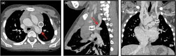
In Patient 1, the plug occluded the left superior caval vein obstructing the drainage of the accessory hemiazygos vein into the left superior caval vein and averted any residual right-to-left shunt (Fig 4). In Patient 3, the accessory hemiazygos vein did not drain into the left superior caval vein, but into the azygos vein, as seen in the most common normal variant.Reference Blackmon and Franco 10
Contrast-enhanced CT 1 year after percutaneous occlusion of the left superior caval vein in Patient 1 showing persistent patency of the accessory hemiazygos vein (red arrow, a and b ) but complete occlusion of the proximal left superior caval vein below the device as shown in the sagittal/oblique view ( b ) and in the coronal view ( c ). Please note that the device completely regained its original unstretched shape and its nominal size.

At CT evaluation, in Patients 1 and 3, measurements of the waist and height of the plugs were equivalent to the nominal ones. The left superior caval vein diameters above the devices were smaller than the diameters immediately after the procedure, leading to a measured waist/left superior caval vein diameter ratio of 2.3 and 1.8 in Patient 1 and Patient 3, respectively.
Discussion
In this study, we report the long-term outcome of percutaneous closure of persistent left superior caval vein in three patients. Despite the limited number of patients, we found that percutaneous closure of persistent left superior caval vein draining into the left atrium by using the Amplatzer® devices is safe and effective. We found no immediate or late complications.
To the best of our knowledge, there are only a few case reports in the literature providing some useful information and tips regarding the technique and the immediate post-procedural outcome;Reference Troost, Gewillig and Budts 1 – Reference Kim, Park, Jung, Kim and Choi 4 however, there are no data in the literature reporting on the long-term safety and efficacy of this approach.
Normally, standard follow-up of these patients consists of oxygen saturation measurements and echocardiography with Doppler evaluation aimed at detecting residual shunts. In our study, however, we wanted to provide more detailed information regarding the fate of the shunt and of the devices; hence, we decided to perform a contrast-enhanced CT in our patients. Avoiding the use of ionising radiation, cardiac MRI would have been the ideal technique for this study; however, it is still hampered by significant artefacts in the presence of a nitinol stent or Amplatzer® devices, complicating a proper angiographic evaluation of small vessels.
The first interesting finding of this study is that if implanted above the accessory hemiazygos drainage a persistent residual shunt may occur. Accessory hemiazygos veins may frequently drain into the left superior caval vein. In this sense, we recommend performing initial angiography in both anteroposterior and lateral views, in order to obtain proper visualisation or to exclude the drainage of the accessory hemiazygos vein into the left superior caval vein. In the presence of accessory hemiazygos veins, the plug must be implanted below its drainage to avoid any persistent residual shunt. In Patient 2, the shunt persisted for more than 10 years. By provoking only a trivial left-to-right shunt without right ventricular overload, we think there is no indication for further procedures, which, in the case of a significant shunt, would have been possible only through a trans-septal atrial approach.
Second, these data show that even at mid-term follow-up, an oversized plug tends to return to its original unstretched shape, regardless of implantation in a growing child or an adult. When an Amplatzer® Vascular Plug type-1 is oversized in comparison with the venous vessel, it immediately assumes a dog-bone aspect that tends to disappear relatively early to regain the nominal size. In Patients 1 and 3, the measured height/nominal height ratio of the plugs decreased, respectively, from 2.2 and 1.4 immediately after the procedure to 1.0 at the CT evaluation, whereas the measured waist/nominal diameter ratio increased from 0.7 and 0.9 to 1.0. In other words, over time, the device forces the compliant venous vessel to contain its original shape and nominal dimensions. This was also evident when comparing the device shape at fluoroscopy, as shown in Figure 3d with the same device shape only 1 year later (Fig 4c). It can be argued that the diameter of the left superior caval vein might increase with time, allowing the plug to regain its nominal shape and size; however, our data are contrary to this, because the left superior caval vein diameters measured during CT above the devices were smaller than the diameters measured immediately after the procedures, both in the adult and the child. We speculate that over time the left superior caval vein occlusion might provoke a preferential drainage of the left systemic venous return into the right superior caval vein through collateral veins, gradually emptying the residual distal left superior caval vein.
Most authors use a device size/vessel diameter ratio between 1.3 and 1.5 to embolise vessels by using the Amplatzer® Vascular Plug devices.Reference Blackmon and Franco 10 Our experience suggests that the limits of this range might be very wide, at least for venous embolisation. The youngest patient – that is, Patient 1 – had a device size/left superior caval vein diameter ratio of 1.7, and the adult patient had a device size/left superior caval vein diameter ratio of 1.2. In both cases, we obtained a complete and definitive occlusion of the left superior caval vein without any complication. We speculate that a device size/left superior caval vein diameter ratio between 1.2 and 1.5 in adults and between 1.5 and 1.7 in children might be reasonable.
Some authors support the use of balloon test occlusion to evaluate the presence of the bridging vein or of collaterals and their haemodynamic tolerance to the sudden closure of the left superior caval vein.Reference Dehghani, Benson and Horlick 2 This was performed in Patient 2 to evaluate left atrial pressure. As this test prolongs fluoroscopy and procedural times as well as increases the total amount of contrast, we do not perform it routinely in our clinical practice if we have to perform high-quality preoperative CT evaluation, as in Patients 1 and 3. Compared with MRI, CT has better spatial resolution, and compared with contrast-enhanced MRI angiography CT angiography injects more volumes at higher velocities, resulting in significant augmentations of systemic venous pressure, hence providing more capacity to recruit and visualise all of the possible collateral veins. Non-invasive diagnostic tests cannot predict post-procedural systemic venous pressure elevation and clinical evidence of left superior caval vein occlusion; however, as we have described in a previous experience, even if caval syndrome occurs, it is transient and clinically well tolerated.Reference Kim, Park, Jung, Kim and Choi 4
Diagnosis of persistent left superior caval vein draining into the left atrium is rare and might be challenging; however, when an asymptomatic patient with a normal heart has low oxygen saturation or recurrent paradoxical embolisms, an anomalous drainage of the systemic veins into the left atrium or the pulmonary vein should always be kept in mind.Reference Dehghani, Benson and Horlick 2 Patients with congenital or progressive pulmonary arteriovenous fistulas, as in Rendu–Osler–Weber disease, may have similar clinical presentations.Reference Dehghani, Benson and Horlick 2 , Reference Lacombe, Lacout and Marcy 11 Contrast-enhanced echocardiography is the first-line diagnostic tool in cyanotic, asymptomatic patients.Reference Troost, Gewillig and Budts 1 – Reference Kim, Park, Jung, Kim and Choi 4 , Reference Gupta and Pearson 12 Of note, contrast-enhanced echocardiography should always be performed by injecting the agitated saline from the left arm for appropriate diagnosis;Reference Gupta and Pearson 12 however, frequent presence of the bridging vein or of interatrial septal defects may be confounding factors for the diagnosis. When contrast-enhanced echocardiography is not conclusive, cardiac MRI is the best diagnostic tool to study the systemic venous return for definitive diagnosis of persistent left superior caval vein draining into the left atrium, devoid of radiation exposure. Owing to the precise capability to study dimensions and positions of small vessels in free-breathing patients, however, contrast-enhanced CT may be an acceptable alternative in our opinion, playing a significant role in the differential diagnosis of pulmonary arteriovenous fistulas and abnormal systemic drainage.Reference Lacombe, Lacout and Marcy 11
In conclusion, percutaneous occlusion of left superior caval vein draining into the left atrium is safe and effective both at early and long-term follow-up. Even if limited by the small number of cases reported, this study provides interesting insights concerning the procedure and the follow-up, suggesting that in order to avoid any residual and persisting shunt, the device has to be positioned below the drainage of the accessory hemiazygos vein; furthermore, the device size may be chosen within a wide range of device size/left superior caval vein diameter ratios according to the age of the patient.
Acknowledgements
The authors thank Jessie Mei Lim for providing English language revision.
Financial Support
This research received no specific grant from any funding agency or from commercial or not-for-profit sectors.
Conflicts of Interest
None.
Ethical Standards
The authors assert that all procedures contributing to this study comply with the ethical standards of the relevant national guidelines on human experimentation and with the Helsinki Declaration of 1975, as revised in 2008, and has been approved by the institutional committees.