
Introduction
Electrochemical migration (ECM)[Reference Kohman, Hermance and Downes1] has been studied as a more environmentally friendly method for fabricating micro- and nanostructures[Reference Fukaya, Aoki, Kimura and Saka2–Reference Aoki, Li and Saka4] used in a variety of applications. ECM is capable of synthesizing micro/nanodeposits with various micro- and nanostructures.[Reference Fukaya, Aoki, Kimura and Saka2–Reference Aoki, Li and Saka4] In addition, ECM uses water, metallic electrodes, and applied voltage, avoiding the use of toxic substances and large amounts of energy.
ECM is an electrochemical phenomenon like plating, electrolysis, and corrosion. ECM occurs when DC voltage is applied to metallic electrodes in water, causing ionic dissolution, accumulation, migration, and deposition.[Reference Yang, Wu and Christou5] At first, metallic ions dissolve and accumulate in the water around the anode. Next, ions migrate to the cathode by an electrical force. Finally, ions acquire electrons and deposit on the cathode. As the deposit grows, conductive filaments are formed and get longer until a short circuit between electrodes occurs, halting further deposit growth.[Reference Fukaya, Aoki, Kimura and Saka2] Even if a few water absorbed from the air exist between electrodes in electronic circuit, ECM can occur and causes the failure of the electronic circuit. Therefore, the focus of conventional research on ECM in aqueous solutions has been to find ways to inhibit ECM from occurring.[Reference Minzari, Grumsen, Jellesen, Møller and Ambat6–Reference Zhong, Chen, Madgyes, Zhang, Gao and Jalab10]
Studies have been carried out to investigate the influences of experimental system configuration,[Reference Fukaya, Aoki, Kimura and Saka2] electrical field,[Reference Nakakura and Saka3] and temperature[Reference Aoki, Li and Saka4] on ECM and ECM deposits growth. However, knowledge about using ECM to fabricate micro- and nanostructures is lacking because the history of the field is short. In this study, the influence of ultrasonic irradiation on ECM is examined, with a goal toward using ultrasonic irradiation to accumulate knowledge and propose the effective way of ultrasonic irradiation as a control factor in ECM deposit growth. The influence of ultrasonic irradiation on both the current during ECM and the morphology of deposits formed are discussed based on the experimental results.
Materials and methods
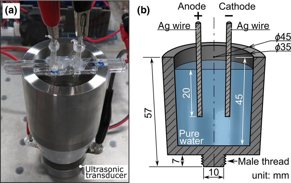
ECM deposits were formed via water drop tests (WDTs) in a 40 kHz ultrasonic bath with pure water. The electrical resistivity was 8 MΩ m, and the purity of the Ag electrodes was 99.99%. A photograph and a schematic of the ultrasonic bath are shown in Fig. 1. The ultrasonic bath consists of a stainless steel bath and an ultrasonic transducer. The stainless steel bath was machined from a round bar having a diameter of 48 mm, with a male thread at the bottom. At the top of the bath wall, two square notches were cut to attach electrodes. The transducer (Shinka Industry Co., Ltd., Kawasaki, Japan, CCH-4538D-40LB) has a female thread at the top center. The stainless bath and the transducer were connected by the threads and strengthened with epoxy adhesives.
(a) Photograph of ultrasonic bath with attached electrodes and ultrasonic transducer and (b) cross-sectional schematic of the stainless bath chamber with dimensions indicated.

Two round Ag electrodes with a 2 mm diameter were inserted into the bath. The electrode pitch was 13 mm from the center to center. A portion of each electrode was covered with a silicon tube to prevent contact at the water's surface. Avoiding such contact prevents deposits from forming on the water's surface independent of any influence from the ultrasonic irradiation. The electrodes and tubes were fixed to an acrylic resin beam, and the beam was placed into the notches on the bath wall. Pure water was poured into the bath to a height of 36 mm. Electrodes were immersed to a depth of 20 mm. DC voltage (40 V) and ultrasonic irradiation were applied for 15 min immediately after water was poured into the ultrasonic bath. DC voltage was applied to the electrodes constantly by a source meter. The current flowing between the electrodes was logged by the source meter. Ultrasonic irradiation was applied via the transducer driven by an ultrasonic oscillator (Shinka Industry Co., Ltd., SK-40-50GTJ). In this study, both continuous and pulsed ultrasonic irradiations were carried out. For pulsed irradiation, ultrasonic waves were applied in intervals of 3 s irradiation followed by 30 s of no irradiation. During both pulsed and continuous ultrasonic irradiations, the output settings on the oscillator were identical; however, as mentioned in the Results and discussion section, we observed minor differences in the oscillator's power consumption as measured by a power consumption meter connected between the oscillator and a commercial power supply.
After each test, the contents of the bath were stirred to disperse deposits uniformly and transferred to test tubes. After 22 h, deposits were collected from the bottom of the test tubes and dropped onto Cu films heated by a hot plate set at 120 °C to evaporate moisture quickly. Deposits on Cu films were observed by a scanning electron microscope (SEM).
Results and discussion
Pulsed ultrasonic irradiation grew filament-like nanostructures from the cathode in water, which can be confirmed enough with the naked eye. During each pulse, some filaments were fractured by ultrasonic irradiation and then fragmented filaments stay under water as deposits; however, since not all of them were fragmented, they grew longer with time until a short circuit occurred when longer deposits eventually formed a bridge (see Supplementary Fig. S1). In contrast, continuous irradiation formed no filaments similar to that formed by pulsed irradiation. We presume that most of the filaments became deposits by fracturing those due to mechanical effect of shock waves induced by cavitation in the ultrasonic irradiation.
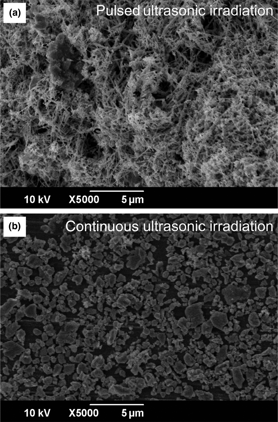
SEM images of deposits gathered from the bottom of the test tubes are shown in Fig. 2. Deposits formed during pulsed irradiation are composed of wires, dendrites, and particles [Fig. 2(a)]. During continuous irradiation, only particles formed [Fig. 2(b)]. The diameter d of those deposits was approximately measured by image-processing of FE-SEM images shown in Fig. 2. The 100 specimens that were randomly chosen were used for the measurement of d. The diameter of wire-deposits synthesized by pulsed irradiation was defined as the value of d. Since the particle-deposits synthesized by continuous irradiation have irregular shapes, the shortest length of particle-deposits was defined as the value of d for the underestimation of differences of d in the pulsed and continuous irradiations. The wire-deposits by pulsed irradiation have d of 140 nm smaller than that of 600 nm in the particle-deposits by continuous irradiation. The deposit colors were different: deposits formed during pulsed irradiation were black, and those formed during continuous irradiation were whitish gray. The number of whitish-gray deposits was relatively few compared to the number of black ones. Interestingly, the differences in ultrasonic irradiation induce the morphological change of deposits, although the irradiation time is considered to just reduce the length of deposits. To discuss the effect of ultrasonic irradiation on the morphological change of deposits synthesized by ECM, the following measured current in ECM was used.
SEM images of ECM deposits formed with (a) pulsed and (b) continuous ultrasonic irradiation.

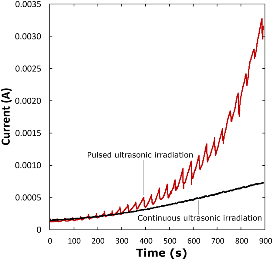
Figure 3 shows the change in current over time during the experiments. Regardless of the type of ultrasonic irradiation used, current increased with time. These increases in currents in pulsed and continuous irradiations are due to increasing metallic ions which are dissolved from anode electrode into the water: in the ECM, the initial pure water has no ions ideally. The metallic ions which work as carriers increase with time during DC voltage supply. On the other hand, the rate at which the current increased during continuous irradiation was lower than that during pulsed irradiation. Whereas the current increase is steady and more linear during continuous irradiation, the current curve follows a sawtooth-like pattern during pulsed irradiation. The cycle of the sawtooth-like curve followed the cycle of pulsed ultrasonic irradiation. Immediately after each ultrasonic pulse ended, the current was always lower than that immediately before the start of the next irradiation pulse. These results suggest that ultrasonic irradiation reduced the current during ECM via WDT.
Relationship between current and time of ECM via WDT with pulsed (the red line) and continuous (the black line) ultrasonic irradiation.

As mentioned in the Materials and methods section, minor differences in the oscillator's power consumption exist. During continuous irradiations, oscillator power consumption was 10 W, as measured by a power consumption meter connected between the oscillator and a commercial power supply. During pulsed irradiations, the power consumption changed, with an upper limit of 11 W during pulses, although the output settings on the oscillator were the same as for continuous irradiations. This power consumption difference may have been caused by a change in resistance due to Joule heating and/or a response characteristic of the experimental system.
Let us consider the effect of ultrasonic irradiation on ECM in terms of the changes in current and deposit morphology. As a mechanical effect, ultrasonic irradiation mechanically fragments the filaments growing from the cathode, and thus, the resistance between the anode and the cathode, which is inversely proportional to current, becomes higher because the distance between the anode and dendrites growing from the cathode gets longer. Such a repetitive cycle of dendrite deposition and fragmentation is consistent with the sawtooth-like curve of current observed during pulsed ultrasonic irradiation (Fig. 3). In addition, the greater number of shock waves occur during continuous ultrasonic irradiation fractures filaments more effectively, thereby preventing the formation of wires and dendrites at the cathode, which is consistent with the particle formation observed [Fig. 2(b)]. The sawtooth-like curve is also seen at the beginning of the experiment before a deposit has yet formed. This fact suggests that a sonochemical effect due to ultrasonic irradiation contributes to changing the current. Some studies report ion reduction and particle synthesis in water-based electrolyte solutions, utilizing ultrasonic irradiation without any additives such as surfactant and reducer.[Reference Sakai, Enomoto, Torigoe, Sakai and Abe11–Reference Chen, Rajaji, Chen and Ramalingam14] The synthesis of Ag micro- and nanostructures from silver nitrate (AgNO3) solutions utilizing ultrasonic irradiation was also reported.[Reference Chen, Rajaji, Chen and Ramalingam14–Reference Wang, Fan, Li, Wu and Zhang17] Considering those reports of a sonochemical effect, it is presumed that this effect contributed to the current reduction and the deposit formation patterns we observed during ECM via WDT. The repetition of ion reduction by the pulsed ultrasonic irradiation, while ion dissolution continued by ECM could result in the sawtooth-like curve shown in Fig. 3. In addition, deposit growth would be inhibited by decreasing ionic concentration. The formation of only particles during continuous ultrasonic irradiation is also explained by this sonochemical effect and is consistent with the morphology differences observed under the two ultrasonic conditions (Fig. 2).
Conclusions
ECM via WDTs was carried out in a 40 kHz ultrasonic bath with either continuous or pulsed ultrasonic irradiation. Deposits formed during continuous irradiation consisted of only particles, whereas deposits formed during pulsed irradiation were composed of wires, dendrites, and particles. Ultrasonic irradiation also reduced the current flowing through the electrodes. Ultrasonic irradiation had a mechanical effect of fragmenting filaments and forming deposits during ECM, but its mechanical effect alone is not sufficient to explain the reduction in current that we observed. We conclude that ion reduction and particle synthesis occurred in these experiments as sonochemical effects, and such effects are consistent with the results we observed. Ion reduction causes current reduction and would inhibit the formation of long and dendritic filaments, thus increasing the percentage of particle formation during ECM.
Supplementary material
The supplementary material for this article can be found at https://doi.org/10.1557/mrc.2019.135.
Acknowledgment
This work was supported by JSPS KAKNEHI Grant-in-Aid for Scientific Research (B) No. 17H03139.