Introduction
Schistosomiasis is an important parasitic disease and it affects more than 218 million people worldwide (WHO, 2017). Blood fluke (Schistosoma mansoni, S. japonicum and S. haematobium) can cause schistosomiasis. In infected individuals, eggs can be transported to the host liver by portal circulation, and deposited in the portal venous system and become trapped in the perisinusoidal spaces of the liver (Chang et al., Reference Chang, Ramalho, Ramalho, Martinelli and Zucoloto2006). These can result in liver granulomatous, inflammation and deposition of scar tissue around the trapped eggs (Boros, Reference Boros1989; Andrade, Reference Andrade2009; Burke et al., Reference Burke, McManus, Ramm, Duke, Li, Jones and Gobert2010).
Schistosoma mansoni morbidity arises from granulomatous CD4+ T cell-mediated response to eggs trapped in the liver and intestinal tissues (Pearce & MacDonald, Reference Pearce and MacDonald2002; Layland et al., Reference Layland, Wagner and da Costa2005). Immunologically, an S. mansoni infection is dominated by two distinct Th phases: an initial Th1 (Interferon gamma, IFN-γ) response that then switches to a stronger Th2 (IL-10, IL-5 and IL13) response (Pearce et al., Reference Pearce, Caspar, Grzych, Lewis and Sher1991; Wynn et al., Reference Wynn, Cheever, Jankovic, Poindexter, Caspar, Lewis and Sher1995). These adaptive responses can be obtained ex vivo upon re-stimulation of antigen-specific CD4+ T cells with soluble egg antigen (SEA), prepared from S. mansoni eggs.
Inflammasomes are oligomeric molecular platforms that are critical regulators of innate immune and inflammatory responses (Lamkanfi et al., Reference Lamkanfi, Mueller, Vitari, Misaghi, Fedorova, Deshayes, Lee, Hoffman and Dixit2009; Kanneganti, Reference Kanneganti2010). Various Nod-like receptor (NLR) family members (i.e. NLRP1, NLRP3 and NLRC4) as well as the pyrin and hematopoietic interferon-inducible nuclear antigens domain-containing protein (PYHIN) family member AIM2 (absent in melanoma 2) can form inflammasome complexes. These multi-protein complexes contribute to the activation of inflammatory Caspase-1 that results in the proteolytic processing and secretion of cytokines, including interleukin IL-1β and IL-18 (Martinon, Reference Martinon2010). Different microbial pathogens, such as parasites, trigger inflammasome activation (Lamkanfi & Dixit, Reference Lamkanfi and Dixit2009). A previous study has shown that S. mansoni activates the NLRP3 inflammasome by triggering Dectin-2 in mouse bone marrow dendritic cells (Ritter et al., Reference Ritter, Gross, Kays, Ruland, Nimmerjahn, Saijo, Tschopp, Layland and Prazeres da Costa2010). The NLRP3 inflammasome was also activated in mouse liver during S. japonicum infection (Meng et al., Reference Meng, Xia, Lu, Wang, Boini, Li and Tang2016). Interestingly, some parasites may engage more than one inflammasome during infection, as seen in the case of malaria infection, where parasites can activate both NLRP3 and AIM2 inflammasomes (Kalantari et al., Reference Kalantari, DeOliveira and Chan2014).
During inflammation, damaged mitochondria generate high levels of reactive oxygen species (ROS) and release calcium and intrinsic apoptotic proteins (Llesuy et al., Reference Llesuy, Evelson, Gonzalez-Flecha, Peralta, Carreras, Poderoso and Boveris1994; Taylor et al., Reference Taylor, Ghio and Piantadosi1995; Kantrow et al., Reference Kantrow, Taylor, Carraway and Piantadosi1997). ROS are not only pro-inflammatory, but they also induce the expression of NF-κB and other transcription factors that regulate inflammation (Schreck et al., Reference Schreck, Rieber and Baeuerle1991; Koay et al., Reference Koay, Christman, Segal, Venkatakrishnan, Blackwell, Holland and Blackwell2001). Studies of NLRP3 activators have shown that ROS act as second messengers to drive inflammasome activation (Fukumoto et al., Reference Fukumoto, Fukumoto and Parthasarathy2013; Heid et al., Reference Heid, Keyel, Kamga, Shiva, Watkins and Salter2013). Prior to Caspase1-mediated proteolytic cleavage of IL-1β, Toll-like receptor (TLR) agonists facilitate the NLRP3 inflammasome pathway through an NF-κB-dependent priming signal, which may represent a rate-limiting step of inflammasome complex formation (Bauernfeind et al., Reference Bauernfeind, Horvath and Stutz2009). Our previous research demonstrated that S. mansoni infection caused mitochondrial dysfunction and triggered both the intrinsic and extrinsic apoptosis pathways in mouse livers eight weeks post-infection (Chen et al., Reference Chen, Xu, Li, Tao, Xiang, Huang, Niu, Zhong and Meng2015). Moreover, the NF-κB p65 subunit was also increased in S. mansoni-infected mice.
Here, we aimed to examine whether ROS and NF-κB (p65) expression elicited other types of inflammasome activation in S. mansoni-infected mouse livers. A previous study indicated that Hepatitis C virus (HCV), Ribonucleic acid (RNA) activated the NLRP3 inflammasome in a ROS-dependent manner in infected hepatic HuH-7 cells (Chen et al., Reference Chen, Wu, Hsu, Pang, Lee, Cheng and Peng2014). To demonstrate whether schistosomal components activate this inflammasome, we investigated inflammasome activation in HuH-7 cells treated with different doses of SEA for 12 and 24 h. In addition to the previous findings that S. mansoni infection induced apoptosis and mitochondrial dysfunction in mouse livers, we report here that the AIM2 inflammasome is also triggered by exposure to schistosomal antigen.
Methods
Parasites and experimental animals
Schistosoma mansoni (the Puerto Rican strain) was obtained from the Biomedical Research Institute (Rockville, MD 20852, USA) and had been maintained in our laboratory. Biomphalaria glabrata snails and eight- to ten-week-old male BALB/c mice were used as the intermediate and final hosts. The life cycle is maintained according to our previous study (Chen et al., Reference Chen, Xu, Li, Tao, Xiang, Huang, Niu, Zhong and Meng2015). BALB/c mice were purchased from the National Laboratory Animal Breeding and Research Centre, Taipei. Each group (including the uninfected and infected groups) for every experiment contained five mice. Each mouse was infected with 100–120 S. mansoni cercariae through percutaneous exposure of the tail. All protocols involving animals were approved by the regulation of the Institutional Animal Care and Use Committees of Tzu Chi University (no. 102068). All experimental procedures were carried out in accordance with the approved guidelines of the National Institutes of Health (NIH) Guide for the Care and Use of Laboratory Animals (Department of Health and Human Services publication no. NIH 85-23, revised 1996).
Isolation of RNA from liver tissue
Total RNA was extracted as we previously described (Chen et al., Reference Chen, Xu, Li, Tao, Xiang, Huang, Niu, Zhong and Meng2015). Total RNA was isolated from the livers of five normal and five infected mice using a Qiagen RNA Mini-kit (Qiagen, Hilden, Germany) according to the manufacturer's protocol. The RNA concentration of each sample was assessed using a Nanodrop ND-1000 spectrophotometer (Thermo Scientific, Waltham, MA, USA).
cDNA synthesis
Reverse transcriptase–polymerase chain reaction (RT–PCR) was performed according to the method previously described (Nagano et al., Reference Nagano, Pei, Wu, Wu, Cui, Boonmars and Takahashi2004; Chen et al., Reference Chen, Xu, Li, Tao, Xiang, Huang, Niu, Zhong and Meng2015), with a slight modification using a RevertAid First Strand cDNA Synthesis Kit (Fermentas International Inc., Ontario, Canada). The synthesized cDNA was stored at −20°C for further analysis.
Real time PCR (qPCR)
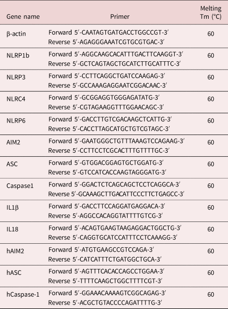
The qPCR assay was performed as described according to our previous study (Chen et al., Reference Chen, Xu, Li, Tao, Xiang, Huang, Niu, Zhong and Meng2015). This assay was performed using a SYBR qPCR kit (LabStar™, Taipei, Taiwan). PCR was performed by an initial single heating cycle (95°C for 30 s) followed by 45 cycles (95°C for 15 s, 60°C for 30 s, 70°C for 30 s). The primer pairs used in this study are listed in table 1. Each qPCR reaction was performed in triplicate for each gene, and the mean values were used for statistical analyses.
Primer pairs of candidate genes used in real-time PCR.

Total protein extraction
The protein samples were prepared according to our previous study (Chen et al., Reference Chen, Xu, Li, Tao, Xiang, Huang, Niu, Zhong and Meng2015). Liver tissues from uninfected and infected mice were washed with sterile phosphate buffered saline (PBS) (137 mm NaCl, 8.1 mm Na2HPO4, 1.5 mm NaH2PO4 and 2.7 mm KCl, pH 7.4) and reacted with 200 µl radioimmunoprecipitation assay buffer (1% Nonidet P40, 0.25% sodium deoxycholate, 150 mm NaCl, 1 mm EDTA, 1 mm phenylmethylsulfonyl fluoride, 1 mm sodium orthovanadate, 1 mm NaF, 10 µg/ml aprotinin, 10 µg/ml leupeptin and 50 mm Tris–HCl, pH 7.4). The cell lysates were incubated for 30 min at 4°C and centrifuged at 8100 ×g for 10 min at 4°C. The protein concentration in supernatant was detected by the Pierce™ BCA Protein Assay Kit (Thermo Scientific, Waltham, MA, USA).
Western blot
The assay was performed and modified as described according to our previous study (Chen et al., Reference Chen, Xu, Li, Tao, Xiang, Huang, Niu, Zhong and Meng2015). The protein concentration of each sample was determined using the Bio-Rad Protein Assay kit (Bio-Rad, Hercules, CA, USA). For each sample, 30 µg protein was loaded onto 10% SDS–PAGE gels. After electrophoresis, the proteins were transferred to a polyvinylidene difluoride membrane (0.45 µm, Merck Millipore, Cork, IRL) and blocked for 1 h with 5% non-fat milk in phosphate buffered saline with Tween-20 (PBST) (PBS with 0.05% Tween-20) followed by incubation with the primary antibody at 4°C. The membranes were blotted against the primary antibody. The primary antibody (polyclonal rabbit anti-AIM2, 1:1000, Abcam, USA; polyclonal rabbit anti-NF-κB (p65), anti-NLRP3, anti-Caspase1, anti-apoptosis-associated speck like protein containing a caspase recruitment domain (ASC) and anti-glyceraldehyde 3-phosphate dehydrogenase (GAPDH), 1:1000, Gene Tex, USA; anti-IL18 and anti-IL1β, 1:1000, Cell Signalling, USA) incubations were conducted at 4°C overnight. The membranes were washed three times with PBST, and incubated with the secondary antibody (Rabbit polyclonal IgG, 1:1000, GeneTex, USA) for 1 h at 4°C. The result was visualized by Immobilon Western Chemiluminescent HRP Substrate (Merck Millipore, Cork, IRL). Western blot images were scanned using a Gel Capture 2950 instrument (CulBio). GAPDH was assessed as an internal control for protein loading and did not vary between samples. Protein expression was analysed using the ImageJ software (Rasband, W.S., ImageJ, US National Institutes of Health, Bethesda, MD, USA, http://imagej.nih.gov/ij/, 1997–2011), and relative expression was normalized as percentage respective to the controls.
Detection of intracellular ROS by fluorescence microscopy
The protocols of collecting liver cells were modified according Allen et al. (Reference Allen, Dey, Nissly, Fraser, Yu, Balandaram, Peters and Hankey-Giblin2017). Liver samples from five uninfected and five infected mice were injected with PBS containing 5% fetal bovine serum (FBS) using a syringe needle (Gibco-BRL, Carlsbad, CA, USA), and the liver cells dissociated and released in Dulbecco's Modified Eagle Medium (DMEM; Gibco) by medium-point forceps. Cells were recovered by centrifugation (250 ×g, 5 min, 4°C) and were resuspended in red blood cell lysis buffer (0.15 M NH4Cl, 1 mm KHCO3 and 0.1 mm Na2EDTA, pH 7.2–7.4). Liver cells were again collected after centrifugation (250 ×g, 5 min, 4°C), resuspended in DMEM and subjected to flow cytometry analysis. To study the effect of S. mansoni infection in mouse hepatocytes, changes in total ROS/superoxide content were evaluated by 2′,7′-dichlorofluorescin diacetate (DCF-DA) and dihydroethidium (DHE) staining. Superoxide was measured using a fluorometric technique (ROS-ID Total ROS/Superoxide detection kit; Enzo Life Sciences Inc., Ann Arbor, MI, USA) that allows real-time measurement of superoxide and total ROS generation in cells. The kit contains a cell-permeable fluorescent dye (green: DCF-DA; orange probe: DHE) that specifically reacts with products of oxidative stress (Arredondo Zamarripa et al., Reference Arredondo Zamarripa, Diaz-Lezama, Melendez Garcia, Chavez Balderas, Adan, Ledesma-Colunga, Arnold, Clapp and Thebault2014), generating the green and orange fluorescent product that can be detected by fluorescence microscopy equipped with standard filter sets (Ex: 490/525 and 550/620 nm). Cells were stained with DCF-DA and DHE at 37°C for 30 min. Subsequently, cells were washed twice with PBS, and the cells were observed.
SEA extracts
Schistosome eggs from the livers of infected mice were isolated and SEA was prepared using modifications of previously described protocols (Schramm et al., Reference Schramm, Gronow and Knobloch2006). To obtain eggs for antigen preparation, mice were infected with 100–120 S. mansoni cercariae. Eight weeks after infection, the mice were sacrificed, liver portal veins were perfused to recover schistosome adult worms and the liver was taken from the mice. Livers were homogenized separately in 0.85% NaCl. Eggs were separated from liver tissue by passing the tissue through a series of sieves with decreasing pore size (420, 177, 105 and 25 µm). The eggs retained on the smallest pore-size sieve were collected in PBS on ice and washed ten times, followed by centrifugation at 370 ×g for 2 min. The centrifuged eggs were washed with PBS and stored at −80°C until further use. SEA was prepared by homogenizing the eggs in 1 ml PBS with a glass homogenizer.
Cell culture
HuH-7 cells were maintained in DMEM (Gibco) supplemented with 10% FBS (Gibco), 2 mm L-glutamine (Gibco) and 1% penicillin-streptomycin (Gibco) at 37°C and 5% CO2. Cells were maintained at sub-confluent densities and split every 2–3 days. For splitting, cells were washed in PBS (Invitrogen) and detached using trypsin (Sigma-Aldrich). HuH-7 cells were incubated with SEA concentrations of 5 µg/ml and 10 µg/ml in complete media for 12 h and 24 h, respectively. HuH-7 cells were stimulated with lipopolysaccharide (LPS; 100 ng/ml, Sigma-Aldrich) to serve as a positive control for inflammatory response. HuH-7 cells treated with media only served as negative controls.
Isolation of RNA from HuH-7 cells
Total RNA was extracted from HuH-7 cells using a Qiagen RNA Mini-kit (Qiagen, Hilden, Germany) according to manufacturer's instructions. The RNA concentration of each sample was assayed using a Nanodrop ND-1000 spectrophotometer (Thermo Scientific, Waltham, MA, USA).
Statistical analysis
Data are collected from three independent experiments and expressed as means ± standard deviation (number of mice in each group = 5). Statistical significance of data was analysed using t-tests. Statistical significance was defined as P < 0.05.
Results
Mice infected with S. mansoni for eight weeks show indications of granulomatous syndrome and inflammasome activation
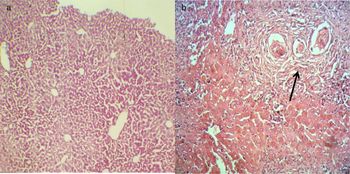
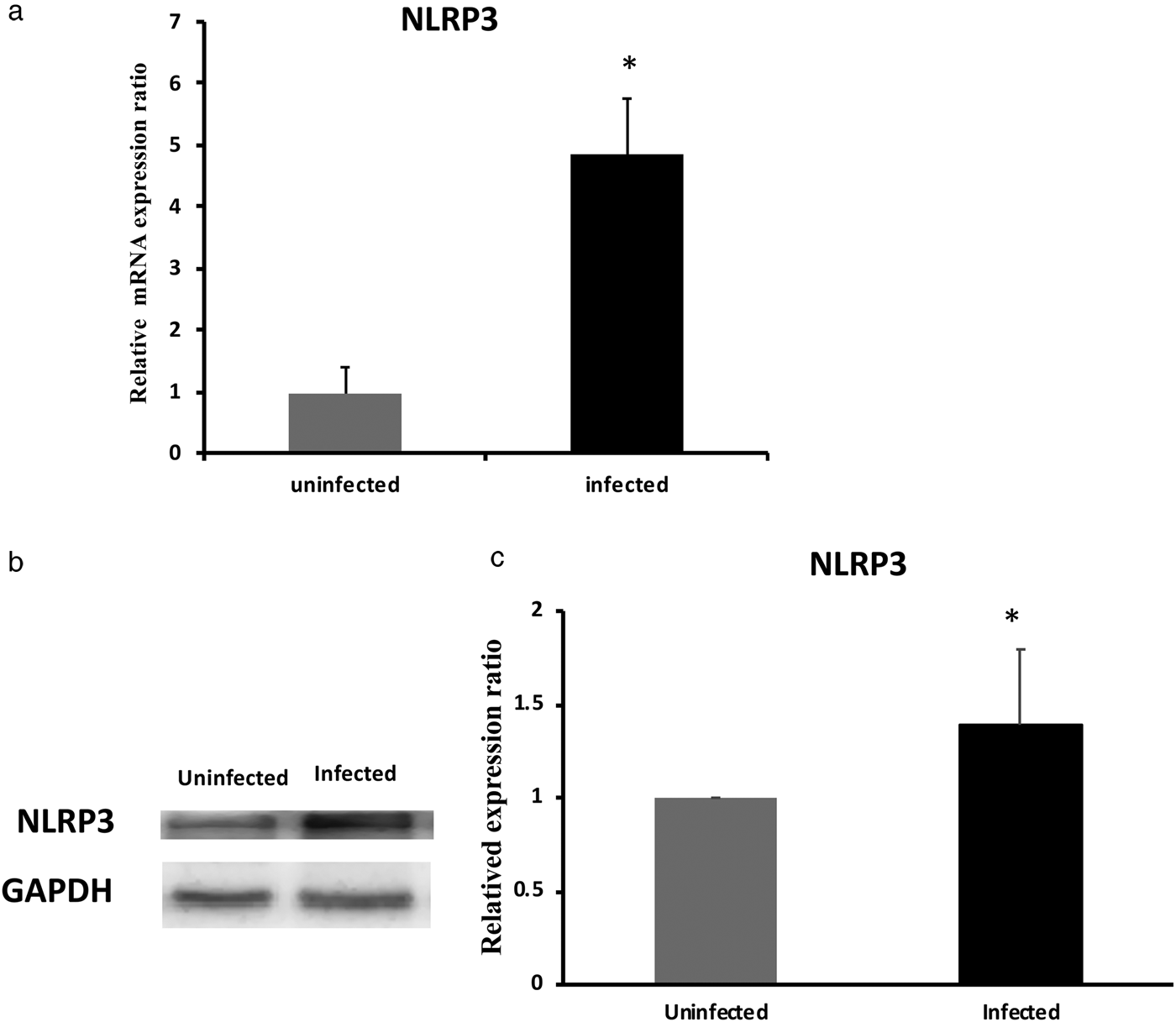
BALB/cByJ mice infected with S. mansoni for eight weeks showed granulomatous syndromes in their livers (fig. 1). The livers with granulomas were then dissected to extract total RNA and protein for subsequent purification and analysis. Furthermore, protein and RNA expression of NLRP3 was significantly increased in the livers of S. mansoni-infected mice compared to the livers of uninfected mice (fig. 2a–c; P < 0.05), indicating activation of the NLRP3 inflammasome.
Morphological differences in infected and uninfected mouse livers. (a) Hematoxylin and eosin (HE) stains of liver tissue of uninfected BALB/c mice. (b) HE stains of liver tissue of BALB/c mice after eight weeks of infection with S. mansoni. Arrow indicates a granuloma, 100×.

NLRP3 is an important inflammasome sensor during acute S. mansoni infection. (a) qPCR results for NLRP3 mRNA expression. (b) Western blot visualization of NLRP3 protein expression; GAPDH is presented as a housekeeping protein control. (c) Densitometric analysis of NLRP3 protein expression in livers from S. mansoni-infected mice and from uninfected controls (P = 0.0016, N = 5; *P < 0.05 vs. uninfected group).

Schistosoma mansoni infection increases total ROS and NF-κB levels in mouse livers
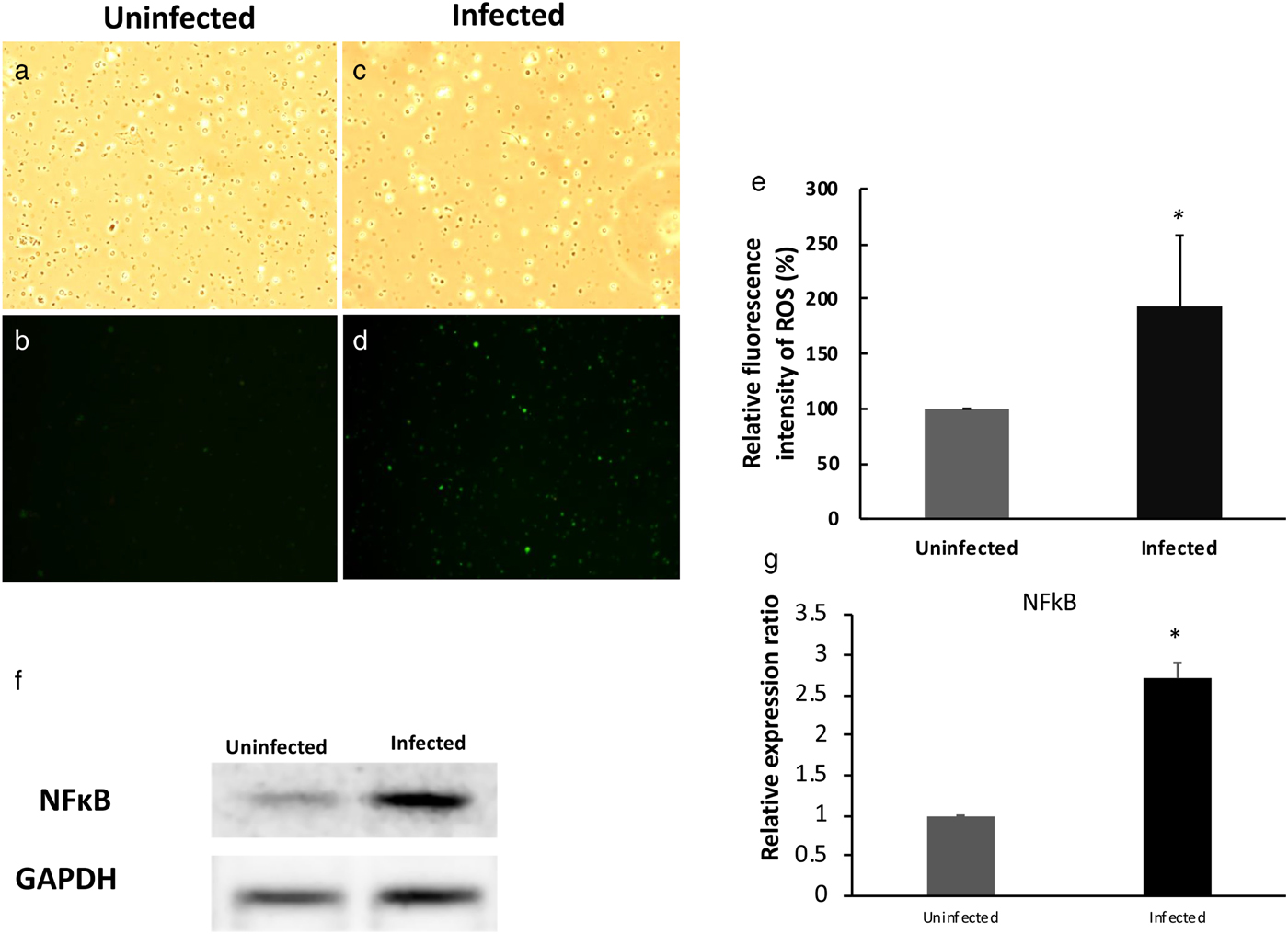
DCF-DA and DHE staining revealed an increase in total ROS levels in liver cells from S. mansoni-infected mice. Total ROS production in mouse liver cells was evaluated by both inverted fluorescence microscope and flow cytometry. Compared with ROS levels in cells from uninfected livers, ROS levels were significantly increased in S. mansoni-infected liver cells (fig. 3). Flow cytometry analysis revealed that the fluorescence intensity of ROS in liver cells from the S. mansoni-infected group was 214% higher than in liver cells from the control group (fig. 3e; P < 0.05). Additionally, total protein was extracted from the livers of infected and uninfected mice and subjected to western blot analysis. The protein level of NF-κB (p65) in livers from the S. mansoni infected group was significantly higher than in livers from the control group (fig. 3f, g; P < 0.05).
Schistosoma mansoni infection increases total ROS level in mouse liver. (a–d) Representative optical (a, c) and fluorescent (b, d) microscopic photomicrographs showing ROS generation in uninfected (a, b) and infected mouse liver cells (c, d); 100× magnification. (e) Quantification of relative ROS levels in liver cells from uninfected and S. mansoni-infected mice. Liver cells from uninfected and S. mansoni-infected mice were stained with DCF-DA and DHE. Observation was performed by fluorescence microscopy (P = 0.002, N = 5; *P < 0.05 vs. uninfected group). (f) Western blot for NF-κB (p65); GAPDH was used as the housekeeping protein control. (g) Densitometric analysis of NF-κB (p65) expression in livers from S. mansoni-infected mice and uninfected controls (P = 0.042, N = 5; *P < 0.05 vs. uninfected group).

Schistosoma mansoni infection increases superoxide levels in mouse liver
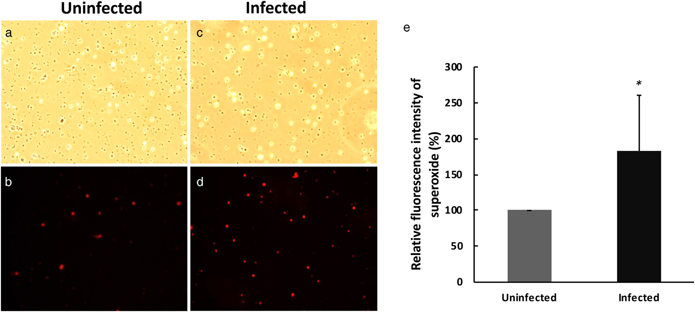
DCF-DA and DHE staining demonstrated a significant increase in the superoxide levels of liver cells of mice infected with S. mansoni cells compared to liver cells from uninfected mice (fig. 4). Flow cytometry analysis revealed that the relative fluorescence intensity of superoxide within cells from the S. mansoni-infected group was 200% greater than in liver cells from the control group (fig. 4e; P < 0.05).
Schistosoma mansoni infection increases superoxide level in mouse liver. (a–d) Representative optical (a, c) and fluorescent (b, d) microscopic photomicrographs of superoxide in uninfected (a, b) and infected mouse liver cells (c, d); 100× magnification. (e) Relative fold changes in superoxide. Liver cells were from uninfected and S. mansoni-infected mice, and stained with DCF-DA and DHE. Observation by fluorescence microscopy (P = 0.017, N = 5; *P < 0.05 vs. uninfected group).

AIM2 serves as critical inflammasome sensor during acute S. mansoni infection
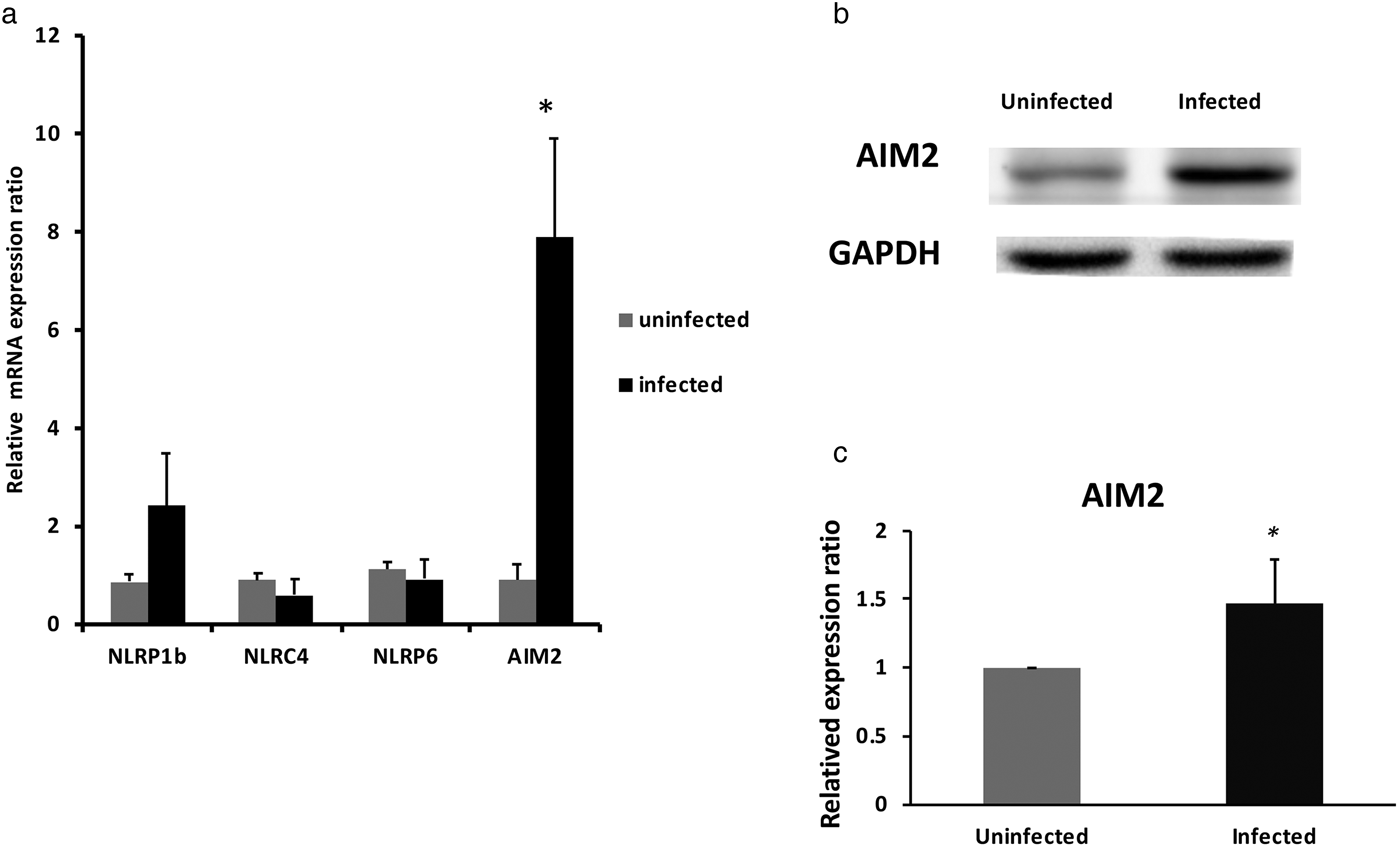
We investigated whether another inflammasome in addition to the NLRP3 inflammasome was involved in S. mansoni infection. RNA expression of inflammasome genes, including NLRP1b, NLRC4 and NLRP6 (of the NLR family) and AIM2 (of the PYHIN family), were analysed in liver samples from S. mansoni-infected mice and uninfected mice (fig. 5). AIM2 RNA expression was significantly increased in the livers of S. mansoni-infected mice (fig. 5; P < 0.05). Western blot analysis demonstrated that S. mansoni infection significantly increased AIM2 protein expression in mouse livers (fig. 5b, c; P < 0.05).
The AIM2 inflammasome sensor is induced during acute S. mansoni infection. (a) mRNA expression of inflammasome genes NLRP1b, NLRC4, NLRP6 and AIM2 from livers of uninfected and S. mansoni-infected mice were analysed by qPCR. Data were normalized against amplification of an internal housekeeping control gene (β-actin); (P = 0.064, 0.121, 0.357, 0.0012, respectively, N = 5; *P < 0.05 vs. uninfected group). (b) Western blot of AIM2 protein from livers of uninfected and S. mansoni-infected mice; GAPDH was used as a housekeeping protein control. (c) Densitometric analysis of AIM2 protein expression in livers of S. mansoni-infected mice and from uninfected controls (P = 0.013, N = 5; *P < 0.05 vs. uninfected group).

Schistosoma mansoni infection induces expression of inflammasome components in mouse liver
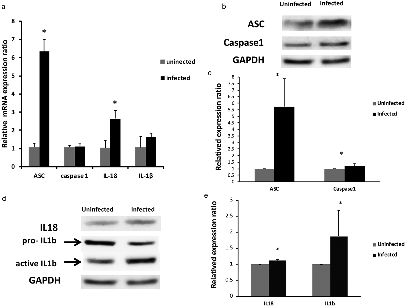
The expression of RNA and protein of inflammasome components, including the adaptor protein ASC, the effector protein Caspase1, IL1β and IL18 was also analysed by qPCR and western blot analysis (fig. 6). The RNA expression of Caspase-1, ASC and IL18 was significantly increased in the livers of S. mansoni-infected mice compared to livers from uninfected mice (fig. 6; P < 0.05). Additionally, protein expression of Caspase-1, ASC, IL18 and IL1β was significantly higher in the livers of S. mansoni-infected mice than in livers from control animals (fig. 6b–e; P < 0.05).
Schistosoma mansoni infection induces the expression of inflammasome components in mouse liver. (a) Gene expression for ASC, Caspase1, IL18 and IL1β was analysed in livers of uninfected and S. mansoni-infected mice. Data were normalized against amplification of an internal housekeeping control gene (β-actin); (P = 1.92 × 10⁻⁵, 0.3012, 0.000288 and 0.175, respectively, N = 5; *P < 0.05 vs. uninfected group). (b) Western blot for protein expression of ASC and Caspase1; GAPDH was used as the housekeeping protein control. (c) Densitometric analysis of ASC and Caspase1 protein expression in livers of S. mansoni-infected mice and from uninfected controls (P = 0.007 and 0.005, respectively, N = 5; *P < 0.05 vs. uninfected group). (d) Western blots for IL18 and IL1β; GAPDH was used as the housekeeping protein control. (e) Densitometric analysis of IL18 and IL1β protein expression in livers of S. mansoni-infected mice and from uninfected controls (P = 0.003 and 0.035, respectively, N = 5; *P < 0.05 vs. uninfected group).

SEA-induced AIM2-inflammasome activation in HuH-7 cells
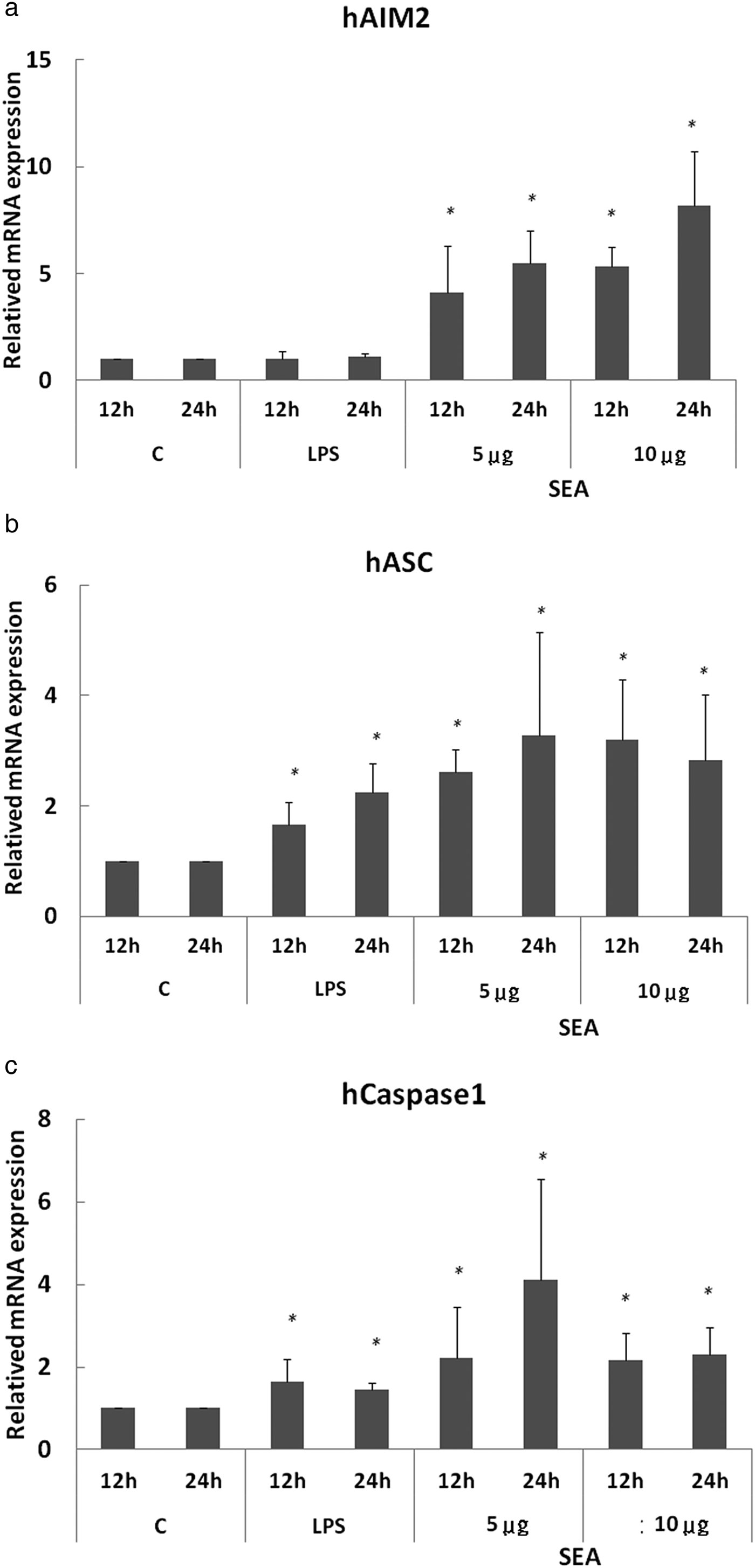
To address whether schistosomal components induce the inflammasome pathway, HuH-7 cells were cultured with the addition of SEA (5 µg/ml or 10 µg/ml) (Duan et al., Reference Duan, Gu and Zhu2014) in complete media for 12 h and 24 h. HuH-7 cells were treated with LPS (100 ng/ml), served as a negative internal control for AIM2 expression (hAIM2) and as positive internal controls for ASC and Caspase1 expression (hASC and hCaspase1, respectively). Additionally, HuH-7 cells treated with only media were used as a control. After 12 h and 24 h, total RNA was isolated from HuH-7 cells, and qPCR analysis was performed. RNA expression of hASC and hCaspase-1, but not that of hAIM2, was increased significantly in the LPS-treatment group (fig. 7a–c; P < 0.05), supporting LPS induction of NLRP3 inflammasome activation (Franchi et al., Reference Franchi, Eigenbrod, Munoz-Planillo and Nunez2009). In agreement with previous reports, treatment with SEA resulted in significant increase in hAIM2, hASC and hCaspase-1 after 12 h (fig. 7a–c; P < 0.05) and 24 h (fig. 7a–c; P < 0.05), compared to untreated and LPS controls. Interestingly, parallel experiments conducted with 12-h treatments of 5 µg/ml and 10 µg/ml SEA, but not 24-h treatments, showed that the RNA expression of hAIM2, hASC and hCaspase1 was significantly increased following co-incubation with SEA. RNA expression of hAIM2, hASC and hCaspase1 was increased after 24-h treatment with either 5 µg/ml or 10 µg/ml SEA.
SEA induces AIM2, ASC and Caspase-1 inflammasome activation in HuH-7 cells. qPCR analyses of HuH-7 cells treated with either 5 µg/ml and 10 µg/ml SEA for 12 h and 24 h, respectively. Cells treated with either LPS or media served as positive and negative controls, respectively. (a) mRNA abundance of AIM2 increased significantly in both 5 µg/ml and 10 µg/ml SEA treatment groups (P = 0.45, 0.03 and 0.0006, respectively, N = 5; *P < 0.05 vs. controls). (b) RNA expression of ASC was significantly increased in LPS and 5 µg/ml and 10 µg/ml SEA treatment groups (P = 0.027, 0.001 and 0.013, respectively, N = 5; *P < 0.05 vs. controls). (c) mRNA abundance of Caspase-1 was significantly increased in LPS and 5 µg/ml and 10 µg/ml SEA treatment groups (P = 0.04, 0.04 and 0.04, respectively, N = 5; *P < 0.05 vs. controls).

Discussion
We had previously demonstrated that eight weeks of infection with S. mansoni caused mitochondrial damage in the liver and promoted both intrinsic and extrinsic apoptosis pathways in mouse liver cells. We also reported that NF-κB (p65) expression was significantly increased in the livers of S. mansoni-infected mice (Chen et al., Reference Chen, Xu, Li, Tao, Xiang, Huang, Niu, Zhong and Meng2015). It has also been shown that ROS promote inflammation and activate NF-κB to regulate inflammation in pathological states (Schreck et al., Reference Schreck, Rieber and Baeuerle1991; Koay et al., Reference Koay, Christman, Segal, Venkatakrishnan, Blackwell, Holland and Blackwell2001). In this study, we demonstrate that the amount of total ROS and superoxide released is significantly increased in mouse livers following S. mansoni infection; these increases were accompanied by an increase in NF-κB (p65) protein expression. These data are in agreement with previous reports that damaged mitochondria generate high levels of ROS during inflammation (Llesuy et al., Reference Llesuy, Evelson, Gonzalez-Flecha, Peralta, Carreras, Poderoso and Boveris1994; Taylor et al., Reference Taylor, Ghio and Piantadosi1995; Kantrow et al., Reference Kantrow, Taylor, Carraway and Piantadosi1997), which may be an important host defence mechanism against intracellular parasites such as Leishmania amazonensis and Toxoplasma gondii (Kawai & Akira, Reference Kawai and Akira2010).
Here, we demonstrate that S. mansoni infection triggers activation of the NLRP3 inflammasome. In addition, the parasite may activate more than one inflammasome during infection. Recently, the malarial parasite, Plasmodium, was shown to be a prominent example of this phenomenon; Plasmodium can dually engage both the NLRP3 and AIM2 inflammasomes during infection (Kalantari et al., Reference Kalantari, DeOliveira and Chan2014). We report that S. mansoni activated both the NLRP3 and AIM2 inflammasomes in mouse liver during infection. Furthermore, the transcription and translation of Caspase1, the bipartite adaptor protein ASC and mature IL18 were significantly increased, and the expression of IL1β protein was also significantly increased (fig. 6).
A mechanism similar to that of the NLRP3 inflammasome (Lamkanfi & Dixit, Reference Lamkanfi and Dixit2012), the AIM2 inflammasome pathway may be triggered by stimulation of HuH-7 cells with different doses of the SEA. AIM2 is a cytosolic protein, which has a C-terminal DNA binding domain and an N-terminal pyrin domain; this structure enables it to respond to dsDNA from both host and pathogens (Rathinam et al., Reference Rathinam, Jiang and Waggoner2010). As a non-specific receptor for cytoplasmic DNA, AIM2 can be activated by and bind to plasmid DNA, DNA from the bacterium Listeria monocytogenes and even synthetic ds-DNA (Jones et al., Reference Jones, Kayagaki and Broz2010). In addition to the NLRP3 inflammasome, DNA-mediated AIM2 inflammasome activation triggers Caspase-dependent cell death and apoptosis. Our previous study demonstrated that S. mansoni infection causes Caspase-dependent apoptosis in mouse liver. This may be evidence that the AIM2 inflammasome can be activated by S. mansoni DNA.
We speculated that up-regulation of AIM2 expression may be partially caused by granulomas. This kind of host innate immune response inflammasome may favour S. mansoni survival and may facilitate the persistence of the associated granuloma in liver tissues. Despite the recognition of different pathogen-associated molecular patterns, the NLRP3 and AIM2 inflammasomes both contain Caspase1 and the ASC adaptor (Schroder & Tschopp, Reference Schroder and Tschopp2010). The similarities in these inflammasomes supports the notion that granulomas caused by S. mansoni can activate the host immune response via both the NLRP3 and AIM2 inflammasomes.
In summary (fig. 8), we found that acute schistosomiasis caused ROS release, upregulated NF-κB expression and promoted inflammasome activation in mouse livers. Evidence from the mouse model and in vitro model of schistosomiasis suggests an important role for SEA. Additionally, inflammasome activation in acute hepatic schistosomiasis may be mediated by the NLRP3 and AIM2 inflammasome pathways triggered by SEA. Collectively, our findings may help provide a new therapeutic target for acute schistosomiasis and a new avenue for studying its pathogenesis. The direct mechanism of AIM2 inflammasome involvement in schistosomiasis remains unknown, and the relationship between S. mansoni dsDNA and AIM2 needs to be examined further. Furthermore, the cross-talk between AIM2 and NLRP3 inflammasomes is also worthy of further study.
Model by which acute schistosomiasis causes ROS release and promotes inflammasome activation in mouse livers. Acute S. mansoni infection causes mitochondrial damage, results in the release of ROS and superoxide, and up-regulates NF-κB (p65) expression. Schistosomal antigen released from S. mansoni eggs triggers both NLRP3 and AIM2 inflammasome activation. NLRP3 and AIM2 inflammasome share a common feature: they both consist of Caspase-1 and the ASC adaptor. Thus, ASC, Caspase-1, mature IL1β and IL18 are all up-regulated by S. mansoni infection. The increase of ROS and NF-κB (p65) may promote inflammasome(s) activation and, thus, IL1β and IL18 mature, respectively. The AIM2 inflammasome is potentially triggered by S. mansoni dsDNA in cells, leading to ASC and Caspase-1 recruitment, and in pro-IL1β and pro-IL18 processing. This cascade, in turn, protects the host during acute infection and is independent of controlling S. mansoni.

Financial support
This work was supported by Tzu Chi University (S. Y. P., grant number TCMRC-P-103013). The funders had no role in study design, data collection and analysis, preparation of the manuscript or decision to publish.
Conflicts of interest
The authors declare that they have no competing interests.
Ethical standards
BALB/c mice were purchased from the National Laboratory Animal Breeding and Research Centre, Taipei. All protocols involving animals were approved by the regulation of the Institutional Animal Care and Use Committees (IACUC) of Tzu Chi University (No. 102068). All experimental procedures were carried out in accordance with approved guidelines of the National Institutes of Health (NIH) Guide for the Care and Use of Laboratory Animals (DHHS publication No. NIH 85-23, revised 1996).