Introduction
Low birth weight is defined as a birth weight <2500 g for a full-term newborn. Low birth weight not only contributes to the increasing incidence of perinatal complications, but is also related to a host of adult chronic diseases, including coronary artery disease, Type II diabetes mellitus, cancer, osteoporosis and various psychiatric illnesses.Reference Calkins and Devaskar1 Although many factors have been associated with low birth weight, on the basis of our current understanding, placenta plays an important role in the occurrence of low birth weight.
MicroRNAs (miRNAs) are a class of noncoding regulatory single-stranded small RNA molecules of about 21–25 nucleotides in length. They bind to the 3′-untranslated region (3′-UTR) of target mRNAs and inhibit translation of the target mRNA or target the mRNA for degradation. Generally, miRNAs are highly conserved across species and are expressed in a stage-specific and tissue-specific manner.Reference Lee and Ambros2 A previous study has shown that eight miRNAs, including miRNA-517a, were highly expressed in the placenta compared with other tissues, such as the liver, brain, heart and so on, and therefore were referred to as placenta-specific miRNAs.Reference Luo, Ishibashi and Ishikawa3
The miR-517a gene is found on chromosome 19q. Grimwood et al. Reference Grimwood, Gordon and Olsen4 previously reported that regions of chromosome 19 are associated with familial hyperlipidemia, insulin resistance and other diseases. More recent studies have shown that abnormal expression of miRNAs can affect the invasive abilities of several tumor cell types through modulation of the expression of specific target proteins.Reference Fang, Zhou and Zeng5–Reference Fang, Yang and Ma7 As there are many biological similarities between trophoblast cells and tumor cells, we hypothesized that the placenta-specific miRNA miR-517a regulates birth weight by affecting the invasive ability of trophoblasts. As has been previously shown, miRNAs are biologically stable and may be used as specific serum markers for certain diseases.Reference Yu, Han and Hu8 Therefore, we proposed that the expression of the placenta-specific miR-517a in maternal serum could also represent a potential serum marker for low birth weight.
Methods
Collection and storage of tissue and serum samples
Human placental samples and maternal serum were collected from full-term patients who gave birth in Shengjing Hospital, which is affiliated with China Medical University, from September 2010 to December 2010. The ages of the patients ranged from 25 to 35 years old and all were without pregnancy complications. The height of the patients ranged from 150 to 170 cm, and the weight gain during pregnancy ranged from 12 to 20 kg. The patients were divided into two groups on the basis of infant birth weight: the low birth weight group (birth weight <2500 g; n = 10) and the normal birth weight group (2500 g < birth weight < 4000 g; n = 20). Informed consent was obtained from each patient, and the study was approved by the Institute Research Ethics Committee of Shengjing Hospital. Fresh placenta tissues were divided into 1 cm3 samples, and were immediately frozen in liquid nitrogen and stored at −80°C. Peripheral blood from the mothers were collected in nonenzymed EP tube simultaneously, with 5 ml per patient. The serum was fully mixed and incubated at 4°C for 2 h before centrifugation at 1600 × g at room temperature for 2 h. The upper solution was then collected and stored at −80°C.
RNA isolation and real-time qRT-PCR
Total RNA was isolated from placenta samples and maternal serum samples using Ambion mirVana miRNA Isolation Kit reagent (Austin, TX, USA) for miRNA analyses. A reverse transcription kit and a SYBR Green fluorescence quantitative PCR detection kit were purchased from the Takara Company. The U6 RNA was used as an internal control. The primers used for miR-517a amplification were designed and synthesized by Junying Company (Shanghai, China), the sequences were as follows: miR-517a RT 5′-GTCGTATCCAGTGCAGGGTCCGAGGTATTCGCACTGGATACGACACACTC-3′, miR-517a-UP 5′-GCGCATCGTGCATCCCTTTA-3′, and miR-517a-Down 5′-TCCAGTGCAGGGTCCGAGGT-3′. The amplification reaction protocol consisted of 40 cycles of 95°C for 1 min, 95°C for 30 s, 60°C for 30 s and 72°C for 30 s in an ABI7500 instrument (Applied Biosystems, USA).
Cell culture and oligonucleotide transfection
The trophoblast cell line HTR8/SVneo was supplied by Department of Anatomy and Cell Biology of Queen's University. Cells were cultured in RPMI-1640 medium (HyClone, USA) supplemented with 10% fetal bovine serum (HyClone) and were maintained in a humidified incubator at 37°C and 5% CO2 (Thermo Scientific, USA). The 2′-O-methyl oligonucleotides were chemically synthesized by Shanghai GenePharma Company (Shanghai, China). The sequences were as follows: hsa-miR-517a: sense 5′-AUCGUGCAUCCCUUUAGAGUGU-3′ and antisense 5′-ACUCUAAAGGGAUGCACGAUUU-3′; negative control: sense 5′-UUCUCCGAACGUGUCACGUTT-3′ and antisense 5′-ACGUGACACGUUCGGAGAATT-3′. Cells were transfected with oligonucleotides after reaching a confluency of 70–80% using the Lipofectamine 2000 reagent (Invitrogen, USA), according to the manufacturer's instructions.
In vitro transwell invasion assays
The HTR8/SVneo cell line was transfected with either miR-517a or negative control oligonucleotides, and after 24 h cells were transferred to the top chamber of a Matrigel-coated invasion chamber (24-well insert, 8-μm pore size) (Corning, NY, USA) in RPMI-1640 medium containing 0.2% fetal calf serum, 104–105 per chamber. RPMI-1640 containing 10% fetal calf serum was added to the lower chamber to function as a chemoattractant. After incubation for 72 h, noninvasive cells were removed from the upper well, and the remaining invasive cells were fixed with 4% paraformaldehyde and stained with Giemsa. Invasive cells were observed using a light microscope (Nikon E800; Japan), and images of three independent fields were captured for each well. Results are reported as the average of three independent experiments.
Identification of miRNA targets
The TargetScan algorithm http://www.targetscan.org/ was used to identify human miR-517a-binding sites.
Statistical analysis
The Student t-test was used to analyze significant differences using SPSS 16.0 software. P-values <0.05 were deemed statistically significant.
Results
The expression of miR-517a is highly expressed in placental tissues and maternal serum associated with low birth weight infants.
Expression of miR-517a in placental tissue from low birth weight newborns (61.79 ± 23.06, ranged from 0.173 to 194.371) was significantly higher compared with that of the normal birth weight newborns (5.01 ± 1.97; P < 0.05, ranged from 0.050 to 36.335). The expression of miR-517a was also significantly higher in the maternal serum associated with the low birth weight infants (25.78 ± 8.69, ranged from 2.342 to 70.006) compared with that of normal birth weight infants (3.21 ± 1.07; P < 0.05; ranged from 0.001 to 12.407; Fig. 1).
The expression of miR-517a. (a) The expression of miR-517a in the placenta; (b) the expression of miR-517a in the maternal serum. NBW = normal birth weight; LBW = low birth weight.

Overexpression of miR-517a inhibits trophoblast invasion
Results from transwell invasion assays showed that the number of invasive cells per 40 × field in the group transfected with a miR-517a mimetic RNA (6.67 ± 0.58) was decreased compared with that of the negative control group (13.33 ± 3.06; P < 0.05; Fig. 2).
Transwell invasion assay. (a) One field of miR-517a mimics group at 10 × magnification; (b) one field of negative control group at 10 × magnification; (c) the number of invasive cells per 40 × magnification. *compared with negative control group, P < 0.05.
Discussion
Recently, metabolic syndrome, which is associated with adult obesity, hypertension and diabetes, became the primary factor affecting human health in developed countries, and the incidence of this syndrome has been increasing each year in China. Development of a better understanding of the mechanism of occurrence of this disorder has great significance for human health and improvement of life quality. Fetal programming, or the idea that fetal intrauterine development determines the occurrence of certain adult diseases,Reference Ong and Dunger9 has been proposed as a potential mechanism to explain the occurrence of adult metabolic syndrome. Several studies have shown that low birth weight can increase the incidence of adult diseases through alterations in a variety of metabolic regulatory pathways. Increased incidence of glucose and lipid disorders results in abnormal regulation of the insulin pathway in low birth weight newborns, and the superimposed effects of all of these biochemical changes can further lead to an increased incidence of adult obesity, cardiovascular disease and type II diabetes.Reference Varvarigou10, Reference Calkins and Devaskar11 Therefore, it is of great concern to characterize the mechanisms associated with low birth weight in newborns, and thus identify potential interventions for metabolic syndrome.
Our previous studies have shown that growth factors, hormonesReference Yang, Weiwei and Jingli12 and imprinted genesReference Xiao-Xiao and Wei-wei13 can regulate birth weight through the placenta. Interestingly, Amiruddin et al.Reference Amiruddin and Yusuf14 found that low placental weight was strongly correlated with low birth weight. All these suggest that the placenta plays an indispensable role in the regulation of low birth weight. Development of the placenta is a complex procedure, requiring differentiation, invasion and proliferation of trophoblasts, and trophoblast invasion plays a key role in this process.
Shallow invasion of uterine spiral arteries in the maternal decidual tissue can lead to poor material exchange in placenta, and poor trophoblast invasion will eventually lead to the occurrence of intrauterine growth restriction and other diseases.Reference Roberts15 Therefore, we speculate that abnormal trophoblast invasion can lead to abnormal placentation, which may be one of the important mechanisms underlying low birth weight.
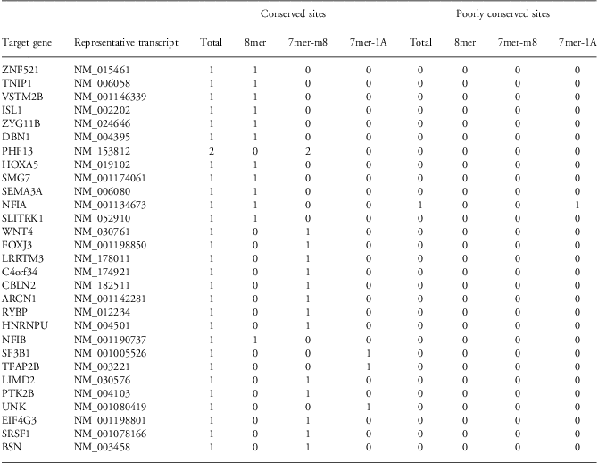
Our results reveal that miR-517a expression was highly expressed in the placentas of low birth weight newborns, suggesting that miR-517a may play a role in placental regulation of birth weight. Abnormal expression of miR-517a in the placenta may be involved in one of the mechanisms that lead to low birth weight in full-term infants. To our delight, we also found that miR-517a was expressed at higher levels in the maternal serum associated with full-term low birth weight newborns compared with controls, a trend similar to that observed in the placenta. This observation leads to the following question: can miR-517a expression levels in maternal serum be used as a maternal biomarker for low birth weight? Additional controlled clinical trials should be carried out to test this hypothesis in future studies. It should be noticed that there was a high variability of expression levels in the low birth weight samples, which may be because of the small number of the low birth weight group, and it may also suggest that mir-517a is not the only one genetic reason responsible for the low birth weight; some other factors may play a part in this procedure. We also found that miR-517a overexpression can inhibit the invasion ability of trophoblasts using transwell assays. On the basis of these observations, we hypothesized that miR-517a inhibits trophoblast invasion, resulting in abnormal placentation and subsequent low birth weight of the full-term newborn. This study is the first to characterize the association of placenta-specific miR-517a expression with full-term low birth weight. Unfortunately, we have not yet identified the downstream mRNAs targeted by miR-517a to regulate trophoblast invasion. However, we have conducted a preliminary target gene screen using TargetScan (http://www.targetscan.org/). This analysis has led to the identification of potential regulatory sites in genes that include SEMA3A, EIF4G3 and others. Importantly, previous studies have shown that high expression of SEMA3A can inhibit tumor cell invasion in prostate cancer.Reference Herman and Meadows16 Therefore, it is possible that miR-517a may modulate trophoblast invasion through regulation of SEMA3A expression. To this end, we plan to characterize the downstream targets of miR-517a in future studies to better understand these regulatory mechanisms.
Acknowlegments
The authors thank Department of Anatomy and Cell Biology of Queen's University for supplying us HTR8/SVneo cell line,Reference Graham, Hawley and Hawley17 and thank Ma Li for technical assistance.
Financial Support
None.
Conflicts of Interest
None.
Ethical Standards
The authors assert that all procedures contributing to this work comply with SFDA/GCP and with the Helsinki Declaration of 1975, as revised in 2008, and has been approved by the Institute Research Ethics Committee of Shengjing Hospital.