Introduction
The Cambrian Evolutionary Radiation (CER) is arguably the most dramatic interval in metazoan history. It featured the terminal Ediacaran–early Cambrian diversification of coelomates that produced behaviorally complex and deep burrows, and the subsequent origin and diversification of modern, higher-level groups of skeletalized metazoans (e.g., Lipps and Signor, Reference Lipps and Signor1992; Zhuravlev et al., Reference Zhuravlev, Liñan, Vintaned, Debrenne and Fedorov2012; Landing et al., Reference Landing, Geyer, Brasier and Bowring2013a). Two major metazoan groups are peculiar in their seeming absence in the CER and later in the Cambrian; these include the phylum Bryozoa Ehrenberg, Reference Ehrenberg1831, and the coelenterate subclass Octocorallia Haeckel, Reference Haeckel1866. The bryozoans have been said to have ‘missed’ the Cambrian radiation and only appeared later in the Early, but not earliest, Ordovician (Taylor and Ernst, Reference Taylor and Ernst2001, Reference Taylor and Ernst2004; Xia et al., Reference Xia, Zhang and Wang2007; Ma et al., Reference Ma, Taylor and Xia2013). On the other hand, octocorals, particularly the sea pens or sea pencils (order Pennatulacea Verrill, Reference Verrill1865), have a tradition of being regarded as a very archaic metazoan group (e.g., Kölliker, Reference Kölliker1880), and pennatulaceans have often been reported in terminal Precambrian (Ediacaran) and Paleozoic fossil assemblages. However, no fossils can be confidently assigned to the Pennatulacea prior to the Mesozoic, a situation that agrees with paleontological, DNA, and molecular studies that indicate a late derivation of pennatulaceans and other octocorals from ancestral anthozoans (e.g., Williams, Reference Williams1997; Antcliffe and Brasier, Reference Antcliffe and Brasier2008; and discussion below).
The origins of the bryozoans and pennatulaceans intersect in reports on the tiny fossils of the colonial organism Pywackia Landing, 2010 (in Landing et al., Reference Landing, English and Keppie2010). Landing et al. (Reference Landing, English and Keppie2010) argued that Pywackia, presently only known only from Gondwanan southern Mexico (Landing et al., Reference Landing, Westrop and Keppie2007a), is the oldest known bryozoan (Figs. 1, 2). This interpretation brought the fossil record of the Bryozoa back ca. 8 m.y. from the Lower, but not lowest, Ordovician (late Tremadocian) of South China (e.g., Xia et al., Reference Xia, Zhang and Wang2007), into the late Cambrian. Pywackia specimens were later compared to the larger axial rods of a species of the modern pennatulacean octocoral Lituaria Valenciennes, 1850 (in Milne-Edwards and Haime, 1850) by Taylor et al. (Reference Taylor, Berning and Wilson2013). This latter interpretation brought the record of the club-like pennatulaceans, or sessilifloraeds, back over 400 million years from the Cretaceous (Antcliffe and Brasier, Reference Antcliffe and Brasier2008) into the late Cambrian.
Zooaria of the bryozoan Pywackia baileyi from the late Cambrian lower Yudachica Member of the Tiñu Formation, section near Río Salinas, Oaxaca State, Mexico. All specimens from sample Tu-4.95 (Landing et al., Reference Landing, Westrop and Keppie2007a, Table 1); scale bars 0.2 mm long unless otherwise indicated. Abbreviations: a, autozooecium; ad, diaphragm within axial zooecium; az, axial zooecium; d, diaphragm; h, hemiphragm; k, keel; n, probable nanozooid; numbers “1” and “2,” autozooid generations 1 and 2. (1) Wave-abraded proximal extremity, NYSM 17263; (2, 3) proximal extremity showing hemiphragm (shelves and diseppiments (trans-zooecial partitions) in abraded zooecium, “n” at end of lines marks probable nanozooid, NYSM 13511; (4) spike-like proximal extremity, with medial ridge bifurcating into initial part of daughter autozooecium, NYSM 13509; (5) apical end, many zooecia bases filled with late diagenetic dolomite, NYSM 13514; (6–8) medial stem fragment with diaphragms; (6) four dissepiments (d), white line is position of Fig. 1.7; (7) wave-abrasion shown by rounded margins of trilamellar autozooecium; resistant layers (L1, L2) separated by less resistant middle layer (ML) that forms deep recess; disordered granular histology evident throughout fragment and best illustrated in upper right part of figure; (8) remnant of lowest diaphragm in Fig. 1.6 perched on black phosphatic fill of lower part of autozooecium, NYSM 13613; (9) medial stem fragment showing prominent basal internal zooecial ridges and typical longitudinal elongation of autozooecia, NYSM 13515; (10, 11) proximal extremity with area at arrow tip (Fig. 1.10) enlarged (Fig. 1.11) to show rare granular-prismatic fabric along left- and right-lower part of Y-intersection of autozooecial walls (arrow, enlarged figure at limit photomontage system resolution), hypotype NYSM 17264; (12) medial stem fragment with short longitudinal section through phosphate-infilled axial zooecium at base (“az,” outlined by black lines), second generation autozooid (2) with low medial zooecial internal ridge (“k”) at upper left corner, NYSM 17488; (13) transverse section with phosphate-infilled axial zooecium (note partial, NE segment of curved, light gray axial zooecium wall marked by white line with “az” at end of line), first generation autozooecia (designated by “1”) with fragments of second generation autozooecia (“2”); (14) longitudinal section through medial stem fragment shows phosphate-infilled axial zooecium outlined by black lines (note short section of axial zooecium not completely phosphate-infilled below lower “az” symbol, first and second generation autozooecia (“1” and “ 2”) with crenulated walls to left and right of axial zooecium, NYSM 17415; (15) longitudinal section through medial stem fragment shows axial zooecium (“az”) with longitudinal crenulated wall outlined by black lines at base of figure, two diaphragms (“ad”) in axial zooecium, wave-abraded autozooecia walls (bud generation “1”) to left and right of axial zooecium, NYSM 13517.

Zooaria of the bryozoan Pywackia baileyi from the late Cambrian lower Yudachica Member of the Tiñu Formation, section near Río Salinas, Oaxaca State, Mexico. Specimens from sample Tu-4.95 (Landing et al., Reference Landing, Westrop and Keppie2007a, Table 1); scale bars 0.2 mm long unless otherwise indicated. Abbreviations as in Figure 1 caption. (1) typical tube-like form of autozooecia near proximal end of zooarium, individual autozooecium marked by three successive dissepiments (“d”), NYSM 13510; (2) proximal end, as Fig. 2.1 shows succession of diaphragms (“d”) and hemiphragms (“h”) in elongate autozooecium near proximal end, note crenulated internal wall of autozooecium; (3) longitudinally broken medial stem fragment shows uncommon example of axial zooecium (“az”) not infilled with phosphate but with phosphate-replaced axial zooecium wall and axial diaphragm (“ad”), broken bases of first and second daughter autozooecia (“1” and “2”) on left, and sections through more complete generation 1 and 2 autozooecia on right, NYSM 17264.
Landing et al.’s (Reference Landing, English and Keppie2010) description and higher-level classification of Pywackia was limited by the space requirements of Geology. They only distinguished Pywackia colonies with bilaterally symmetrical autozooecia from those of two skeletalized colonial coelenterate groups: hydrozoans with generally radially symmetrical zooids and tabulate corals, which often have intercorallite pores and septa that are lacking in Pywackia. In addition to the much younger age (Mesozoic) of the oldest confidently identified pennatulaceans (Antcliffe and Brasier, Reference Antcliffe and Brasier2007, Reference Antcliffe and Brasier2008), an interpretation of Pywackia specimens as pennatulacean axial rods, which range from elongate whip-like forms with circular to quadrate cross-sections and rarely to morphologically complex axial rods with deep calyx-like pits, was considered unlikely and not discussed by Landing et al. (Reference Landing, English and Keppie2010).
Ascertaining the biological affinity of Pywackia has important implications for reconstructing the evolutionary history of the phylum Bryozoa and the cnidarian class Pennatulacea. This report details the reasons to assign Pywackia to the Bryozoa, and possibly to the order Cryptostomata Vine, Reference Vine1884. It describes the homologies and putative synapomorphies between Pywackia and bryozoans and emphasizes the differences between the mineralized skeletons of bryozoans and pennatulaceans. The morphologic terminology applied to bryozoans and pennatulaceans in this and other reports is discussed in the Appendix. The first use of significant morphologic terms discussed in the Appendix is marked by an asterisk (*) in the text.
Systematic paleontology
Specimens with NYSM numbers are in the New York State Museum Paleontology Collection (NYSM) and comprise the only specimens known to be reposited in a collection.
Class Stenolemata Borg, Reference Borg1926
Order Cryptostomata? Vine, Reference Vine1884
Remarks
Restudy of Pywackia and an appreciation that its stem-like zooaria* are better interpreted as a result of the close packing of autozooids* into a 14-hedron means that assignment of this bryozoan genus to the cryptostomes is more tentative than in Landing et al. (Reference Landing, English and Keppie2010). Pywackia is considered to be a very early ‘stony bryozoan,’ or a stem group stenolaemate, derived from unmineralized ctenostome-grade ancestors. The skeletalized autozooecia have a combination of hemiphragms and diaphragms and axial zooids as in some cryptostomes (e.g., Blake, Reference Blake1983a). Well-preserved autozooecia are deeper than Landing et al. (Reference Landing, English and Keppie2010) reported, but still relatively shallow, as in cryptostomes.
Family undetermined
Genus Pywackia Landing in Landing et al. (Reference Landing, English and Keppie2010)
Type species
Pywackia baileyi Landing in Landing et al. (Reference Landing, English and Keppie2010), upper Cambrian Cordylodus andresi Zone, type section of Tiñu Formation (lower Yudachica Member) near Río Salinas, Oaxaca State, Mexico.
Other species
The genus is presently monospecific.
Diagnosis
Emended from Landing in in Landing et al. (Reference Landing, English and Keppie2010). Probable cryptostome with stem-like, likely unjointed, roughly cylindrical zooaria with autozooecia, axial zooid, and probable nanozooecia; zooaria largely consist of closely packed autozooecia with hemiphragms and diaphragms; histology microgranular to rarely granular-prismatic. Proximal autozooecia* elongated roughly parallel to zooarium axis near proximal end*; autozooecia on medial stem* radiate away from zooarial longitudinal axis. Absence of extrazooecial structures means that the generally proximally–distally elongated autozooecia appear somewhat disordered as they form a 14-hedron; general longitudinal polarity or somewhat helical arrangement of autozooecia. Axial zooecia with widely spaced diaphragms and longitudinal crenulation occur at least in lower part of zooarium; possible nanozooids at intersections of autozooecial walls. Autozooecial walls trilamellar with thin, erosionally nonresistant inner layer. Proximal extremities range from spike-like forms with flat to low triangular cross-sections and low longitudinal ridges and furrows, but more commonly pointed to sharply rounded with autozooecia that extend to proximal extremity tip. Holdfast unknown, may be absent. Autozooecia form 6-sided zooarium; bud to form 12-sided zooarium with broadly elliptical to circular cross section. Apical end rounded. Autozooecia with roughly linear to helical arrangement and apertures that range from oval to 4–7-sided polygons with slightly convex, to straight, to weakly concave sides; autozooecial basal surface with internal zooecial ridge on midline; longitudinal crenulation on inner autozooecium walls; autozooecial walls imperforate, trilamellar with less resistant (possibly organic-rich) median lamina.
Occurrence
Presently known only from the type locality.
Description
Proximal extremity a trilobed, elongate spike with low expansion angle (5–16°), lacks autozooecia (Fig. 1.4), but more commonly sharply rounded or forms point with autozooecia appearing at base (Figs. 1.1, 1.2, 2.1, 2.2). Middle zooarium tubular, stem-like (Fig. 1.9), end zooarium a rounded apical tip (Fig. 1.5); zooarial cross-section generally circular to broadly elliptical (Fig. 1.13). No evidence of ball-and-socket joints, branching, or holdfast. On medial stem and at distal tip, autozooecia apertures rounded to prismatic (rectangular to 7-sided) and radiate away from zooarium axis (Figs. 1.9, 1.13–1.15, 2.3); at proximal end, autozooecia tube-like, elongated parallel to zooarium axis (Figs. 1.2, 2.1, 2.2); likely with weakly calcified frontal walls (Fig. 2.1, 2.2); series of hemiphragms and diaphragms in autozooecia (Figs. 1.6, 1.8, 2.1, 2.2). Interzooecial walls trilamellar with less resistant median lamina (Fig. 1.7) and granular/rarely granular-prismatic histology of outer and inner layers (Fig. 1.7, 1.11). Tiny polymorphs (possible nanozooecia) at wall intersections (Fig. 1.2, 1.3). Internal zooecial ridge, often bilobed, on basal surface of autozooecia (Fig. 1.9). Axial zooecium with widely spaced diaphragms (Figs. 1.15, 2.3), may not extend complete length of zooaria. Autozooids bud to form second-generation autozooids that lengthen and broaden zooarium (Figs. 1.12–1.15, 2.3).
Remarks
The diagnosis of Pywackia is emended from Landing et al. (Reference Landing, English and Keppie2010) to emphasize the close packing of autozooecia as the growth mode of the stem-like zooaria and the absence of interzooecial skeletal structures. The emendation emphasizes the tube-like autozooecia that are elongated parallel to the zooarial axis near the proximal extremity. By contrast, more distal autozooecia have a radial orientation along the zooarium.
Pywackia baileyi Landing in Landing et al., Reference Landing, English and Keppie2010
2010 Pywackia baileyi Landing in Landing, Keppie and English, p. 549, 550, figs. 2A–L, 3A–D.
2013 Pywackia baileyi; Taylor, Berning, and Wilson, figs. 1.1–1.3, 2.1–2.11, 4.1
Types
Holotype NYSM 13515; paratypes NYSM 13508–13514 and13516–13520.
Remarks
Pywackia is presently monospecific; the emended diagnosis, description, and discussion of P. baileyi are the same as for the genus.
Geologic setting and age of topotype Pywackia collection
Approximately 150 fragments of Pywackia zooaria were first reported from three samples low in the lower member (lower Yudachica Member, lower Cordylodus andresi Zone) of the Tiñu Formation in southern Mexico (Landing et al., Reference Landing, English and Keppie2010; Table 1). A ca. 491 Ma date on the lower Tiñu Formation is based on U-Pb zircon data on an approximately coeval interval in the late Cambrian of the Avalonian paleocontinent (Davidek et al., Reference Davidek, Landing, Bowring, Westrop, Rushton, Fortey and Adrain1998; Landing et al., Reference Landing, English and Keppie2010). An approximate age of 483 Ma on the Chinese upper Tremadocian bryozoans discussed in this report (Xia et al., Reference Xia, Zhang and Wang2007) is based on earliest Ordovician ages in Landing et al. (Reference Landing, Bowring, Fortey and Davidek1997, Reference Landing, Bowring, Davidek, Rushton, Fortey and Wimbledon2000).
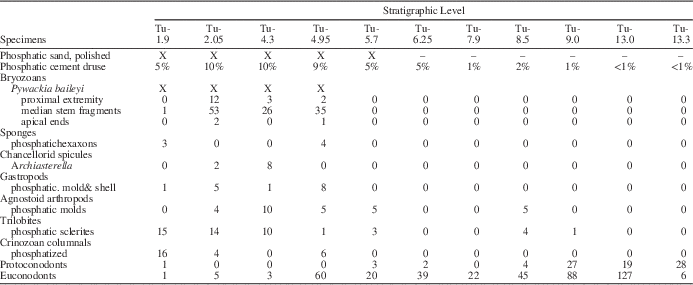
Stratigraphic distribution of Pywackia baileyi (indicated by X). Table shows association of Pywackia zooaria with phosphatic pebbles, higher phosphatic cement content of limestone beds (% of 6 kg sample disaggregated by formic acid), other secondarily phosphatized sclerites, and originally phosphatic (proto- and euconodonts) remains. Samples from upper Cambrian Cordylodus andresi Zone, lower Tiñu Formation, lower Yudachica Member, Río Salinas section, Oaxaca State, southern Mexico. Number after hyphen (i.e., -1.9, -2.0, -4.3, -4.95) is meters above middle Proterozoic–Tiñu Formation nonconformity.
The Pywackia-bearing samples are from fossil grainstone to calcisiltite beds in the lower part of the type section of the Tiñu Formation (Robison and Pantoja-Alor, Reference Robison and Pantoja-Alor1968). This section nonconformably overlies middle Proterozoic gneiss near Río Salinas, Oaxaca State, and includes two type 1 depositional sequences: an upper Cambrian Yudachica Member and a Lower Ordovician Río Salinas Member (Robison and Pantoja-Alor, Reference Robison and Pantoja-Alor1968; Landing et al., Reference Landing, Keppie and Westrop2006a, Reference Landing, Westrop and Keppieb, Reference Landing, Westrop and Keppie2007a, Reference Landing, English and Keppie2010; Streng et al., Reference Streng, Mellbin, Landing and Keppie2011). Pywackia zooaria were reported from 2.05 m, 4.3 m, and 4.95 m above the base of the Yudachica Member (Landing et al., Reference Landing, English and Keppie2010). One more Pywackia specimen is now reported at 1.9 m (Table 1). The lower 8.5 m of the Yudachica Member were deposited in an open-shelf environment. Evidence includes wave-deposited, thin limestone tempestites and weakly burrowed, medium gray shale with a dysoxic olenid and agnostoid biofacies (e.g., Landing et al., Reference Landing, Westrop and Keppie2007a; Schovsbo, Reference Schovsbo2001).
Taphonomy and taphofacies of Mexican Pywackia
Pywackia and phosphatic taphofacies
Formic acid disaggregation shows that Pywackia-bearing samples are limited to limestones with higher concentrations of a granular, light to dark gray, porous (‘punky’ or ‘druzy’) phosphatic cement. The Pywackia-bearing samples yielded almost all of the phosphate-replaced or -infilled fossils at Río Salinas (from sponge hexaxons to crinozoan columnals; Table 1). Thus, the Pywackia zooaria are limited to a taphofacies with a distinctive diagenetic history. This history included the following: (1) phosphatic cementation of lower limestones in the Yudachica Member, and (2) the phosphatic replacement and infill of associated calcareous skeletons. The Pywackia type collection occurs in a phosphatic facies and with secondarily phosphatized calcareous skeletal debris (e.g., Landing et al., Reference Landing, Myrow, Benus and Narbonne1989; Brasier, Reference Brasier1990), features that are consistent with an interpretation that the Pywackia zooaria were originally calcareous (Landing et al., Reference Landing, English and Keppie2010). Other phosphate-replaced bryozoan zooaria occur in slowly deposited shelf successions, and are best known from the European lower Paleozoic (e.g., Dzik, Reference Dzik1992; Federov et al., Reference Federov, Krusanov and Federov1997).
Significance of pre-final burial taphonomy
Detailed study of colonial marine skeletal fossils traditionally relies on polished slabs or thin sections. However, thin-section preparation is largely unnecessary for the Pywackia zooaria as pre-final burial breakage and wave abrasion produced oblique, transverse, and longitudinal sections through the stem-like colonies. The preservation of the topotype Pywackia collection allows direct examination of the three-dimensional relationship of skeletal structures and shows the locations of asexual budding in some specimens (Fig. 1.12, 1.13). Wave abrasion destroyed many exterior walls and, consequently, exposed fine details on the interior walls of long, tube-like structures (e.g., Figs. 1.9, 2.2). Three-dimensional preservation reveals the external and interior surfaces of Pywackia specimens and allows determining just how wave-abrasion has modified individual specimens.
The outer edges of the walls of Pywackia zooaria and associated black phosphatic sand and granules are highly polished (e.g., Fig. 1.4, 1.7; Table 1). The shiny appearance of both types of clasts is regarded as evidence of a complex taphonomic history (detailed below). This history included: early burial, phosphatization, and manganese mineral staining/coating of Pywackia zooaria; excavation and wave abrasion with sclerite polishing and local removal of manganese coats; fracture of the phosphatized Pywackia zooaria; and final burial of the Pywackia zooaria, phosphatized sclerites, and phosphatic pebbles (Landing et al., Reference Landing, English and Keppie2010). This taphonomic history is recognizable through the lower 6.25 m of the Río Salinas section (Landing et al., Reference Landing, Westrop and Keppie2007a, p. 913), and compares closely with the taphonomic models for secondary phosphatization of calcareous sclerites in the early Cambrian (e.g., Brasier and Hewitt, Reference Brasier and Hewitt1979; Landing et al., Reference Landing, Myrow, Benus and Narbonne1989; Brasier, Reference Brasier1990, Reference Brasier1992). Post-phosphatization excavation and wave abrasion is also evident in the polishing and rounding of associated phosphatized trilobite, agnostoid arthropod, gastropod, and crinozoan debris.
Pywackia zooaria colors and taphonomy
Two dominant colors are represented by topotype Pywackia fragments. Most specimens are medium to dark brown and comparable to the thermal color alteration of associated, originally phosphatic conodont elements (Landing et al., Reference Landing, English and Keppie2010; e.g., Fig. 1.2, 1.14).
Other Pywackia zooaria are infused with a light purplish color (Figs. 1.12, 2.3), and a few have surfaces that are dark purple or almost black (Fig. 1.1, 1.10). This purplish to black color is reminiscent of the manganese coloration of shell fragments that have been buried in sediment at about the level of the transition from deeper anoxic/strongly dysoxic to shallower, oxic environments (e.g., van der Zee et al., Reference van der Zee, van Raaporst, Helder and de Heij2003).
The dark purple to black, manganese-based color is absent on the edges of higher-standing interzooecial walls of a few Pywackia specimens (Fig. 1.1, 1.10). This local absence of a dark coloration and the light brownish color of the top of the interzooid walls are consistent with exhumation and abrasion of a manganese coating from these specimens. The very low interzooecial walls of these multicolored (brown and purple/black) specimens suggests wave-induced abrasion of the interzooecium walls removed the dark surface coloration (Fig. 1.1, 1.10).
Taxonomic consequence of abrasion and fracturing
Post-phosphatization wave abrasion likely explains the wearing down of the upper edges of Pywackia zooarium walls. It also erased any original microtopography from the tops of the Pywackia autozooecia walls.
As discussed below, the wearing down of the autozooecia walls was so severe on most of the zooaria that the autozooecia appear to be shallow, open pits (Fig. 1.9). This abrasion was taxonomically useful, however, as it reveals the histology of the zooaria (Fig. 1.7), the internal, transverse skeletal structures (hemiphragms and diaphragms) on the proximal zooecia (Fig. 2.2), and shows how autozooecia budded to produce two generations that radiate away from an axial tube (Figs. 1.17, 2.3). This abrasion allowed observations that conclusively led to evaluation of Pywackia as Earth’s oldest-known bryozoan.
Histology of Pywackia zooaria
Erosionally polished Pywackia walls generally have a fine-grained, non-laminated microcrystalline structure composed of blocky, randomly oriented crystallites with uniform diameters of about 1.0 µ (Fig. 1.7). These crystallites have a phosphatic composition as shown by qualitative elemental analyses completed during scanning microscopy. More rarely, the crystallites are not disordered, but occur in a brick-like arrangement perpendicular to the walls (Landing et al., Reference Landing, English and Keppie2010, fig. 2c; Fig. 1.11). These two histologies are best described, respectively, as granular and granular-prismatic (Landing et al., Reference Landing, English and Keppie2010). Pywackia walls are not composed of a replacement, moldic, space-filling botryoidal phosphate.
Landing et al. (Reference Landing, English and Keppie2010) noted that a calcareous, granular or granular-prismatic microstructure is primary in some bryozoans. It is widespread in the Cystoporata Astrova, Reference Astrova1964 (Utgaard, Reference Utgaard1983, p. 358), and occurs locally in the generally laminated Cryptostomata Vine, Reference Vine1884 (Blake, Reference Blake1983b, p. 550). The interpretation of a phosphate-replaced, originally calcareous composition in Pywackia (Landing et al., Reference Landing, English and Keppie2010) comports with the phosphatization of the limestones that yield its zooaria, and with the phosphatized sclerites of associated calcareous taxa (Table 1).
No histologic comparison can be made of the finely granular phosphatic histology of Pywackia zooaria with other metazoan groups with originally phosphatic or organo-phosphatic sclerites. For example, phosphatic linguloid valves differ from Pywackia as their histology consists of: (1) alternating organic and dense phosphatic layers, (2) phosphatic lamellae inclined at low to high angles to the valve surface, or (3) layers composed of phosphatic fibers oriented normal to the valve surface (e.g., Williams and Rowell, Reference Williams and Rowell1965). Other originally phosphatic groups such as Cambrian tommotiids and protoconodonts have microgranular phosphatic laminae that parallel sclerite surfaces (e.g., Landing, Reference Landing1984), a histology shared with the operculum-like sclerites of mobergellids (Bengtson, Reference Bengtson1968; Landing, unpublished data). The carbonate hydroxylapatite minerals in the axes and holdfasts of Recent gorgonian octocorals occur in strings of submicron-sized spheres or as tiny needle-like fibers within gorgonin layers and do not compose solid phosphatic structures (Macintyre et al., Reference Macintyre, Bayer, Logan and Skinner2000). Thus, gorgonian axes and holdfasts are compositionally and histologically distinct from the longitudinally oriented calcite crystallites that comprise the axis of pennatulaceans (Franc and Chassagne, Reference Franc and Chassagne1974; Wilson et al., Reference Wilson, Andrews, Brown and Cordes2002) or the phosphate-replaced, granular walls of Pywackia.
In conclusion, no extinct or living metazoan with a phosphatic sclerotome has the Pywackia zooarium histology. This, however, is not the case for the Pywackia histology, which is interpreted as a replacement of microgranular carbonate by phosphatic minerals, and for which a bryozoan interpretation is most plausible.
Trilamellar walls in Pywackia and other bryozoans
The edges of many of the vertical (interautozooidal) walls in Pywackia are trilamellar as they expose a cross-section through a ‘sandwich’ largely composed of two granular or, rarely, granular-prismatic phosphatized layers. These relatively thick layers are separated by a much thinner recessed layer that forms the midline of many interautozooecial walls (Fig. 1.7).
A trilamellar wall structure with an inner lamina of organic-rich or an organic-calcite crystal mixture occurs in some members of the diverse Class Stenolemata Borg, Reference Borg1926. The trilamellar wall structure is best illustrated in some Recent stenolaemates (e.g., Boardman, Reference Boardman1983, figs. 30.3b, 30.4, 32.1–32.4, 33.1). The thick walls of the ‘sandwich’ are produced cooperatively by adjacent autozooids and are separated by an organic-rich organic membrane or cuticle.
It is important to emphasize that Pywackia walls, as they are preserved, are similar to this particular type of trilamellar stenolemate wall. By comparison, the interzooidal walls of other stenolemates are composed of an undifferentiated laminae or a granular microstructure forms a single-layered interzooecial wall that lacks an organic lamina (e.g., Boardman and Cheetham, 1973; Boardman, Reference Boardman1983). As discussed in the next section, phosphate diagenesis and replacement can also produce a trilamellar wall in sclerites that originally had a uniform calcareous composition without an inner organic lamina.
Phosphate diagenesis models of Pywackia zooecia walls
By one interpretation, phosphatization has preserved an originally trilamellar Pywackia wall that was originally a ‘sandwich’ of two calcareous layers separated by an organic lamella. Thus, the two calcareous layers were replaced by phosphate that preserved an original, fine-grained, granular histology (Fig. 1.7). An inner organic lamina would be incompletely impregnated by phosphate and consequently less resistant to etching or weathering (Fig. 1.7).
An alternative explanation for the less-resistant interior lamina is that it was not organic, but simply a surface of contact between adjacent autozooecial walls formed by two autozooids. The triple layering then only reflects a phosphatic replacement of the thick calcareous walls, and that this phosphatic replacement effectively sealed off the interior of the sclerite from a comparably dense phosphatization. This three-layered structure of diagenetically altered, calcareous fossils is common. For example, Taylor (Reference Taylor1966) noted that the inner and outer surfaces of the terminal Precambrian vase-shaped fossil Wyattia Taylor (Reference Taylor1966, p. 199) were replaced by dolomite, while the middle part of the wall was sealed by this diagenesis, and consists of recrystallized calcite.
Comparable examples of three-layered, phosphatic sclerites include lower Cambrian fossils with phosphatized outer and inner surfaces and a calcite core (e.g., Bengtson et al., 1990, figs. 166A–D, 168A, B). Even more similar to the phosphatized three-layered walls of Pywackia zooaria are lower Cambrian sclerites with dense phosphatic inner and outer layers separated by a void-bearing, phosphate-replaced central layer (Bengtson et al., 1990, figs. 167A, B, 173H, I, L, 174E). This type of secondary phosphatization of calcareous skeletons occurred throughout the Phanerozoic, and especially during the Cambrian (Brasier, Reference Brasier1992; Porter, Reference Porter2004).
Transverse structures and long autozooecia in Pywackia
Tube-like structures elongated parallel to the longitudinal axis of the zooaria comprise the proximal ends* of many Pywackia zooaria (Figs. 1.2, 2.1, 2.2). Abrasion of the outer surface of these long proximal tubes exposed two types of transverse skeletal structures. These transverse structures, hemiphragms and diaphragms (discussed below), are present in the tube-like autozooecia of many trepostome and stenolaemate bryozoans and show that Pywackia had long autozooecia (e.g., Fig. 2.2).
Hemiphragms
Some of the abraded Pywackia zooaria have short transverse ‘shelves’ that extend only part way across the interior of the autozooecia. These shelves occur in the lower and middle (older) parts of the tubes (Figs. 1.2, 2.2), and are comparable in form and position to autozooecial structures known as “hemiphragms” in stony bryozoans (Bassler, Reference Bassler1952, p. G11). Hemiphragms are a skeletal division in tube-like stenolaemate autozooecia that mark a position beyond which the zooid’s tentacles extend during feeding and behind which the feeding tentacles are retracted (Boardman, Reference Boardman1983, p. 83, fig. 40.6). As a bryozoan autozooecium grows and elongates, hemiphragms are formed as the aperture extends (Fig. 2.2) and mark successive positions of the base of the everted feeding tentacles (e.g., Boardman, Reference Boardman1983). Successive hemiphragms are common and, locally, regularly spaced along the interior of proximal Pywackia autozooecia (e.g., Landing et al., Reference Landing, English and Keppie2010, fig. 2G; Fig. 2.2).
This distribution of hemiphragms shows that Pywackia’s autozooecia are not only elongate, but are elongated roughly parallel to the longitudinal axis near the proximal end of the zooarium (Fig. 2.2). Earlier reconstructions of Pywackia autozooecia as shallow and cup-like are incorrect (Landing et al., Reference Landing, English and Keppie2010; Taylor et al., Reference Taylor, Berning and Wilson2013, p. 986, 988).
Diaphragms
Pywackia fragments help show that most specimens in the topotype collection are not representative of zooarial morphology because of wave abrasion. Most medial and apical stem fragments suggest a slender zooarium with a linear to slightly helicoid arrangement of autozooecia with apertures that open radially from the longitudinal axis of the zooarium (Fig. 1.9).
One specimen shows how wave abrasion changed the apparent morphologies of the zooaria. It has four autozooids with transverse partitions that overlie a ‘punky’ black phosphatic fill (Fig. 1.6). Such transverse partitions are common in stenolaemate and trepostome autozooecia and are termed “diaphragms.” In one part of this zooarium, so much of the colony has been worn away that a diaphragm sits on a pedestal of black phosphatic fill (Fig. 1.8).
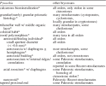
In most medial and distal stem fragments of Pywackia, the outer zooarium and the distal parts of autozooecia were worn away so severely that only cup-like bases of the autozooecia remain; these are the “modules”* of Taylor et al. (Reference Taylor, Berning and Wilson2013; see Fig. 1.9, 1.12). However, the fortuitous preservation of a medial stem with diaphragms shows not only that autozooecia were not ‘cup-like,’ but were much longer. In addition, the presence of diaphragms in proximal and medial autozooecia is a strong argument for assigning Pywackia to the Bryozoa because many stony bryozoans have diaphragms (Landing et al., Reference Landing, English and Keppie2010). Of course, diaphragms are transverse skeletal structures that are not limited to bryozoans, and occur in tabulate corals and calcareous sponges. However, their association with other morphologic and histologic features also known in stony bryozoans (Table 2) is consistent with the assignment of Pywackia to the Bryozoa. Thus, the combination of successive hemiphragms and diaphragms in a calcareous zooarium with tube-like autozooecia (Figs. 1.2, 2.1, 2.2) is typical of trepostome and stenolaemate skeletons, and includes a number of homologies, or putative synapomorphies (Sober, Reference Sober1988, p. 116), between Pywackia and most bryozoan orders (Table 2).
Homologies or putative synapomorphies (marked by *) and other characters common to Pywackia and non-phylactolaemate bryozoans. “Some cheilostomes1” have trilamellar walls with a middle cuticle (organic layer) (e.g., Cheetham and Cook, Reference Cheetham and Cook1983, fig. 1b); “Some cheilostomes2” have “transverse partitions” that are otherwise comparable to diaphragms (e.g., Cheetham and Cook, Reference Cheetham and Cook1983, figs. 89, 90).
Pre-abrasion shape of Pywackia zooarium
The preservation of successive diaphragms (i.e., at least two) indicates not only long autozooecia on the medial stem of Pywackia, but demonstrates that the slender colony shape suggested by the available Pywackia zooaria is misleading. A wider zooarium existed before erosive beveling of the radially oriented autozooecia of the middle stem. Even if only one diaphragm was produced with autozooid growth along the zooarium (Landing et al., Reference Landing, English and Keppie2010; Fig. 1.6, 1.8), the pre-abrasion width of the medial stem was likely at least twice as wide as that of the abraded specimens.
Autozooids: largest of three types of zooids in Pywackia
Pywackia autozooecial apertures range from 0.15–0.39 mm long and 0.27–0.59 mm wide (Taylor et al., Reference Taylor, Berning and Wilson2013). These measurements are “consistent with a bryozoan affinity” (Taylor et al., Reference Taylor, Berning and Wilson2013, p. 986), and bracket the size of the largest zooids, or autozooids.
This range in autozooecial aperture sizes in Pywackia reflects, in part, wave abrasion and beveling of the upper part of the autozooecia, original ranges in size of older autozooids, as well as the intercalation of older and younger (daughter) autozooecia (discussed below) (Figs. 1.14, 2.3). Many Pywackia zooaria show relatively uniformly sized autozooecial apertures (e.g., Fig. 1.5, 1.6, 1.9), but others differ in the sizes of autozooecia of the same generation (Fig. 1.1, 1.14). However, aperture size in stony bryozoans can vary significantly and does not detract from a bryozoan assignment of Pywackia. Landing et al. (Reference Landing, English and Keppie2010) cited some of the many illustrations of irregular autozooid size and distribution in massive stenolaemate bryozoans (e.g., Shimer and Shrock, Reference Shimer and Shrock1944, pl. 96, figs. 10, 16, 17; Boardman and Cheetham, Reference Boardman and Cheetham1987, fig. 29D). Other examples of dissimilar zooecia sizes in bryozoans include Hall, 1887, pl. 61, fig. 18, for the Devonian Trematopora (Orthopora) bispinulata [Hall, Reference Hall1881]; Sarichebal, Reference Sarichebal1960, figs. 69a, 70b, 84a, 85a, 89a, 100 s, for the Late Ordovician Escharopora Hall, Reference Hall1847 (Fig. 3); and Dzik, Reference Dzik1992, fig. 7, for several Middle Ordovician genera. Shimer and Shrock’s (Reference Shimer and Shrock1944) figures show differences in length and width measurements of about 25%, thus meaning an ~50% difference in apertural cross-sectional areas. Greater differences in autozooecia size are shown in Hall’s (1887) and Dzik’s (Reference Dzik1992) figures, with length and width measurements of apertures that differ by 50% and show differences in apertural cross section areas of 75% in single autozooid generations.
Proximal end and elongate proximal autozooecia (also termed “heterozooids”) in the Late Ordovician bryozoan Escharopora Hall, Reference Hall1847, resemble comparable features in the late Cambrian bryozoan Pywackia. (1) Curved proximal tip with highly elongate, proximal autozooecia (heterozooecia) marked by elongate longitudinal ridges and short cross ridges (diaphragms or “vestigial walls”) in E. angularis Ulrich, Reference Ulrich1893, redrafted from Bassler (Reference Bassler1952, fig. 98.5) by EL. (2) Transition from very elongate proximal autozooecia to oval autozooecia in E. suberecta (Ulrich, Reference Ulrich1893), redrafted from Sarichebal (Reference Sarichebal1960, fig. 146b) by EL.
Autozooecial aperture sizes in Pywackia overlap those of a number of calcareous hydrozoans, but hydrozoan colonies are distinguished by dimorphic zooid pores in milleporinids and stylasterinids and vertical skeletal pillars in spongiomorphs (Oliver and Coates, Reference Oliver and Coates1987). Pywackia autozooecia sizes are much smaller than typical diameters of tabulate coral apertures (~0.5–5.0 mm; Oliver and Coates, Reference Oliver and Coates1987, p. 178). The autozooecia apertures in the pennatulacean Lituaria sp. of Taylor et al. (Reference Taylor, Berning and Wilson2013, p. 988, 1.3–2.3 mm long and 0.7–2.0 mm wide) are much larger than those in Pywackia and other bryozoans. The apertural area of the largest Lituaria sp. autozooecium is almost 20× that of the largest Pywackia autozooecium.
Bryozoan autozooidal budding in Pywackia
Some bryozoans produce new autozooids by budding of a stolon (i.e., the non-mineralized ctenostomes, some cheilostomes) and/or budding of an axial zooid (Pywackia, as discussed below; some cryptostomes and cystoporates). Most Paleozoic stenolemates and younger bryozoans increase colony size by autozooidal budding.
Bryozoan autozooidal budding is recorded by medial stem fragments of Pywackia zooaria that were broken into coin-like wafers and abraded by waves (Fig. 1.13). The walls between the autozooids have been largely worn away in these fragments. However, six shallow depressions that radiate away from the zooarium axis comprise the oldest autozooecia (“generation 1”) of the Pywackia colony. These generation 1 autozooids comprised six individuals of a six-sided zooarium (Landing et al., Reference Landing, English and Keppie2010; Fig. 1.13, generation “1”).
In one wafer-like fragment, the six generation 1 autozooecia abut several rounded, wave-abraded ridges that are oriented transverse to the zooarium’s longitudinal axis (Fig. 1.13). The transverse ridges are interpreted as abraded inter-zooid walls. Shallow depressions marginal to the ridges are the record of generation 2 autozooids formed by budding of the generation 1 autozooids (Fig. 1.3, generation “2”). This centrifugal arrangement of two generations of autozooids indicates that Pywackia had two bryozoan methods of asexual growth; autozooid budding to form generation 2, and by axial zooid budding to form generation 1 (discussed below). The lateral expansion of the Pywackia colony by autozooid budding is similar to that of Paleozoic stenolemate bryozoans (e.g., Boardman, Reference Boardman1971, Reference Boardman1983; Boardman and Cheetham, Reference Boardman and Cheetham1987).
Autozooid budding is also shown by a medial stem fragment with an attached piece of a generation 2 autozooecium (Fig. 1.12). Very similar budding was illustrated by Taylor et al. (Reference Taylor, Berning and Wilson2013, fig. 2.1), who figured a Pywackia apical end with six generation 1 autozooecia and the basal end of a broken, generation 2 zooecium shown in the upper right part of their figure. This generation 2 zooecium originated from and rests on the wall between two generation 1 autozooecia. Finally, a number of longitudinally broken Pywackia zooaria examined in this study show generation 2 autozooecia located among and above larger generation 1 autozooecia (Figs. 1.14, 2.3). These are generation 2 autozooecia and not oblique views through adjacent generation 1 autozooecia because these Pywackia zooaria are 12-sided, not six-sided, in cross-section. Thus, the zooaria are composed of generation 1 and daughter generation 2 autozooecia.
Orientation of early and later budded autozooecia
As noted above, earlier produced autozooecia near the proximal extremity of the Pywackia zooarium are elongated roughly parallel to the longitudinal axis of the zooarium (Fig. 2.1, 2.2). Later produced autozooaria along the abraded stem and distal tip radiate away from the colony axis (Figs. 1.4, 1.5, 1.9, 1.14, 1.15, 2.3) and continue their elongation with the addition of diaphragms (Fig. 1.8).
The first generation of radiating autozooids includes six individuals (Landing et al., Reference Landing, English and Keppie2010), which can be counted when medial stem fragments are examined under the microscope and rolled around the longitudinal axis. This six-fold symmetry is also shown by transverse sections through the zooarium (Fig. 1.13). Larger medial stem fragments show twelve individuals when the zooaria are turned over. This bauplan of the Pywackia zooarium with chambered autozooecia and the replacement of long autozooecia near the proximal extremity by radially oriented zooaria along the medial stem is present in the growth of many stenolaemate bryozoans (e.g., Boardman, Reference Boardman1983, p. 118).
Longitudinal features in Pywackia and bryozoan autozooecia
The interior surfaces of Pywackia autozooecia are not smooth. Landing et al. (Reference Landing, English and Keppie2010) described a bilobed “keel” on the basal midline of the autozooecia that does not extend up the proximal and distal walls of the autozooecia (Fig. 1.9). As keels are external features on autozooecia (Boardman, Reference Boardman1983), a better term is “internal zooecial ridge.” The internal zooecial ridge is not invariably bilobed in Pywackia, but can be a simple ridge as noted by Taylor et al. (Reference Taylor, Berning and Wilson2013; e.g., lowest medial autozooecium in Fig. 1.9). In addition, a longitudinal wavy crenulation is present on autozooecial inner walls (Figs. 1.9, 1.14, 2.2).
The ribbed microtopography on the inner surfaces of Pywackia autozooecia is shared with genera in several stenolemate bryozoan orders, and does not detract from an assignment of Pywackia to the Bryozoa. Thus, the internal zooecial ridge in Pywackia is comparable in position to coarse ridges on the exterior of rhabdomesinid cryptostomates that extend into the autozooecium (e.g., Hemiulrichostylus Bassler, Reference Bassler1952, and Ulrichostylus Bassler, Reference Bassler1952). Even more similar to Pywackia’s internal zooecial ridge is a well-defined longitudinal ridge in the basal part of the autozooecium in the cyclostome Hederopsis Bassler, 1937 (Bassler, Reference Bassler1953, fig. 42.8). More elaborate intra-autozooecial ridges occur in the actinotrypidid cryptostomate Actinotrypa Ulrich, Reference Ulrich1889, which has 8–10 septa-like ridges that extend along the outer zooecium (vestibule) nearly to the aperture (Bassler, Reference Bassler1953, p. 145).
Relatively few high-resolution optical or scanning microscopy studies document the surface microsculpture of the interior of stenolaemate autozooecia. A study of Cinctipora Hutton, Reference Hutton1873, species shows that their autozooecial interior surfaces show considerable variation in one zooarium. The interior walls in Cinctopora autozooecia range from smooth to transversely terraced, and can have 20–25 longitudinal grooves (Boardman et al., Reference Boardman, McKinney and Taylor1992, p. 60, figs. 8, 9, 102a) that resemble the crenulation in Pywackia. The microtopography of some cheilostome autozooecial walls ranges from smooth, to subtly transversely ribbed, to low growth ridges that parallel the aperture (Sandberg, Reference Sandberg1983, fig. 128.1, 128.2, 128.4).
Axial zooids: second polymorph in the bryozoan Pywackia
Landing et al. (Reference Landing, English and Keppie2010) described and illustrated an axial zooecium in longitudinally fractured Pywackia zooaria, and they interpreted the axial zooecium as the tube-like skeleton of the original budding individual. They used this structure as additional evidence for an assignment of Pywackia to the Bryozoa. The axial zooecium shows the characteristic transverse partitions (diaphragms) of other bryozoan axial zooecia, and is more slender than adjacent autozooecia (e.g., Blake, Reference Blake1983a; Lutaud, 1983) (Figs. 1.14, 1.15, 2.3).
Most axial zooecia in Pywackia zooaria are phosphate-filled and hard to distinguish from the rest of the Pywackia zooarium. However, longitudinally broken stem fragments show the outline of the infilled axial zooecium by darker or lighter colors of the phosphate-replaced axial zooecium wall (Figs 1.12, 1.14). Several transverse sections of Pywackia zooaria show the outline of the phosphate-infilled axial zooecium wall with its circular cross section (Fig. 1.13), which is light gray in color and best seen when the specimens are moistened with water.
In two specimens, the axial zooecium is not phosphate-infilled and only the zooecium wall and the axial diaphragms are phosphate-replaced (Figs. 1.15, 2.3). Interestingly, the inner walls of the latter two axial zooecia show longitudinal crenulations that are similar to the crenulations on the interior wall of the Pywackia autozooecia.
Although axial zooecia are not uncommon in the Pywackia topotype collection, they are somewhat difficult to see under light magnification, and when completely phosphate filled, are invisible by scanning microscopy. Thus, the note that “our SEM investigations (have not) revealed their existence” (Taylor et al. Reference Taylor, Berning and Wilson2013, p. 985) reflects the fact that phosphate-infilled axial zooecia typically have no discernable relief on longitudinally fractured Pywackia zooaria. However, this report includes illustrations of open and partially to completely phosphate-infilled Pywackia axial zooecia with internal diaphragms (Figs. 1.14, 1.15, 2.3).
Axial zooecia were limited, until this late Cambrian report on the possible cryptostomate Pywackia, to younger Paleozoic rhabdomesinid cryptostomates (Taylor et al., Reference Taylor, Berning and Wilson2013). This latter group has forms such as Orthopora Hall, Reference Hall1886, that may have axial zooecia that proximally lie along the axial region and then apically diverge to the exterior surface (Blake, Reference Blake1983b, p. 574). This adventitious occurrence of the axial zooecium with its transformation into a zooecium that opens laterally serves to demonstrate that bryozoan skeletons are not morphologically invariant. It also may explain why the axial zooecium does not appear on all transverse sections through Pywackia zooaria (Landing et al., Reference Landing, English and Keppie2010, fig. 3a).
Axial cavities also occur in post-Paleozoic bryozoans. Some examples include Mesozoic cyclostomates with axial cavities with closely spaced diaphragms that resemble the axial zooecium in Pywackia, for example, Pergensia Walford, Reference Walford1894 (Jurassic; Bassler, Reference Bassler1953, figs. 20.1b, 22.6c) and Cavaria von Hagenow, Reference von Hagenow1851 (Upper Cretaceous) (Fig. 1.15).
Upward zooarial growth in Pywackia by budding from an axial zooid resembles growth of the loosely organized zooaria of some much younger stoloniferan cheilostone bryozoans. In the latter, a stolon with transverse septa (i.e., diaphragms) buds laterally to produce autozooids. These daughter autozooids secrete basal septa and diaphragms that isolate them from the stolon (Lutaud, Reference Lutaud1983). Evidence for a similar budding history is present in Pywackia, in which the daughter autozooid apparently sealed itself off from the axial zooid by secreting a relatively thick ridge of calcareous material that forms the base of the autozooecium. The locus of this budding zone and its subsequent closure, by this interpretation, is likely marked by the internal zooecial ridges in Pywackia autozooecia (Fig. 1.9).
Nanozooids: third polymorph in the bryozoan Pywackia
Tiny, triangular depressions (~0.03 mm diameter) occur at the intersections of some autozooecial walls in Pywackia (Fig. 1.2, 1.3). Landing et al. (Reference Landing, English and Keppie2010) suggested that these are tiny zooecia, and compared them to the zooecia occupied by nanozooids in stenolemates (e.g., Boardman, Reference Boardman1983, p. 100–103). Nanozooids in modern stenolaemates have one long tentacle, a reduced alimentary canal, no reproductive organs (Borg, Reference Borg1926, p. 188, 232–239), and seem to serve a primarily cleaning/protective function (Silén and Harmlin, 1974). The nanozooids described by Borg are much larger (up to 0.1 mm) than the possible nanozooids in Pywackia. However, the smaller size of the suggested Pywackia nanozooids may be due to two factors. The first is that the autozooecial walls in the tiny Pywackia zooarium are much thinner than those of the specimens examined Borg (Reference Borg1926), and a nanozooecium could not be wider than the common ~0.05 mm wall thickness in Pywackia. Taphonomic factors also would reduce the size of the Pywackia nanozooecia as they occur on the tops of autozooecial walls and their size would have been diminished by abrasion.
The possible nanozooecia are not continuous with the embayed and eroded segments of the less-resistant (organic-dominated?), middle lamina of the autozooecia walls. In fact, the interpreted nanozooecia seem to be isolated on sharp, seemingly less abraded sections of walls. The possible nanozooecia are about three times as wide as the thickness of the eroded areas of the middle lamina, and thus seem to be distinct from the middle lamina of the autozooecia walls. For these reasons, these depressions seem to be primary, not taphonomic structures produced by abrasion or erosion of the less-resistant inner lamina. The caveat of ‘possible’ or ‘suggested’ nanozooids is used in this report because it is also possible that these triangular depressions mark the sites where generation 2 autozooecia budded from the growing edges or corners of earlier formed autozooecia as in other stenolaemates (e.g., Boardman and Cheetham, Reference Boardman and Cheetham1987, p. 524).
The presence of these “smallest modules” was accepted by Taylor et al. (Reference Taylor, Berning and Wilson2013, p. 986), but the significance of these bryozoan-like structures in the higher-level taxonomy of Pywackia was not further discussed. They were regarded as representing “the smallest modules at the end of a continuous range of size variation” (Taylor et al., Reference Taylor, Berning and Wilson2013, p. 987). However, the possible nanozooids have a distinctive size that is 10% of the diameter of the autozooecia and axial zooecia, and the nanozooids do not show any size variation that would allow them to be interpreted as part of a continuous size spectrum. The probable nanozooecia differ from the axial zooecium and autozooecia in their position in the zooarium as they only occur at the intersections of autozooecia walls. Finally, the apertural cross section of the nanozooecia is triangular, whereas the apertural cross sections of the axial zooecia are circular, and the autozooecia are circular to elliptical to polygonal.
The occurrence of possible nanozooids with axial zooids and autozooecia demonstrates a typical bryozoan zooid polymorphism in Pywackia. Polymorphism is not restricted to bryozoans, and occurs in a number of colonial metazoans. However, this polymorphism is regarded as yet another homology or putative synapomorphy with stenolaemates and other bryozoans in the early-appearing bryozoan Pywackia (Table 2).
Proximal ends in Pywackia and other bryozoans
Bryozoans and substrate relationships
An expanded colony base (or holdfast) cemented to firm substrate is a common but not invariant feature in bryozoans (e.g., Boardman and Cheetham, Reference Boardman and Cheetham1987). Bryozoans range from sessile to mobile in habit, with sessile forms not restricted to attachment by holdfasts. Non-holdfast attachments in sessile bryozoans include (1) calcified or non-calcified runners on permanent substrates; (2) substrate bioimmuration (encrusting); (3) large, calcified ‘roots’ in soft substrates; (4) endolithic boring; (5) hair-like cuticular roots, which are unlikely to be fossilized, in ephemeral substrates; (6) calcareous plates or tubes, sometimes articulated by cuticle; and (7) large, sedentary or tumbled colonies. Habit (8) is the most peculiar benthic life mode—small mobile colonies that move by coordinated action of avicularia on their undersurface (e.g., Cook, Reference Cook1979; McKinney and Jackson, Reference McKinney and Jackson1989; Hageman et al., Reference Hageman, Bock, Bone and McGowran1998, Reference Hageman, James and Bone2000).
No holdfast is known in Pywackia. The genus may have had a holdfast that was not recovered either attached to or in association with the zooaria, or this early bryozoan lacked a holdfast. The Mexican collections show that Pywackia proximal extremities included two end members; a spike-like basal extremity with longitudinal ridges and furrows (Fig. 1.4) and a tapered end in which autozooecia appear near the proximal tip of the zooarium (Figs. 1.1, 1.2, 2.1. 2.2). The lower part of both proximal extremity end members is sharp and triangular in cross section, and there is no evidence that a holdfast was attached (Figs. 1.4, 2.1)
An absence of a known holdfast does not detract from assignment of Pywackia to the Bryozoa: the genus may or may not have had a holdfast, and, if not, bryozoans show a wide variety of substrate relationships. Blake (Reference Blake1976) documented that a holdfast may be unnecessary for some bryozoans. He illustrated Permian zooaria of the rhabdomesine Rhabdomeson Young and Young, 1874, that had been detached from a holdfast. The broken zooaria healed the break, and even show growth reversals in what must have been recumbent stem-like fragments. The possibility of a recumbent life habit opens up the life mode of Pywackia from an erect posture on the sea floor (Landing et al., Reference Landing, English and Keppie2010; Taylor et al., Reference Taylor, Berning and Wilson2013) to include a possible habit as a primarily recumbent form that could survive overturning and continued to live in a recumbent position (Fig. 4).
Reconstructions of Pywackia zooaria with erect (left) or recumbent (right) habits; dead zooarium in background. Black scale bar (lower right) is 2.0 mm long.
Tapered proximal ends in Bryozoa
Just as a cemented, expanded colony base is not invariant in bryozoans, the tapered proximal ends as in Pywackia are consistent with the morphology and life habit of Paleozoic bryozoans. Tapered proximal ends occur in Bryozoa, but have been regarded as exceptions, with occurrences limited to middle and late Paleozoic taxa with jointed, or supposedly jointed, colonies (Taylor et al., Reference Taylor, Berning and Wilson2013, p. 287). Indeed, jointed rhabdomesinad cryptostomates may have pointed proximal ends (e.g., Bactropora Hall and Simpson, Reference Hall and Simpson1887, [Devonian–lower Carboniferous] with the proximal end “extremely pointed” [Bassler, Reference Bassler1953, p. G132, fig. 91.4b] and Orthopora Hall, Reference Hall1886 [Lower Devonian]). Pointed proximal ends also occur in arthrostylid and ptylodictylid cryptostomates (e.g., Nematopora Ulrich, Reference Ulrich1888 [Blake, Reference Blake1983b, p. 583]; Coscinella Hall and Simpson, Reference Hall and Simpson1887 [Devonian, Bassler, Reference Bassler1953, fig. 98.5]; Escharopora Hall, Reference Hall1847 [Upper Ordovician, Bassler, Reference Bassler1953, figs. 98.4b, 98.5]; see Fig. 3). Thus, pointed proximal ends similar to that in Pywackia occur in a prominent Paleozoic bryozoan order.
There is no evidence that some proximally pointed ptylodictilines, as exemplified by curved specimens of Escharopora (Fig. 3.1), are always only in the upper part of a zooarium that had a broad attached base (e.g., Karklins, Reference Karklins1983, p. 499), even though Palmer and Palmer (Reference Palmer and Palmer1977) noted that some Escharatopora had a holdfast. If not jointed to a large base, then forms such as other Escharopora and the much smaller bryozoan Pywackia could, speculatively, have been comparable in their life habit to some solitary, erect to recumbent ceratiform corals (Fig. 4).
Unjointed zooaria from two other bryozoan orders also have pointed proximal ends. These include fenestellid cryptostomates (e.g., Protoretepora de Koninck, Reference de Koninck1876 [Carboniferous–Permian; Bassler, Reference Bassler1953, fig. 86.1a]) and sulcoretepirid cyclostomates (e.g., Ptilocella Simpson, Reference Simpson1897 [Devonian; Bassler, Reference Bassler1953, fig. 104.3a]).
Pywackia zooaria were likely unjointed because ball-and-socket joints have not been found (Landing et al., Reference Landing, English and Keppie2010), although wave-abrasion may have obscured evidence for jointing in the topotype collection. However, as noted above, proximally pointed zooaria may occur in bryozoans with unjointed zooaria. In addition, the ceratiform zooaria in genera traditionally assigned to bryozoan families with joints may not have been attached to a holdfast (Fig. 3.1). As bryozoan colonies do not have invariant attachment structures (discussed above), life habits in Pywackia, and possibly Escharopora, as epifaunal, detached colonies are possible (Fig. 4).
Zooids on tapered bryozoan proximal ends
Similarities of the proximally pointed zooaria of Escharopora and Pywackia include narrow longitudinal ridges and furrows on the proximal ends of Escharopora and the spike-like Pywackia proximal extremities (Figs. 1.4, 3.1). In well-preserved Escharopora specimens, the proximal extremities show that progressively more closely spaced partitions (likely diaphragms) appear distally in the furrows (Fig. 3.2). These diaphragms show, at the very least, that early-formed zooids have longer living chambers. In addition, the ridges and furrows are confluent distally with interzooecial walls and oval autozooecia (Fig. 3.2). Similarly, the spike-like apical ends of Pywackia show that the longitudinal ridges divide and become confluent with the walls of the earliest autozooecia (Landing et al., Reference Landing, English and Keppie2010, fig. 2B; Taylor et al., Reference Taylor, Berning and Wilson2013, fig. 1.3; Fig. 1.4).
A continuation of longitudinal furrows and ridges into the lowest autozooecia of Pywackia and Escharopora (Fig. 3.1, 3.2) suggests that the proximal ridge-and-furrow topography was taphonomically exposed, perhaps by abrasion of a weakly calcified, very elongate frontal wall in the proximal autozooecia. Indeed, frontal walls of Paleozoic stenolaemates may be thin and weakly calcified, and can be so long that they extend the length of the zooarium (Boardman, Reference Boardman1983, p. 66–70). Similarly, the sharply rounded to angular Pywackia proximal extremities with exposed hemiphragms and diaphragms in the early autozooecia (Fig. 2.1, 2.2) also suggest that a weakly/less-strongly calcified frontal wall was abraded away.
Disordered zoooid packing, and nonbranching in Pywackia
Regularity of zooid size, shape, and distribution in many Bryozoa
The somewhat irregular, oval to polygonal outlines, and variable sizes of the autozooid apertures in Pywackia are striking, and are dissimilar to the regular distribution and size of autozooecia in many stony bryozoans. This may seem to detract from an assignment of Pywackia to the Bryozoa.
However, the actual problem that exists is explaining the regularity, not irregularity, of autozooid and autozooecium size and distribution in most stony bryozoans. Close packing of autozooids in foliose, stem-like, or massive zooaria should lead to irregular distributions of autozooids with significant size and shape differences. These irregularities would result from space constraints with the close packing of autozooids with non-homogeneous internal structure as enhanced by interzooid competition.
The regular spacing and distribution and relatively uniform size of autozooids that is present and expected in most stony bryozoans is easily illustrated by specimens of Ordovician and younger stony bryozoans (e.g., Taylor et al., Reference Taylor, Berning and Wilson2013, fig. 3.1, 3.2). However, the latter illustrations feature Late Ordovician and middle Silurian rhabdomesine cryptostones that have a variety of extrazooidal skeletal features that are not present between the autozooecia in Pywackia. These skeletal features can be regarded as effectively serving as rigid structural units that help establish and maintain a uniform spacing and distribution of newly budded autozooecia.
For example, the linear arrangement and lateral and longitudinal separation of autozooecia in Nematopora Ulrich, Reference Ulrich1888 (Taylor et al., Reference Taylor, Berning and Wilson2013, fig. 3.1), mirrors the presence of significant amounts of granular/laminated dark matter at autozooecial boundaries and as wedge-shaped areas between the zooecia (e.g., Blake, Reference Blake1983b). Similar extrazooecial features occur between the thick-walled, quincuncially arranged autozooecia of Rhabdomeson progracile Wyse, Jackson, and Bancroft, 1995 (Taylor et al., Reference Taylor, Berning and Wilson2013, fig. 3.2), which has peg-like, calcareous rods (styles) at the corners of its autozooecia.
14-hedron Pywackia zooarium
Pywackia zooaria from the late Cambrian are dominated by autozooecia and lack any of the extrazooecial structures of Ordovician and younger stem-like zooaria that seemingly serve to establish and maintain regularity in zooecia size, shape, and distribution. The absence of these extrazooecial structures in the Pywackia zooarium permits it to be regarded as having a particularly simple morphology in the evolution of stony bryozoans. The stem-like arrangement of skeletalized zooecia may have served to elevate its feeding zooids above the sea floor (Landing et al., Reference Landing, English and Keppie2010; Fig. 3). However, Pywackia’s tiny size and consequent life in (probably) dominantly low-energy, laminar-flow conditions on the sea floor of the lower Tiñu Formation means its zooarium lacked the hydraulic efficiency of the more structurally complex zooaria of Ordovician and younger stony bryozoans. The relatively irregularly distributed and closely packed feeding individuals in Pywackia may have had a high degree of inter-individual competition and may not have functioned efficiently with the movement of water and food along the zooarium.
The stem-like Pywackia zooarium is a more-or-less tetrakaidekahedral (14-hedron) form that can be regarded as the product of close packing of soft spheres or bubbles (i.e., autozooids) with budding and zooarium growth. Thompson (Reference Thompson1917), in particular, provided a useful summary of several centuries of experimentation and showed that a 14-hedron with irregularly polygonal cells results from the close packing of soft spheres in a rod-shaped press.
By this interpretation, the initially soft (non-mineralized) autozooids of Pywackia are produced by axial zooid budding and are ‘wrapped’ into a stem. The natural tendency by closest packing is to organize the internally non-homogeneous cells (autozooids) into more-or-less irregular 14-hedra (Thompson, Reference Thompson1917), which, in the case of this colonial organism, has its cells (autozooids) open to the environment. The 14-hedron arrangement of open autozooids persists into generation 2 with budding of generation 1 autozooids. The result is a three-dimensional, stem-like structure that somewhat resembles the incomplete (i.e., also open-ended) cells in a two-dimensional, sheet-like, hexagonally close-packed honeycomb produced by bees.
The arrangement and form of autozooecia in the 14-hedron zooarium of Pywackia is somewhat irregular. Thompson (Reference Thompson1917) summarized experiments that showed that cells in a 14-hedron must be irregular, and range from roughly oval to polygons with four to seven sides. These shapes resemble the variable outlines of the autozooecia in Pywackia zooaria (Fig. 1.1, 1.2, 1.5, 1.9). The irregularly convex or concave wall outlines in Pywackia are surfaces of minimal curvature and maximum space partitioning (e.g., Thompson, Reference Thompson1917). The strong irregularity in autozooecial outlines in Pywackia zooaria reflect the close packing of two levels of cells (autozooids) that form the first and second generations of Pywackia autozooecia.
There is a clear polarity of elongate Pywackia autozooids parallel to the zooarium axis (Fig. 1.1, 1.2), although a tendency to a helicoid arrangement may be present (Fig. 1.9). All larger (first generation) autozooecia on the Pywackia proximal extremity, medial stem, and distal end (Figs. 1.5, 1.9, 2.1, 2.2) show this decided elongation parallel to the zooarium axis. The overall shape of stem-like structures and a visible polarity of their Pywackia cells do not provide evidence that strongly limits phylogenetic or taxonomic interpretation. However, given the fact that Pywackia zooaria lack the ‘spacers’ (dark matter, thickened walls, styles, etc.) that are considered responsible for the regular distribution of the autozooecia of later bryozoans with a derived morphology, the somewhat irregular distribution of autozooecia in Pywackia is not an argument against an assignment of this morphologically simple genus to the Bryozoa.
14-hedra and nonbranching Pywackia zooaria
Branching in Pywackia seems unlikely given how the zooarium grew. The autozooids were initially produced by axial zooid budding and later by autozooid budding. Once the 14-hedron-stem shape was initiated, the possibility of branching at the tip of the growing stem does not seem possible, as Pywackia lacked the extrazooecial deposits and structures that separate autozooecia in later bryozoans. Budding of initially soft autozooids likely meant that new zooids stayed in contact with adjacent zooids and could not diverge to form the bases of a branch. Both the somewhat peculiar appearance of the Pywackia zooarium and the absence of branching in its tiny zoorium (up to 7 mm long based on lengths of fragmentary proximal extremities, medial stem, and distal tips) reflect the absence of extrazooecial structures that occur in derived Ordovician and younger stony bryozoans.
Pywackia and early bryozoan evolution
Pywackia as an early stenolaemate bryozoan
The Tiñu Formation bryozoans provide an insight into the origin of mineralized (stony) bryozoans. The early occurrence and morphology of Pywackia in the late Cambrian are consistent with phylogenetic analyses (e.g., Taylor and Ernst, Reference Taylor and Ernst2001, Reference Taylor and Ernst2004, fig. 16.2, Taylor et al., 2010) that derive mineralized bryozoans from ctenostome-grade bryozoans, unmineralized taxa in which stolon budding forms a loosely integrated colony.
Despite Pywackia’s relatively early age, it has a variety of morphologic features that are concordant with a long list of homologies or putative synapomorphies (Sober, Reference Sober1988) that demonstrate it is a stenolaemate bryozoan (Table 2). Pywackia zooaria are regarded as having the morphology of a hypothetical stem-group stenolaemate with a very simply constructed zooarium. This zooarium consists of a stem-like 14-hedron composed of at least one or two generations of autozooids with an axial zooid and no extrazooecial structures. Pywackia shows that calcareous biomineralization and the specialized zooid polymorphs of later bryozoans, including the younger late Tremadocian trepostomes and cryptostomes of South China (Xia et al., Reference Xia, Zhang and Wang2007; Ma et al., Reference Ma, Taylor and Xia2013), appeared at least eight million years earlier in the history of bryozoan evolution (Fig. 5).
Ranges of fossil anthozoans and bryozoans, showing great range extension (dashed line) if Pywackia is considered a pennatulacean and modest range extension with determination that Pywackia is a bryozoan. Phylogeny of pennatulaceans after Williams (Reference Williams1997) as reanalyzed by Antcliffe and Brasier (Reference Antcliffe and Brasier2008). Figure modified from Antcliffe and Brasier (Reference Antcliffe and Brasier2008, fig. 8).
Calcareous biomineralization accompanied a key structural transition in bryozoan evolution. The muscles that everted and retracted the feeding tentacles changed from those that compressed and relaxed an unmineralized ctenostome-grade zooid’s flexible wall and fluid-filled body cavity. This rise of the stony bryozoans was apparently accompanied by the appearance of a muscle that attached to a newly inflexible (mineralized) body wall and retracted the feeding apparatus (Taylor and Ernst, Reference Taylor and Ernst2001). Pywackia appears to represent an early stage in stony bryozoan evolution because its mineralized, inflexible body wall would have been appropriate for attachment of a retractor muscle that pulled feeding tentacles below the level of the hemiphragms in its autozooecia (e.g., Boardman, Reference Boardman1983; Boardman and Cheetham, Reference Boardman and Cheetham1987). The loss of the frontal wall by wave abrasion in proximal Pywackia autozooecia (Figs. 1.2, 2.1, 2.2) suggests that the frontal wall was not strongly mineralized along the proximal extremity.
Autozooidal budding was an evolutionary advance from ancestral ctenostome-grade bryozoans (Taylor and Ernst, Reference Taylor and Ernst2001; Taylor et al., Reference Taylor, Berning and Wilson2013). By the interpretation herein (also Landing et al., Reference Landing, English and Keppie2010), it allowed production of an elongate, stem-like zooarium in which feeding could take place somewhat above the sediment-water interface. Thus, Pywackia zooarial growth was more complex than in its probable ctenostome-grade ancestors in that budding was no longer limited to a stolon, and its seeming homolog, the axial zooid. Pywackia colony growth possibly included an overall more rapid rate of zooarium size increase by the budding of numerous autozooecia that quickly lengthened and broadened the stem-like zooarium.
A wider, elongate, possibly erect zooarium (Fig. 4) was unlike that in Pywackia’s probable ctenostome-grade ancestors. Budding limited to a stolon in a recumbent or erect ctenostome-grade zooarium would produce an elongate, very thin, and fragile (brittle when mineralized) stem. One way to reach a higher level above the sea floor with a stronger and thicker mineralized zooarium would have been the adoption of autozooecia budding to form a second autozooecial generation (Figs. 1.13, 1.14, 2.3).
Without an ability to produce extrazooidal structures (e.g., calcareous laminae, stereom, styles), Pywackia’s budding formed 14-hedron stems composed of close-packed, open autozooecia in which branching was not possible in its tiny, disordered-appearing zooaria. This growth mode seems to make the genus distinct from Ordovician and younger taxa with branching, stem-like zooaria.
Other and earlier Cambrian bryozoans
Discovery of the late Cambrian stem group stenolaemate Pywackia is an artifact of preservation and sampling. There is no reason why other taphonomic windows will not expand the distribution of Cambrian bryozoans geographically and temporally. Pywackia has a number of features regarded as homologies or putative synapomorphies with later stenolaemates. While regarded as a stem group stenolaemate, it is morphologically too complex to be the ancestor of the Bryozoa. At the very least, Pywackia shows that a colonial habit and characteristic stenolaemate features (Table 2) had appeared by the late Cambrian. The early occurrence of colonial Pywackia also demonstrates that a solitary cornulitid-like organism that ultimately became an ancestrula, as suggested by Dzik (Reference Dzik1991), was not the ancestor of Ordovician bryozoans.
The discovery of Pywackia from low-diversity, dysoxic facies of the Tiñu Formation (discussed above) suggests Pywackia may be only one of a number of bryozoans that lived in other late Cambrian habitats. The fossil record of bryozoans may extend further back into the Cambrian and sampling of the correct (e.g., phosphatized) taphofacies may yield far older skeletalized bryozoans even in the early Cambrian part of the Cambrian Evolutionary Radiation.
The possibility of older Cambrian bryozoans exists because other skeletalized lophophorates such as brachiopods and possibly phoronids are widely distributed in sub-trilobitic successions of the upper part of the lowest Cambrian Terreneuvian Series (e.g., Landing et al., Reference Landing, Westrop and Keppie2007a, Reference Landing, Geyer, Brasier and Bowring2013a). Unillustrated, colonial like forms comprised of silicified, polygonal tubes that suggest bryozoan zooaria are reported from the middle Cambrian Burgess Formation of western Canada (Larwood and Taylor, Reference Larwood and Taylor1979, p. 211), and could prove to be a bridge between Pywackia and possibly older mineralized ancestors in the Cambrian Evolutionary Radiation.
Pywackia and the “oldest” bryozoans in China
Although repeatedly refered to as the oldest unequivocal bryozoans (Xia et al., Reference Xia, Zhang and Wang2007, p. 1308; Taylor et al., Reference Taylor, Berning and Wilson2013, Ma et al., Reference Ma, Taylor and Xia2013), early Ordovician bryozoans from South China are about 8 m.y. younger than Pywackia (Fig. 5). The Chinese bryozoans indicate a morphologically disparate stenolaemate fauna with at least two bryozoan orders had appeared by the late Tremadocian. This fauna includes two branching genera referable to the cryptostomid trepostomes and two species of massive (nodular and hemispherical or dome-shaped) trepostomes, which indicates an interval of unknown length that featured the evolutionary differentiation of the cyclostones and trepostomes (e.g., Xia et al., Reference Xia, Zhang and Wang2007, fig. 6). By comparison with Pywackia, the Chinese forms have zooecia that are much more complex (taxa with styles, thickened exozone walls, regular autozooecia spacing, branching, massive colonies), and this may imply a rapid evolution and morphologic differentiation from the late Cambrian to the late Tremadocian.
If Pywackia is regarded as an early cryptostome (Landing et al., Reference Landing, English and Keppie2010), the late Cambrian appearance of a cryptostome genus is older than the proposed Ordovician appearance of the cyclostomes and trepostones (Xia et al., Reference Xia, Zhang and Wang2007, fig. 6). The great age of Pywackia suggests that phylogeny of the bryozoan orders should be reevaluated, perhaps with cryptostomates at the base of the stony bryozoan radiation. Alternatively, an interpretation of the stenolaemate radiation with cryptostomes as an advanced clade (Taylor and Ernst, Reference Taylor and Ernst2004) requires much deeper/older roots of the radiation than the late Cambrian age of the likely cryptostome Pywackia.
Sparking the Ordovician radiation
The appearance of Pywackia in the late, but not latest, Cambrian was roughly coeval with appearance of the oldest known cephalopods, polyplacophorans, and the initial diversification of euconodonts (Landing et al., Reference Landing, English and Keppie2010, and sources therein). With the possible exception of euconodonts, these ‘new’ higher-level taxa were likely unimportant parts of late Cambrian communities. However, cephalopod macropredators and the reappearance of animal-produced reefs with bryozoans were key elements in the later Ordovician radiation (e.g., Webby, Reference Webby2002; Taylor and Ernst, Reference Taylor and Ernst2004). By the late Tremadocian, the South China bryozoans show that a key component of the animal-produced biostromes and reefs of the Ordovician radiation were at least locally abundant (e.g., Xia et al., Reference Xia, Zhang and Wang2007, fig. 5).
Pywackia: not a pennatulacean octocoral
After its report as an early bryozoan (Landing et al., Reference Landing, English and Keppie2010), Pywackia was reinterpreted as a pennatulacean octocoral, with its zooarium regarded as an originally phosphatic axial rod* by Taylor et al. (Reference Taylor, Berning and Wilson2013). They compared the Pywackia zooarium to the much larger calcareous rod of a modern pennatulacean Lituaria Valenciennes in Milne-Edwards and Haime (1850) (Fig. 6).
Modern Lituaria colonies and sclerites; (1) type species L. phalloides Valenciennes in Milne-Edwards and Haime (1850); (2) sclerites of L. habereri Balss, Reference Balss1910; (3, 4) L. hicksoni Thompson and Simpson, Reference Thompson and Simpson1909, showing complete corallum and enlarged view of distal end with open autozooid with eight tentacles and retracted autozooids (verraculae) Redrawn from Köllicker (1915, figs. 6–9, respectively) by MDB. Sizes of complete corallum not specified in Köllicker (1915); Fig. 1.1, 1.3, 1.4 with estimated scale bars added based on descriptions of Lituaria by d’Hondt (1984); size of L. habereri spicules in Fig. 5.2 from Köllicker (1915, p. 8).
Lituaria and other club-like pennatulaceans, commonly termed sea pens, sea pencils, or sea pansies, belong to the suborder Sessiliflorae Kükenthal, Reference Kükenthal1915 (e.g., Bayer, Reference Bayer1956; Williams, Reference Williams1997; Fig. 6). By comparison, the suborder Subselliflorae Kükenthal, Reference Kükenthal1915, has basally linked autozooids and often forms branching, leaf-like or foliate colonies. This reinterpretation of Pywackia extended the range of sessilifloraeds a remarkable 420 m.y. from their oldest confidently established fossil record in the latest Cretaceous (Maastrichtian) to the late Cambrian (Antcliffe and Brasier, Reference Antcliffe and Brasier2007; Fig. 5).
Morphologic distinctiveness of Lituaria sp. axial rod
The axial rod of Lituaria differs from that of other pennatulaceans in having cup-like autozooecia (e.g., d’Hondt, Reference d’Hondt1984), rather than being a smooth, solid, rod-like structure with rounded or quadrate cross section (e.g., Wilson et al., Reference Wilson, Andrews, Brown and Cordes2002). The Lituaria sp. axial rod has a long tube (central chord, which holds the budding zooid) that is contiguous with and opens into the midline of the autozooecia bases (Figs. 7.1, 6.2). This central chord originates in a furrow on the proximal extremity of the axial rod and continues under the autozooecia on each side of the Lituaria corallum (Fig. 7.2).
Axial rod of the modern pennatulacean Lituaria sp., scanning electron microphotography of specimen RMS ZRC.CNI.0935, peritidal sand flat near Changi Ferry Terminal, Singapore; (1) photomontage shows furrows of distal extremity confluent with hollows of central chord under midline of autozooecia; (2) autozooecium with linear opening to central chord (c. ch., or stolon) on basal midline; (3) characteristic longitudinal grain of microgranular calcite of pennatulacean axial rods is present in Lituaria sp. Specimen in Raffles Museum, Singapore (RMS), photographs modified with permission from Paleontological Society, with thanks to P.D. Taylor, B. Bering, and M.A. Wilson from Taylor et al. (Reference Taylor, Berning and Wilson2013, fig. 5.1, 5.5, 5.7).
Lituaria sp. differs from the type and other Lituaria species in having elongate, not rounded, autozooecia that are relatively large in terms of the axial rod diameter. Its autozooecia are not randomly or helically aligned, but are aligned and elongated parallel to the axis of the rod (compare Lituaria specimens in Kükenthal, Reference Kükenthal1915, and d’Hondt, Reference d’Hondt1984; Figs. 5.1, 5.3, 6.1, 6.2). In short, the morphology of the Lituaria sp. axial rod cannot be extrapolated as an example of a general physical similarity between Pywackia and the axial rods of other pennatulaceans.
Colony shape is not evidence of taxonomic relationship
One of the arguments used in a non-bryozoan assignment of Pywackia was that the “striking morphological similarities (of the Lituaria sp. skeleton) to Pywackia allows an alternative (phylogenetic and taxonomic) hypothesis to be proposed” (Taylor et al., Reference Taylor, Berning and Wilson2013, p. 987). Thus, the stem-like zooaria and somewhat irregular autozooecia in Pywackia and Lituaria sp. indicate homologies rather than a general similarity that result from the wrapping of autozooids of Pywackia into a 14-hedron and the production of a stem-like form by the sequential budding of autozooids in Lituaria sp. (Figs. 1.9, 6.1).
Other than being elongate, the two zooaria differ in their radial symmetry. The Lituaria sp. colony has the quadrate cross-section of many pennatulaceans. The Pywackia zooarium symmetry is more complex with a triangular proximal spike (Fig. 1.4) or proximal end tip (Figs. 1.1, 2.1) and a 6- or 12-sided medial stem depending on whether one or two autozooecia generations are present. Actually, the 14-hedron, stem-like shape of the nests of the Recent wasp Stenogaster Rohwer, Reference Rohwer1910 (Williams et al., 1919, fig. 103), also has a striking similarity to Pywackia zooaria, but shape/general topological similarity is no indication of homology or taxonomic relatedness, particularly when a number of homologies or putative synapomorphies exist between Pywackia and other stony bryozoans (Table 2).
Budding in Pywackia and Lituaria sp
Zooid budding and zooaria growth of Pywackia and Lituaria sp. are so distinct as to preclude assignment of Pywackia to the pennatulaceans. As noted above, budding in octocorals takes place from a central chord, or primary oözoid, in pennatulaceans, which remains in contact with autozooecia frequently by a complex system of tubules (solenia) that also connect adjacent autozooecia (Bayer, Reference Bayer1956, Reference Bayer1973).
The central chord of Lituaria sp. occupies a longitudinal, pipe-like cavity on the form’s axial rod. After budding, the central chord remains in continuity with daughter autozooecia along an open slot (Fig. 7.1, 7.2). A longitudinal ridge or two may lie to the left/right of the longitudinal slit at the base of Lituaria sp. autozooecium (Fig. 7.2). However, the Lituaria sp. adventitious ridge(s) do not lie almost precisely on the autozooecium midline and do not wall off the Lituaria sp. budding individual.
Examination of the 151 Pywackia zooaria from the Tiñu Formation (Table 1) indicates a fundamental developmental difference with Lituaria sp. specimens: no Pywackia autozooecium shows any holes or slots that indicate a continuing connection of the autozooids with the axial zooid. Thus, Pywackia and some other stony bryozoans are similar in the post-budding isolation of the axial zooid from the daughter autozooids. A thickened wall between the axial zooecium and daughter autozooecia is recorded by the median internal zooecial ridge on the basal surface of Pywackia zooaria (Fig. 1.9, discussed above). Taylor et al. (Reference Taylor, Berning and Wilson2013, p. 988) noted that the “longitudinal slots” in Lituaria sp. “recall some Pywackia specimens.” In answer, longitudinal slots in the inner or lower walls of Pywackia autozooecia have never been documented or illustrated. This fundamental difference in the relationship of the axial budding individual to the daughter zooids in Pywackia and Lituaria sp. reflects the higher-level taxonomic distance (bryozoan vs. cnidarian octocoral) of the genera.
Another fundamental difference exists between Pywackia and Lituaria sp. and other sessilifloraeds. Pywackia and other stony bryozoans have a second mode of asexual budding: autozooid budding that leads to the production of daughter autozooids. Bryozoan autozooid budding produces multiple generations of autozooids that overlie each other and enlarge the zooarium (Fig. 1.12, 1.13). An ability to increase colony size by budding of the increasingly numerous, large feeding individuals is not present in Lituaria sp. or other pennatulaceans, which rely on a specialized primary individual, or oözooid (Bayer, Reference Bayer1956, Reference Bayer1973), located on or close to the axial rod to produce a variety of daughter polymorphs (Fig. 7.1, 7.2).
Histologic differences
Pennatulacean axial rods are dominated by calcite, not phosphate minerals. The calcite occurs as very fine grains oriented in lines along the length of the axis (Fig. 7.3), a feature termed “woody fiber texture,” which also occurs in some gorgonians with organic holdfasts and axial structures. Reich and Kutscher (Reference Reich and Kutscher2011) illustrated wood fiber texture in axial rods of two Late Cretaceous species not assigned to a pennatulacean suborder and in two Recent subsellifloraed pennatulacean genera. Similarly, a calcitic axial rod with a woody texture occurs in the Recent sellifloraed Veretillum cynomorium Pallis, 1766 (Franc and Chassagne, Reference Franc and Chassagne1974; Ledger and Franc, Reference Ledger and Franc1978). This same linearly oriented, microgranular composition occurs in Lituaria sp., and is also evident in magnified autozooecial wall pictures (Taylor et al., Reference Taylor, Berning and Wilson2013, fig. 5.7) in which the granular texture is evident (Fig. 7.3). Although the mineralogic composition was not reported, the axial rod of Lutuaria sp. of Taylor et al. (Reference Taylor, Berning and Wilson2013), as other pennatulaceans, is likely to be dominantly a high-Mg calcite.
The granular/rarely granular-prismatic histology of Pywackia is unknown in Mesozoic–Recent pennatulaceans, but occurs in stenolaemate bryozoans (discussed above). This feature alone sets Pywackia apart from pennatulaceans.
More importantly, the granular/locally granular-prismatic histology of Pywackia, which also occurs in bryozoans, is best interpreted as phosphatic replacement of an original histology as discussed above. Even if Pywackia were originally phosphatic, this would imply that its assignment to the pennatulaceans is unlikely as all known pennatulaceans have dominantly calcitic axial rods and spicules.
Taylor et al. (Reference Taylor, Berning and Wilson2013) suggested that the present phosphatic composition of the Pywackia zooarium demonstrated an original phosphatic mineralogy. They followed Macintyre et al. (Reference Macintyre, Bayer, Logan and Skinner2000) in suggesting that this phosphatic composition reflected an original phosphatic skeleton in the earliest coelenterates.
Their argument is weakened by the fact that the primary phosphate minerals reported by Macintyre et al. (Reference Macintyre, Bayer, Logan and Skinner2000) are deposited on the dominantly organic axial structures of modern gorgonians, not the calcitic axes of pennatulaceans. Macintyre et al. (Reference Macintyre, Bayer, Logan and Skinner2000) followed the traditional and outmoded belief (discussed below) that earliest Cambrian metazoan skeletons were dominantly phosphatic, and that a phosphatic composition was essentially primordial in metazoan biomineralization.
Autozooecial walls and transverse structures
The trilamellar walls of Pywackia and a number of other stenolaemates are dissimilar to the Lituaria sp. autozooecial walls. The walls of the latter consist of microcrystalline, longitudinally oriented woody textured calcite (Fig. 7.3).
Transverse partitions (diaphragms and hemiphragms) are present in the axial zooecia and autozooecia of Pywackia and other stony bryozoans. However, to our knowledge no transverse, shelf-like skeletal structure similar to bryozoan diaphragms or hemiphragms has been described below the aperture of the pennatulacean autozooid, and such transverse structures are similarly not present in Liruaria sp. autozooecia (Fig. 7.2). The only transverse structures in octocorals are funnel-shaped tabulae composed of partly fused spicules in a Recent tubiporid stoloniferan genus (Bayer, Reference Bayer1956, p. F184).
The spinose/serrate outer edges of the autozooecia walls of the pennatulacean Lituaria sp. (Fig. 7.2) were noted to be a “notable difference” between Pywackia and Lituaria sp. by Taylor et al. (Reference Taylor, Berning and Wilson2013, p. 988; Fig. 3.2). This “difference” is merely a taphonomic feature without taxonomic significance because the upper autozooecial walls of Pywackia are abraded, and any original structures, except for possible nanozooecia, are absent.
Absence of associated spicules
Evidence against interpretation of the Pywackia zooarium as a phosphatized pennatulacean axial rod (Taylor et al., Reference Taylor, Berning and Wilson2013) is further suggested by the acid-disaggregation of samples from the Yudachica Member. The mineralized skeleton of pennatulaceans consists of a calcitic axial rod, and commonly includes calcitic spicules. The latter range from needles to rods to flat plates (Fig. 6.2). The spicules may occur in the axis or stalk, may support zooarial fronds or autozooids, or provide protection by forming an operculum-like dome over a retracted autozooid (e.g., Bayer, Reference Bayer1956).
Formic acid-disaggregated samples from Río Salinas were sieved on a Tyler Equivalent 400 (37 μm) mesh sieve that retained silt-sized particles, which might have been the size of a tiny pennatulacean’s spicules. Despite ~500 hours of picking samples from the lower Yudachica Member, no spicules were found. If Pywackia was an originally phosphatic pennatulacean (Taylor et al., Reference Taylor, Berning and Wilson2013), some of these spicules should have been recovered. Alternatively, if Pywackia was a calcitic pennatulacean known from phosphatized zooaria at Río Salinas, some of its spicules should have been phosphatized and recovered, just as the associated phosphatized, originally calcitic sclerites of other metazoans (Table 1).
Two caveats are associated with an absence of pennatulacean spicules of any composition from the Tiñu Formation as being consistent with Pywackia’s interpretation as a bryozoan. Indeed, not all pennatulaceans have spicules, just as not all Lituaria species have spicules (Köllicker, 1915). Spicules are absent to rare in the genotype species L. phalloides Pallas, 1766, but abundant in L. habereri Balss, Reference Balss1910 (Kükenthal, Reference Kükenthal1915, p. 8). Kükenthal’s (Reference Kükenthal1915) brief description of Lituaria noted flat, “bisquit-like” sclerites in species with spicules (Fig. 5.2). Unfortunately, the absence, presence, or possible form of spicules in Lituaria sp. were not discussed by Taylor et al. (Reference Taylor, Berning and Wilson2013).
The second caveat involving an absence of spicules in Pywackia-bearing samples could have a basis in pennatulacean evolution. Pennatulaceans probably lost spicules at least three times in their established Mesozoic–Recent phylogeny (Dolan et al., Reference Dolan, Tyler, Yesson and Rogers2013). Thus, an absence of pennatulacean spicules from the Tiñu Formation samples does not rule out a remarkable 420 million-year extension of the group’s range into the late Cambrian (Fig. 5). However, this absence of evidence is readily overcome by the stenolaemate homologies of Pywackia (Table 2).
Polymorphism
Polymorphism does not distinguish Pywackia from pennatulacean octocorals. The complex pennatulacean colony has a variety of polyps with separate functions that include budding (the oözooid), feeding (autozooids commonly with eight tentacles; Fig. 6.4), cleaning (siphonozooids with one tentacle), and burrowing (a proximal polyp) (Bayer, Reference Bayer1973). The distinction between pennatulaceans and bryozoans such as Pywackia is that, with exception of the Lituaria autozooecia, pennatulacean polymorphs do not occupy biomineralized and fossilizable theca-like structures. Rather, pennatulaceans rely on calcitic spicules and a cylindrical or quadrate axial rod for protection and support.
Proximal ends in pennatulaceans and Pywackia
Relatively little is known about pennatulacean axial rods, with their proximal ends poorly documented or illustrated (e.g., Reich and Kutscher, Reference Reich and Kutscher2011). Illustrated proximal ends show slightly expanded to laterally bent tips in axial rods with a circular cross-section (Wilson et al., Reference Wilson, Andrews, Brown and Cordes2002), and range into longitudinally ridged proximal ends in Lituaria sp. (Taylor et al., Reference Taylor, Berning and Wilson2013, fig. 5.1; Fig. 7.1). The Lituaria sp. proximal end seems to resemble the spike-like proximal ends in Pywackia (Fig. 1.4). However, the range of variation in Lituaria sp. proximal ends is presently undocumented, and it is unknown whether the variation in Lituaria sp. proximal ends includes forms similar to the more common, rapidly tapered proximal ends with autozooecia in Pywackia (Figs. 1.1, 2.1).
Establishing a range of variation in Lituaria sp. proximal ends is important as pennatulaceans burrow into soft substrates with a muscular basal polyp that envelops the proximal end (e.g., Antcliffe and Brasier, Reference Antcliffe and Brasier2008). Pywackia has been reconstructed to resemble Lituaria sp. as a pennatulacean that “lived with its proximal end buried in the sediment, and the distal polyp-bearing part oriented vertically in the water column” (Taylor et al., Reference Taylor, Berning and Wilson2013, p. 989). This reconstruction seems unlikely for several reasons. The reconstruction assumes that the spike-like proximal ends and abruptly tapered, autozooecia-bearing proximal ends of Pywackia (e.g., Figs. 1.1, 1.4, 2.1, 2.2) were somehow attached and confluent. However, most proximal ends of Pywackia zooaria have the polyp-bearing part extending to a pointed proximal tip (Fig. 1.10, 1.11) and, as a result, could not have been confluent/attached to a proximal spike (Fig. 1.4). Taylor et al.’s (Reference Taylor, Berning and Wilson2013) reconstruction of an erect life habit means that burial of a Pywackia zooarium with a sharply rounded/pointed proximal tip would consign the apical autozooids to a non-functional role/death, as they would be entombed in dysoxic mud that characterized the depositinal environment of the lower Tiñu Formation. With such a dearth of information, a life habit of Pywackia as a recumbent zooarium or as an erect colony attached by chitinoid rootlets at the sediment–water interface are equally speculative and satisfying (Fig. 4).
Discussion
Pywackia and bryozoan homologies
Taylor et al. (Reference Taylor, Berning and Wilson2013) argued that almost every morphologic feature that they observed obviated Landing et al.’s (Reference Landing, English and Keppie2010) conclusion that Pywackia is a late Cambrian bryozoan. Restudy of Pywackia allows Taylor et al.’s (Reference Taylor, Berning and Wilson2013) arguments to be evaluated, and shows that some of their observations support the conclusion that Pywackia is a stem-group stenolaemate. In addition, the zooaria of Pywackia cannot be regarded as pennatulacean axial rods similar to that of Taylor et al.’s (2013) Lituaria sp.
A reevaluation of the higher-level taxonomy of Pywackia is allowed by the reconstruction of the effects of a taphonomic history that included replacement of originally calcareous zooaria by phosphatic minerals. This preservation allowed acid separation and recovery of Pywackia zooaria that can be studied in three dimensions: an interpretation of structures from two-dimensional thin sections is not required, and the examination of three-dimensional features down to such tiny features as the crenulation of autozooecial and axial zooecia walls is possible. The summaries by Landing et al. (Reference Landing, English and Keppie2010) and this report merely bring the oldest-known fossil record of bryozoans back ca. 8 m.y. from the younger specimens in the upper Tremadocian of China (Zia et al., Reference Xia, Zhang and Wang2007; Ma et al., Reference Ma, Taylor and Xia2013), and restores the confidently established oldest fossils of the penantulaceans to a much younger, Mesozoic interval (Fig. 5).
Color re-illustration of Pywackia zooaria helps develop a list of primary histologic and morphologic features (coloniality, axial zooid and autozooid budding modes, skeletalized polymorphs, and intrazooidal skeletal structures) that are only found in bryozoans, particularly stenolemates (Table 2). These features are regarded as homologous or putative synapomorphies because they pass a test of homology that includes similarity in position, structure, and development; occurrence of the likely homologues in single organisms; and congruence with other presumed homologues (Patterson, 1982). Some of these homologous features (e.g., coloniality) occur in ctenostones and stony bryozoans. Others, such as a budding zooid that produces autozooids, occur in ctenostomes and as an axial zooid in Pywackia, in younger Paleozoic rhabdomesinid cryptrostomes, and perhaps in Mesozoic cyclostomates with an axial cavity with diaphragms (discussed above). The seeming scattershot appearance of some features that otherwise meet the test of homology in taxa in different stony bryozoan orders may reflect a conserved developmental subroutine that remained available for use (e.g., Wray, 1998). An assignment of Pywackia to the octocorals, and particularly to the pennatulaceans (Taylor et al., Reference Taylor, Berning and Wilson2013), runs counter to the many characteristic bryozoan features in Pywackia (e.g., hemiphragms, diaphragms, tube-like autozooecia, autozooecium size, axial zooecium, probable nanozooid). Pywackia can be confidently removed from classification as an octocoral.
Pywackia is a bryozoan
Arguments for the bryozoan assignment of Pywackia require an appreciation of the effects of taphonomy on the topotype collection. In particular, wave abrasion of phosphatized zooaria reduced the length and changed the appearance of most autozooecia on the medial stem to cup-like pits. These cup-like structures are only the bases of the largest-sized (autozooecia) of the polymorphic zooecia. The walls of these large autozooecia range from nearly worn away (Fig. 1.1, 1.10) to much higher structures (e.g., Fig. 1.9, 1.14) that may show diaphragms comparable to those of stenolaemates and trepostomes (Fig. 1.8). On the medial stem, the autozooecia radiate away from the longitudinal axis (Figs. 1.9, 1.13–1.15, 2.3) while on proximal ends the autozooecia are aligned approximately parallel to the longitudinal axis (Fig. 2.1, 2.2). Wave abrasion of the proximal extremities exposed longitudinal sections through tube-like autozooecia with characteristic bryozoan hemiphragms and diaphragms.
The verification of Pywackia as a bryozoan requires countering an approach by Taylor et al. (Reference Taylor, Berning and Wilson2013) that seems to emphasize invariant skeletal morphology and the required presence of particular types of structures and their distribution. Thus, concluding that Pywackia is not a bryozoan because it lacks a known holdfast and that the zooarium does not branch (Taylor et al., Reference Taylor, Berning and Wilson2013), does not encompass the diversity of bryozoan zooaria morphologies and attachment modes that do not include a relatively massive holdfast. Many, but not all, bryozoans have “expanded colony bases cemented to firm substrates” (Taylor et al., 2013, p. 988), but species of forms such as Escharopora do not have holdfasts, while some Permian rhabdomesines continued to grow when broken from their holdfasts (discussed above). Indeed, Pywackia may have had a holdfast that simply has not been recovered. Alternatively, as other bryozoans, it may have had another method of substrate attachment, such as cuticular rootlets that would be unlikely to be preserved, or even lived unattached and recumbent on the substrate (Fig. 4).
Taylor et al.’s (Reference Taylor, Berning and Wilson2013, p. 988) arguments against referring Pywackia to the Bryozoa assumed that a zooarium as tiny as Pywackia’s (~7 mm long) should branch. This requirement extrapolates a branching habit of larger Ordovician and younger, stem-like stony bryozoans with prominent extrazooecial skeletal structures to Pywackia’s 14-hedron structure, which is limited to autozooecia clustered around an axial zooid. Pywackia’s growth mode would have precluded branching, and its zooarium lacked the extrazooecial “spacers” that maintain morphologic regularity and allow branching in Ordovician and younger stony bryozoans.
The zooidal polymorphism of the bryozoan Pywackia cannot be disputed. Its zooecia do not exhibit a continuous size variation as claimed by Taylor et al. (Reference Taylor, Berning and Wilson2013). The largest zooecia, the autozooecia, are long tube-like forms with transverse elements (hemiphragms and diaphragms). The autozooecia radiate away from the zooarial axis on the medial stem or parallel the axis at the proximally. We agree with Taylor et al.’s (Reference Taylor, Berning and Wilson2013, p. 986) conclusion that the range of aperture sizes (~0.3–0.6×0.15–0.4 mm) in Pywackia autozooecia is “consistent with a bryozoan affinity.” Autozooecia may differ in size along the Pywackia zooarium, but this commonly reflects depth of wave abrasion of the zooaria (e.g., Fig. 1.1) or likely is an effect of the close packing of generation 1 and generation 2 autozooids (Figs. 1.14, 2.3). However, the sizes of most autozooecia in planar and longitudinal sections are about the same in many zooaria (Fig. 1.9, 1.17).
The two other polymorphs include common axial zooids with a circular cross-section, diaphragms, longitudinal crenulation, and axial location in the zooarium. In addition, tiny possible nanozooids with triangular cross-sections are known from triangular depressions at the intersections of autozooidal walls. No evidence of a continuous size gradation exists between the skeletons of autozooids, axial zooids, and possible nanozooids in this late Cambrian bryozoan as claimed by Taylor et al. (Reference Taylor, Berning and Wilson2013). The three Pywackia polymorphs only overlap the sizes of homologous structures in bryozoans.
Taylor et al. (Reference Taylor, Berning and Wilson2013, p. 986, 987) acknowledged the presence of diaphragms and hemiphragms in Pywackia, and noted that Landing et al. (Reference Landing, English and Keppie2010, fig. 2G, H) had illustrated these transverse structures. However, Taylor et al. (2013) did not discuss the significance of these transverse structures in reconstructing the higher-level taxonomy of Pywackia. We agree that comparable “complete transverse partitions (diaphragms), partial transverse partitions (hemiphragms)…. do occur in a range of bryozoans” (Taylor et al., Reference Taylor, Berning and Wilson2013, p. 987) and have again illustrated these structures in Pywackia. Similarly, in concluding “a non-tubular morphology of the modular units” as an argument against assigning Pywackia to the bryozoans, Taylor et al. (Reference Taylor, Berning and Wilson2013, p. 989) did not use these transverse structures to illustrate tubular zooecia comparable to those of many stony bryozoans.
Taylor et al. (Reference Taylor, Berning and Wilson2013, p. 987) claimed that bryozoan diaphragms and hemiphragms have “regular morphologies and distributions within species” and asserted that the irregularity of these structures in Pywackia argued against a bryozoan affinity. This approach suggests a nearly invariant structure of these features in bryozoans, which has been countered above by examples of intra-individual and -specific variation in diaphragms and hemiphragms in Ordovician stony bryozoans (e.g., Ernst and Carrera, Reference Ernst and Carrera2012).
Pywackia autozooecia walls are trilamellar, and may be interpreted as phosphate-replaced walls produced by adjacent autozooids, as in some stenolaemates, and have an erosionally less- resistant, middle organic lamellae. The replaced wall mineralogy of Pywackia zooaria provides no evidence of original phosphatic biomineralization (and thus a non-bryozoan assignment) as proposed by Taylor et al. (Reference Taylor, Berning and Wilson2013, p. 989). In addition, the microgranular histology of Pywackia walls is comparable to the granular histology in many stenolaemates, and is distinct from the lineated calcitic histology of the Lutiaria sp. axial rod to which they compared Pywackia.
Even minor morphologic details as internal zooecial ridges and longitudinal crenulations in Pywackia autozooecia allow comparison with comparable structures in the autozooecia of modern stenolaemates (e.g., Boardman et al., Reference Boardman, McKinney and Taylor1992). It cannot be said that “the low profile keels (the “internal zooecial ridges” herein) and longitudinal lineations” in the Pywackia autozooecia “have no obvious analogues among bryozoans” (Taylor et al., Reference Taylor, Berning and Wilson2013, p. 987).
It is important to note that the internal zooecial ridges completely seal the base of the autozooecia from the budding individual represented by the axial zooecium in Pywackia. By comparison, Taylor et al.’s (Reference Taylor, Berning and Wilson2013, p. 988) “longitudinal keels” on the base of the autozooecia of the pennatulacean Lituaria sp. flank a longitudinal gap that connects the autozooid throughout its life with the budding individual. The observation that the “elongate gaps” on the base of Lituaria sp. autozooecia can be regarded as “recalling some Pywackia specimens” (Taylor et al., Reference Taylor, Berning and Wilson2013, p. 988) is not verified by thorough study of the Pywackia topotype collection: no specimen of the 151 available shows a pennatulacean-like gap at the base of literally thousands of completely acid-excavated autozooecia (e.g., Fig. 1.9).
The asexual growth mode of Pywackia is compatible with that of its ancestral ctenostome-grade bryozoans and their probable descendants, the stenolaemates. The axial zooid in Pywackia can be regarded as a homologue to the ctenostome (and cheilostome) stolon. The proximity of the axial zooecium to generation 1 autozooecia that apparently walled themselves off from the axial zooid is structurally comparable to that in some cheilostomes (discussed above). With initial axial zooid budding to produce six radially oriented autozooecia that compose the medial stem and distal tip, these generation 1 autozooecia then budded, just as in many stenolaemates, to produce a second generation. Indeed, evidence exists in Pywackia of the production of generation 2 autozooecia on the growing edges or corners of earlier formed autozooecia just as in other stenolaemates (e.g., Boardman and Cheetham, Reference Boardman and Cheetham1987, p. 524; Taylor et al., Reference Taylor, Berning and Wilson2013, fig. 2.1; Figs. 1.12, 1.13, 2.3).
Aspects of asexual reproduction comparable to that of stenolaemates are well preserved in the topotype Pywackia collection, but no evidence exists for the initial growth of sexually produced individuals. However, it cannot be claimed that Pywackia proximal ends “should preserve a founding zooid (ancestrula) with the bulb-like proximal protoecium that is typical of stenolaemates” (Taylor et al., Reference Taylor, Berning and Wilson2013, p. 987). The absence of evidence for these features in no way weakens a bryozoan assignment of Pywackia. If sexually produced Pywackia zooaria developed as in other stenolaemates, the tiny protecium may simply have been abraded away prior to final burial, while the ancestrula, if not abraded away, may not yet have been recognized in the collection.
An alternative explanation that should be considered may relate to the probable derivation of stenolaemates from unmineralized ctenostones (e.g., Taylor and Ernst, Reference Taylor and Ernst2004). Bryozoans do not necessarily have a protoecium and ancestrula, and gymnolaemates, such as ctenostomes, can have “primary zooids” that are commonly smaller and simpler than autozooecia but have basal, lateral, and transverse walls similar to daughter autozooecia (Cheetham and Cook, Reference Cheetham and Cook1983, p. 192; Boardman and Cheetham, Reference Boardman and Cheetham1983, p. 304). Pywackia is interpreted as a member of stem-group stenolaemates and may have retained a primary zooid similar to and not readily distinguishable from daughter autozooids. Thus, a standard stenolaemate protoecium and ancestrula may not have been part of its zooarium. In addition, an alternative interpretation may reflect a conserved feature in Pywackia that was derived from ancestral ctenostomes. Ctenostomes propagate by budding of autozooecia from a tubular stolon that may produce a succession of single autozooecia or clusters of autozooecia that may be erect as in modern forms (Bassler, Reference Bassler1953). Pywackia, as an early, stem-group stenolaemate, may have retained an asexual method of budding that differed from that known in ctenostomes and is also unknown in later stenolaemates. This budding may have produced an axial zooid bud as the progenitor of a Pywackia zooarium. This speculation cannot be precluded in such an early stenolaemate, and would mean the asexual production of zooaria that lack a protecium and ancestrula.
Other arguments used by Taylor et al. (Reference Taylor, Berning and Wilson2013) against assignment of Pywackia to the Bryozoa include the “striking resemblance” between the small Pywackia zooarium and the significantly larger axial rod of the modern pennatulacean Lituaria sp. (the L. sp. specimen in their fig. 4.2 is about six times the size of the Pywackia specimen in their fig. 4.1), as well as the seeming lack of a holdfast in Pywackia. The “striking resemblance” derives from the somewhat similar appearance of stem-like colonies with calyx-like depressions. Pywackia is a 14-hedron with a somewhat disordered distribution of irregularly shaped autozooecia that comprise a 6- or 12-sided “stem.” By comparison, the Lituaria sp. axial rod has a relatively linear arrangement of autozooecia that comprise a 4-sided colony (Fig. 5.3). The longitudinally ribbed Lituaria proximal end (Fig. 5.1) is used for burrowing by the proximal zooid (Antcliffe and Brasier, Reference Antcliffe and Brasier2008), and resembles the relatively uncommon, longitudinally ribbed proximal spike in Pywackia (Fig. 1.3). However, Pywackia differ strongly in their proximal ends and most specimens do not end in a ribbed spike, but rather have a sharply rounded or abruptly pointed proximal end (Figs. 1.1–1.3).
Similarity in an overall stem-like shape does not provide evidence for a taxonomic assignment. Indeed, with tighter packing and a modification of their egg-case apertures, the stem-like nests of the Recent colonial wasp Stenogaster Rohwer, Reference Rohwer1910 (e.g., Williams, 1919, fig. 103), would closely resemble abraded Pywackia because they would form 14-hedra. However, given the fact that Pywackia zooaria lack the spacers (dark matter, thickened walls, styles, etc.) that are considered responsible for the regular distribution of the zooecia of later stony bryozoans, the somewhat irregular appearance of autozooecia in Pywackia is consistent with its assignment to a morphologically simple stem stenolaemate.
Differences between the stem-like zooaria of Pywackia and that of later stony bryozoans were noted by Taylor et al. (Reference Taylor, Berning and Wilson2013) as evidence against Pywackia being a bryozoan. In particular, the claim that “a clear polarity (is) lacking in the modules of Pywackia” (Taylor et al., Reference Taylor, Berning and Wilson2013, p. 987) as in other stem-like bryozoan zooaria is misleading. In fact, the outlines of most Pywackia autozooecia are largely elongated parallel to the zooarium axis, albeit sometimes showing a somewhat helical arrangement (e.g., Figs. 1.1, 1.2, 1.9, 2.2).
A counter to the interpretation of Pywackia as anything but a bryozoan must somehow discount the homologies or putative synapomorphies between Pywackia and other bryozoans. The many probable homologies or putative synapomorphies between Pywackia and later bryozoans include the following: coloniality, polymorphism, tube-like autozooecia with apertures that overlap in size (~0.3–0.6×0.15–0.4 mm) with those of bryozoans, autozooecia with diaphragms and hemiphragms, autozooids that bud, axial zooids with diaphragms, probable nanozooids, bilamellar wall structure with a medial organic layer, and a granular/rarely granular-prismatic original calcareous histology (Table 2). The Pywackia zooarium is built on probable stony bryozoan synapomorphies.
The myth of primordial phosphatic composition
The argument against a bryozoan assignment of Pywackia was influenced by an unsupported conclusion that its zooaria were likely originally, not secondarily, phosphatic and the persistence of beliefs that early metazoan biomineralization was dominantly phosphatic, not calcareous (e.g., Cook and Shergold, 1986). Thus, noting that carbonate hydroxylapatite is the dominant mineral in a number of gorgonian (but not pennatulacean) octocoral axial rods, Taylor et al. (Reference Taylor, Berning and Wilson2013) suggested that this opened the possibility that the Pywackia zooarium was originally phosphatic. They also followed Mcintyre et al. (Reference Macintyre, Bayer, Logan and Skinner2000) in suggesting that this phosphatic component reflected an original phosphatic skeleton in the earliest coelenterates.
This argument is weakened by the fact that the primary phosphate minerals reported by Macintyre et al. (Reference Macintyre, Bayer, Logan and Skinner2000) are deposited on the dominantly organic axial rods of modern gorgonian, not calcitic pennatulacean, octocoral axes. Macintyre et al. (Reference Macintyre, Bayer, Logan and Skinner2000) followed the traditional and outmoded belief that earliest Cambrian metazoan skeletons were dominantly phosphatic and that a phosphatic composition was essentially primordial in metazoan biomineralization (e.g., Cook and Shergold, 1986).
Landing et al. (Reference Landing, Myrow, Benus and Narbonne1989) first argued that early metazoan skeletons were dominantly calcareous, and that an earliest Cambrian skeletal fossil assemblage had approximately the same mineralogical composition, in terms of relative abundance of calcareous and phosphatic sclerites and taxa, as many modern assemblages. This dominant calcareous mineral was specified as aragonite (Landing, Reference Landing1992), a composition verified by later work (Kouchinsky et al., Reference Kouchinsky, Bengtson, Runnegar, Skovsted, Steiner and Vendrasco2012; references therein). This diversification of skeletalized metazoans took place within the earliest epoch (Terreneuvian) of the Cambrian, across the Terreneuvian-Fortunian boundary, and the subsequent informal Stage 2, or the formally proposed “Laolinian Age” (Landing et al., Reference Landing, Peng, Babcock, Geyer and Moczydłowska-Vidal2007b, Reference Landing, Geyer, Brasier and Bowring2013a).
The associated phosphatized sclerites in the Tiñu Formation are primarily from taxa that have been dominantly calcareous through the Phanerozoic (Table 1), and their present phosphatized composition cannot be used to suggest a primordial phosphatic composition. Thus, a primary phosphatic lamina in several trilobite species (Tiegler and Towe, 1975) coupled with the completely phosphatized trilobite sclerites from the Tiñu Formation (Table 1) cannot be used to argue that the oldest trilobite exoskeletons were phosphatic.
Pywackia is a colonial organism recovered from a strongly phosphatized upper Cambrian rock succession. It is not “difficult to determine whether the Pywackia skeleton was originally phosphatic” (Taylor et al., Reference Taylor, Berning and Wilson2013, p. 989), in part, because all other associated originally calcareous and siliceous sclerites are phosphatized in the Pywackia-bearing samples (Table 1). In addition, the Pywackia zooaria show the microgranular and rare granular prismatic histology of non-phosphatized stenolaemates, and its current composition is best interpreted as the phosphatic replacement of a calcareous histology. As discussed above, no known metazoan has an originally phosphatic granular/granular-prismatic histology.
Taylor et al. (Reference Taylor, Berning and Wilson2013, p. 989, fig. 2.10, 2.11) argued that broken sections through autozooecial walls in Pywackia show dense, phosphatic inner and outer layers and a “central layer containing voids…. that supports the interpretation that the skeleton comprises biogenic phosphate.” Why an original biogenic phosphate composition leads from these observations in Pywackia was unexplained by Taylor et al. (Reference Taylor, Berning and Wilson2013). The alternative explanations, as discussed above, of trilamellar phosphatic wall structures, may reflect diagenetic history and more intense external wall phosphatization of calcareous walls with or without an internal organic or organic-rich layer as known in some stenolaemates.
Original mineralogy and histology are important in evaluating the suprageneric taxonomy of Pywackia. Mesozoic–Recent pennatulacean axial rods are distinct from Pywackia as they have a longitudinally aligned fabric composed of microgranular calcite as in Lituaria sp. (Taylor et al., figs. 5.5–5.7; Fig. 6.3). Even if Pywackia were primarily phosphatic, this would indicate a pennatulacean assignment is improbable, as pennatulaceans have dominantly calcitic axial rods and spicules. Only the gorgonian octocorals, not pennatulacean octocorals, have microcrystalline phosphatic minerals associated with an organic (gorgonin) axis (discussed above).
Mineralogy and temporal distribution of Pywackia
Pywackia’s original calcareous composition helps explain why this supposed pennatulacean (Taylor et al., Reference Taylor, Berning and Wilson2013) has not been found in the 420 m.y. interval from the late Cambrian to the latest Cretaceous (Fig. 5). The lacuna is probably largely a collecting artifact (e.g., chance samples from phosphatic upper Cambrian limestones) and temporal changes in phosphatic preservation, particularly as the incidence of phosphatization fell after the late early Cambrian (Porter, Reference Porter2004). If unphosphatized, tiny Pywackia zooaria likely would not be recovered in fossil assemblage preparation. However, Pywackia and related bryozoans may be present in similar taphofacies at least along the late Cambrian, mid-latitude Gondwanan margin (e.g., paleogeography in Landing, Reference Landing1996; Landing and Westrop, Reference Landing and Westrop2004; Landing et al., Reference Landing, Geyer, Brasier and Bowring2013a, Reference Landing, Westrop and Bowringb).
An originally calcareous composition of Pywackia zooaria, by itself, does not help in specifying the genus’ assignment to the bryozoans or pennatulaceans. However, Pywackia’s granular/granular-prismatic histology, and the pennatulacean lineated microgranular histology distinguish pennatulacean axial rods from all bryozoans. It also means that even if Pywackia were termed a “pennatulacean,” this secondarily phosphatized fossil says nothing about early the coelenterate biomineralization nor indicates a primordial phosphatic composition.
Implications for pennatulacean history and phylogeny
Pywackia joins a number of pre-Mesozoic organisms that have been reported to be octocorals or pennatulacean octocorals, but subsequently prove not to be referable to this supposedly primitive grouping. The most widely distributed, supposed pennatulaceans are the soft-bodied “Petalonae” of the late Ediacaran (Fig. 5). However, these Ediacaran forms lack axial rods, have a fractal modular structure, added branches distally instead of proximally with growth, and are thus unrelated to pennatulacean octocorals (Narbonne, Reference Narbonne2004; Antcliffe and Brasier, Reference Antcliffe and Brasier2008; Narbonne et al., Reference Narbonne, Laflamme, Greentree and Trusler2009). After the Ediacaran, there is a long interval without any purported pennatulaceans, except for the interpretation of the bryozoan Pywackia as a pennatulacean (Taylor et al., Reference Taylor, Berning and Wilson2013) (Fig. 5).
Antcliffe and Brasier (Reference Antcliffe and Brasier2008) summarize the problematical taxa that have been regarded as pennatulacean octocorals from the middle Cambrian Burgess Shale of British Columbia (Ausich and Babcock, 1998; Sprinkle and Collins, Reference Sprinkle and Collins1998) and Swedish material from the lower Middle Ordovician (Lindström, 1978) and Lower Silurian (Bengtson, Reference Bengtson1981). However, none of these reports provide definitive evidence for an octocoral assignment (Antcliffe and Brasier, Reference Antcliffe and Brasier2008). In this post-Ediacaran interval, only the leaf-shaped Thaumaptilon walcotti Conway Morris, Reference Conway Morris1993, from the Burgess Shale has been suggested to be a Charnia-like rangeomorph pennatulacean (Conway Morris, Reference Conway Morris1993). However, other than its overall shape, Thaumaptilon seems not to be referable to either group (Antcliffe and Brasier, Reference Antcliffe and Brasier2007, Reference Antcliffe and Brasier2008) and has even been imaginatively grouped with the ctenophores (Dzik, Reference Dzik2002).
It is only in the Mesozoic that fossil pennatulaceans make their first definitive appearance, with Prographularia Frech, Reference Frech1890, a possible axial rod in the Triassic of Germany (Orlov, Reference Orlov1962). Significantly, the sellifloraed pennatulaceans (Fig. 5), to which Lituaria sp. of Taylor et al. (Reference Taylor, Berning and Wilson2013) belongs and they compared Pywackia with, have a fossil record that confidently extends back only to the latest Cretaceous (Maastrichtian) after a derivation in the earlier Cretaceous (Williams, Reference Williams1997; Antcliffe and Brasier, Reference Antcliffe and Brasier2008; Fig. 5).
Williams (Reference Williams2011, p. e22747) noted that the “phylogenic relationships of pennatulaceans must be considered far from resolved.” There have been few molecular studies of pennatulacean phylogeny, but Dolan et al (Reference Dolan, Tyler, Yesson and Rogers2013) provided a clear basis for discussion. Unfortunately, Dolan et al.’s (2013) analysis used mitochondrial genes and did not include taxa of the Veretilidae Herklots, Reference Herklots1858 (which includes Lituaria) or other morphologically basal forms in the suborder Sessiliflorae. Thus, their synthesis may reflect a degree of long-branch attraction. These analyses agree with earlier morphological work (e.g., Williams, 1995), that the non-foliate Pennatulacea (suborder Sessiliflorae) are paraphyletic, but contrast with other morphological syntheses (e.g., Williams, Reference Williams1997) that the foliate Pennatulacea (suborder Subsessiliflorae) are polyphyletic.
Dolan et al. (Reference Dolan, Tyler, Yesson and Rogers2013) also failed to determine any morphological genus as monophyletic, which suggests that there may be a large degree of morphological homoplasy in the Pennatulacea, though again this could easily be the result of limited taxon sampling. The gene sampling of Dolan et al. (Reference Dolan, Tyler, Yesson and Rogers2013) is also rather narrow and included only two mitochondrial genes, thus they indicated a need for wider sampling. Even so, it is generally agreed from morphological (Williams, Reference Williams1997) and molecular (Dolan et al., Reference Dolan, Tyler, Yesson and Rogers2013) analyses that the ancestral form of pennatulaceans was a sessiliflorid without polyp leaves. Furthermore, among the Sessiliflorae, the Veretilidae (with Lituaria) is probably the most basal (Williams, Reference Williams1997; Antcliffe and Brasier, Reference Antcliffe and Brasier2008). This conclusion agrees with arguments first made by Koch (Reference Koch1878) that the veretilids represent the transitional form between ancestral octocorals and the more-derived sea pens.
McFadden et al. (Reference McFadden, France, Sánchez and Alderslade2006, 2010) used two mitochondrial protein coding genes, one of which, ND2, was used in the Dolan et al. (Reference Dolan, Tyler, Yesson and Rogers2013) study to examine the relationships between octocoral orders. They found that the Pennatulacea is a sister group to the sea fan (Gorgoniacea Lamouroux, Reference Lamouroux1816) family Ellisellidae Gray, Reference Gray1859. McFadden et al. (Reference McFadden, France, Sánchez and Alderslade2006) suggested that this relationship might be supported by similarity in microstructure and by the orientation of microcrystalline spicules on the axes of some ellisillid gorgonians and pennatulaceans, an idea first suggested by Bayer (Reference Bayer1955). There is, therefore, great support for an inference that the Pennatulacea comprises highly derived octocorals with a very specific axial growth program for secondary polyps and possibly also with highly derived and easily recognizable spicule microstructures shared with a number of highly derived gorgonians.
Conclusions
Reevaluation of the microfossil Pywackia demonstrates that its topotype collection from the upper Cambrian of Oaxaca State, southern Mexico, consists of secondarily phosphatized remains of a calcareous colonial organism. A bryozoan assignment of Pywackia is supported by a number of homologies or putative synapomorphies with bryozoans: (1) coloniality, (2) zooid polymorphism with distinctively sized and shaped individuals (autozooids, axial zooids, probable nanozooids in Pywackia), (3) tube-like autozooecia, (4) dissepiments and hemiphragms in autozooecia, (5) axial zooecia with diaphragms, (6) axial zooecium walled off from base of daughter autozooecia, and (7) autozooid budding. Other features in Pywackia that are also known in bryozoans, and which do not in any way preclude a bryozoan assignment, include the following: (1) autozooecia of different sizes, reflecting pre-burial abrasion but also recording apertural size variation in autozooecia of the same generation as in other bryozoans, as well as the admixture of first and second generation autozooecia along the Pywackia zooarium; (2) absence of a known calcified holdfast; (3) an axial zooecium which may diverge laterally and ‘disappear’ to become an autozooecium; (4) thickened internal zooecial ridges and crenulation of the interior walls of autozooecia; and (5) straight to curved proximal end of zooarium.
Pywackia zooaria are not lillipudian axial rods of pennatulaceans because their microgranular/rarely granular-prismatic histology contrasts with the longitudinal grain of pennatulacean axial rods. In addition, the budding individual (axial zooid) is walled off from Pywackia autozooecia, while the functionally similar individual (stolon or oözoid) remains in contact with pennatulacean autozooids. This latter distinction is seen by the thickened median internal zooecial ridges in Pywackia and the basal longitudinal slot/opening along the basal midline of the autozooecium in the pennatulacean Lituaria sp.
Taphonomic alteration of Pywackia zooaria has served to disguise the genus’ characteristic bryozoan features. This alteration included the abrasion of most autozooecia and their reduction largely to shallow pits. In addition, phosphatic infilling of most of the axial zooecia means that they are visible by light microscopy and not by scanning electron microscopy.
Pywackia’s mode of autozooid budding led to the formation of 14-hedron stems. These early stony bryozoan stem-like colonies appear more disordered than those of later bryozoans that produce extrazooecial skeletal structures that effectively serve to maintain an even and ordered spacing of autozooecia. Pywackia is interpreted to be an early stem-like stenolaemate that extends the range of the Bryozoa back into the late Cambrian (Fig. 5). Late Tremadocian bryozoans from China have been repeatedly termed the “oldest bryozoans,” but really show that characteristic branching and massive bryozoan zooecia appeared at most 8 m.y. after the late Cambrian appearance of stem-like stenolaemates. The exclusion of Pywackia from the pennatulacean octocorals means that a ca. 420 m.y. range extension of sea pens, which contravenes known fossil and molecular/DNA evidence, should not be further considered.
Acknowledgments
This report is dedicated to D. B. Blake and the late Frank K. McKinney for their contributions to the study of bryozoans and their helpful and supportive suggestions on the Landing et al. (Reference Landing, English and Keppie2010) study. D. B. Blake provided additional comments on this report. E. Ernst, J. Pachut, and G. Williams constructively reviewed the manuscript. S. Hageman and K. Huber of the Journal are thanked for helpful instructions. EL’s fieldwork in Mexico (2005) was done with $900 donated to EL’s State Museum account with the closing of the Rockland County (NY) Gem and Mineral Club. With thanks to our late colleague Martin D. Brasier.
Appendix
Morphologic terminology
Bryozoan and pennatulacean autozooids and autozooecia
“Autozooid” is the correct designation for a large, typically feeding individual in octocorals and bryozoans (e.g., Bayer, Reference Bayer1956, Reference Bayer1973; Boardman et al., 1983), and is used herein for the large feeding individuals in the pennatulacean octocoral Lituaria sp. of Taylor et al. (Reference Taylor, Berning and Wilson2013). As “autozooid” is the appropriate term in bryozoans (including the bryozoan Pywackia; Landing et al., Reference Landing, English and Keppie2010), “autozooecium” can be used without confusion to include the skeletal structures occupied by bryozoan autozooids and the cup-like structures occupied by autozooids on the Lituaria sp. axial rod.
“Module” is a standardized, often interchangeable component, particularly in architecture, but is not considered appropriate for description of the large zooecia of the Pywackia zooarium (c.f. Taylor et al., Reference Taylor, Berning and Wilson2013). This is because Pywackia has three distinctive types of zooecia (autozooecia, axial zooecia, nanozooecia; Landing et al., Reference Landing, English and Keppie2010; this report), none of which would be interchangeable in constructing its zooarium. “Module” has been applied to the fractule morphologic units of Ediacaran rangeomorphs (e.g., Narbonne, Reference Narbonne2004; Narbonne et al., Reference Narbonne, Laflamme, Greentree and Trusler2009), and is avoided herein because it has taxonomic connotations involving the soft-bodied “Petalonamae” or to the traditional but nontenable interpretation of Charnia-like rangeomorphs as pennatulaceans (e.g., Williams, Reference Williams1997; Antcliffe and Brasier, Reference Antcliffe and Brasier2008).
Pywackia zooarium
The Pywackia colonial skeleton should not be termed a “rod” (e.g., Taylor et al., Reference Taylor, Berning and Wilson2013, fig. 1) because “axial rod” and “rod” are terms for the pennatulacean skeletal axis and have taxonomic connotations (e.g., Wilson et al., Reference Wilson, Andrews, Brown and Cordes2002; Reich and Kutscher, Reference Reich and Kutscher2011). In popular terminology, “sea rods” also include modern holaxonian octocorals (e.g., Meinkoth, Reference Meinkoth1981, p. 361, 362), and the use of “rod” could imply that Pywackia is a non-pennatulacean octocoral. As numerous structures in Pywackia are limited to bryozoans (detailed above), the term “zooarium” is used herein for the skeleton of its colony.
Terminology of Pywackia fragments
Pywackia remains from the Río Salinas section are fragments of stem-like zooaria. Three types of fragments occur. The first is a “proximal extremity,” which ranges from rare, spike-like fragments with low, rounded longitudinal ridges and furrows confluent distally with cup-like autozooecia (Fig. 1.4) to more common, pointed to sharply rounded fragments with autozooecia (Figs. 1.1, 1.2, 1.12, 2.1, 2.2). The second type is the “medial stem”—fragments that are commonly covered with wave-abraded autozooecia (Figs. 1.6, 1.9, 1.14, 1.15, 2.3). The third type of skeletal fragment includes “apical ends” with autozooecia and a rounded profile in lateral view, and are comparable to the growing end in living stony bryozoans (Fig. 1.5, Table 1).