Alphaviruses are small, positive-stranded, enveloped RNA viruses of the genus Alphavirus of the family Togaviridae. Mammalian alphaviruses are mosquito-transmitted, and are able to multiply in mosquitoes as well as mammalian hosts. Although there are many members of the Alphavirus genus, the molecular biology of three viruses has been intensively studied in the laboratory and they have been patented as viral vectors: these are Semliki Forest virus (SFV), Sindbis virus (SIN) and Venezuelan equine encephalitis (VEE) virus. By historical accident, SFV has been studied and SFV vectors developed mainly in Europe, whereas most work on SIN and VEE has been carried out in the USA.
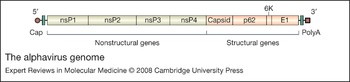
The molecular biology of these viruses has been investigated in detail, mainly because they have a simple genome coding for only nine proteins of unique sequence (Fig. 1). They also grow rapidly and to high titre in a variety of cell lines. The cell line BHK-21 is usually the preferred choice for growth of viruses and virus vectors, and routinely yields about 109 infectious vector or virus particles per 106 cells. The enveloped virion contains two main envelope proteins – E1 and E2 – in all alphaviruses; these form heterodimeric spikes in the case of SIN and VEE, and heterotrimeric spikes with a third envelope protein, E3, in the case of SFV. The capsid (C) protein within the envelope surrounds the RNA and forms the icosahedral nucleocapsid. The envelope is formed by budding from internal cellular membranes as well as the plasma membrane. Also contained within the virion are variable amounts of a smaller protein known as 6K (Ref. Reference Strauss and Strauss1).
The alphavirus genome. The 11.2 kb positive-sense RNA genome of Semliki Forest virus is capped (5′ brown circle) and polyadenylated (3′ brown square). Turquoise rectangles represent the 5′ and 3′ untranslated regions. The genome has two open reading frames: the first (grey) encodes the four nonstructural (ns) proteins that form the replicase complex; the second (orange) codes for the structural proteins (p62 is the precursor of E2 and E3).

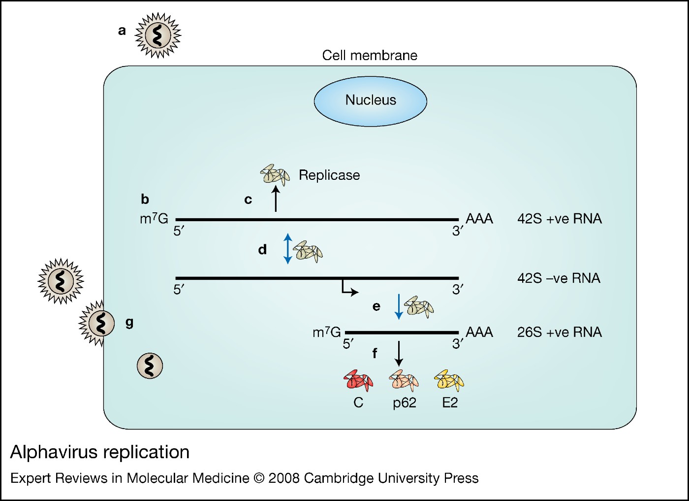
The virion RNA, designated 42S (or 49S for SIN), is approximately 11 kb in size, and has the 5′ cap structure and 3′ polyA tract of typical mammalian mRNAs (Fig. 1). Almost the entire genome is translated after infection to give a single protein that is subsequently cleaved to form the structural proteins (virion or virion-associated) and nonstructural proteins (RNA polymerase components). The structural proteins are amplified by synthesis of a subgenomic RNA species designated 26S, which is transcribed from the negative strand from a subgenomic promoter (Ref. Reference Willems5) (Fig. 2). It is this amplification that is utilised in the construction of alphavirus vectors. Insertion of a multiple cloning site (MCS) in place of the structural protein reading frame in the 26S region allows the cloning and high-level expression of heterologous genes, which are expressed from the 26S promoter (Ref. Reference Liljestrom and Garoff2).
Alphavirus replication. The figure indicates steps in the replication and expression of Semliki Forest virus (SFV) within a mammalian cell. (a) Virus entry. (b) Full-length 42S positive-stranded RNA genome is released into the cytoplasm of the cell. (c) Viral replication begins with translation of the first open reading frame coding for the four nonstructural proteins (nsP1–4) that form the viral replicase complex. (d) Replicase catalyses formation of both a 42S negative-stranded RNA intermediate and a 42S positive-stranded genomic RNA. (e) Replicase also catalyses transcription from an internal 26S promoter to form a subgenomic 26S positive-stranded RNA that (f) codes for the SFV structural proteins. Newly synthesised genomic RNA and capsid proteins form nucleocapsids in the cytosol and associate with glycosylated spike proteins at the plasma membrane, where (g) virus particles are released from the cell by budding.

Biosafety and containment of alphaviruses and their vectors
SFV, the virus on which the first alphavirus vector system was based (Ref. Reference Liljestrom and Garoff2), was first isolated from mosquitoes in Uganda in 1948 (Ref. Reference Smithburn and Haddow3), and many isolates have been made since in central and east Africa. Its primary hosts appear to be small rodents and birds, although it can be transmitted to humans (Ref. Reference Mathiot4). The original isolate (labelled L10) was virulent for mice, and SFV infection was reported to have been responsible for a laboratory death in 1979 (Ref. Reference Willems5). However, most infections in Africa appear to be innocuous, among Europeans as well as native Africans (Ref. Reference Mathiot4).
In the USA, SFV is classified by the Subcommittee for Arbovirus Laboratory Safety (SALS) of the Office of Health and Safety as a virus requiring level 3 containment, with the caveat that most activities can be carried out at level 2 (Ref. 6). Level 2 containment is recommended for all activities associated with SIN and SFV by the European Union (EU) (Ref. 7), and for SIN in the USA (Ref. 6). Wild-type VEE requires level 3 containment in both the USA and EU, but can be utilised as an attenuated vaccine strain (TC-38) or as a debilitated vector at level 2 (Ref. 6).
Pathogenicity of the wild-type viruses
With regard to biosafety, it is important to ensure that alphavirus-based vectors do not induce the host tissue and organ damage that occurs with the wild-type viruses. Wild-type virus could be generated by reversion in the case of replication-competent vectors, or by recombination between vector and helper viruses in the case of recombinant virus-like particles (VLPs) (see later).
Although SIN strains can cause disease in humans, including rash, fever and arthritis (Ref. Reference Laine, Luukkainen and Toivanen8), and encephalitis in mice, laboratory strains used for the construction of vectors cause encephalitis in neonatal mice only, and infect only neurons in the central nervous system (CNS) (Ref. Reference Polo9). VEE can cause lethal encephalitis in humans, but the recombinant VLP vector system based on this virus is derived from an attenuated strain (Ref. Reference Pushko10).
As mentioned above, a lethal case of SFV encephalitis was described following laboratory infection (Ref. Reference Willems5), but in an outbreak in the Central African Republic no deaths occurred, although fever, arthralgia and other symptoms were recorded in European patients (Ref. Reference Mathiot4). By contrast, in mice the original isolate of SFV (Ref. 3; L10) is highly virulent, causing lethal encephalitis after about 5–7 days following peripheral inoculation (intraperitoneal, intravenous or intramuscular) of a low dose (about 102 infectious units). In adult mice, this lethal infection is characterised by an initial viraemia that follows virus multiplication in peripheral tissues, but which is contained by an immune response. Infection of the CNS, however, leads to neuronal necrosis that is lethal. However, most SFV vectors are derived from an SFV strain termed the prototype strain, which was derived originally from L10 but has been somewhat attenuated by passage in cell culture. This strain, and the SFV4 virus derived from the first SFV infectious clone based on it, kill only a proportion of mice when administered peripherally although all mice succumb following intranasal infection (Ref. Reference Atkins, Sheahan and Liljeström11). Infection of the CNS involves infection of neurons and inflammatory demyelination triggered by infection of oligodendrocytes (the myelin-producing cells in the CNS).
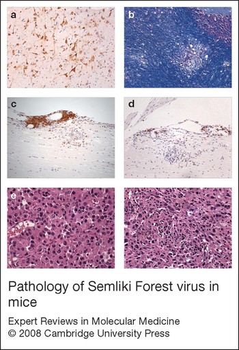
A second strain of SFV used in vector construction is the A7[Reference Chikkanna-Gowda74] strain, derived by cell passage from the original A7 strain (Ref. Reference Bradish, Allner and Maber12). This is a separate SFV isolate that is avirulent in mice when given by any route. Like L10 it produces a viraemia following peripheral administration and infects the CNS, but in this case the infection is not lethal and the mice are resistant to virulent L10 SFV challenge (they show inflammatory demyelination in the CNS but little neuronal necrosis). Examples of the neuropathology of SFV infection are shown in Figure 3a and 3b.
Pathology of Semliki Forest virus in mice. (a) Semliki Forest virus (SFV) antigen (brown) in neurons and neuronal processes in the midbrain, 5 days following infection with SFV4 (anti-SFV immunohistochemistry, ×20). (b) Demyelinated plaque in the cerebellum, 14 days following infection with A7-SFV (luxol fast blue/cresyl violet, ×20). (c, d) B cells (brown) in the leptomeninges and spinal cord in EAE lesions following treatment with SFV10-E empty particles (c) and SFV10-E-IFN-β (d) (anti-B220 immunohistochemistry, ×20). The mice were sacrificed 22 days after disease induction. Treatment with SFV10-E-IFN-β caused a marked reduction in the numbers of B cells. (e, f) K-BALB tumours from a naive, control mouse (e) and a vaccinated mouse 15 days following treatment with SFV4 (f) (haematoxylin and eosin, ×40). Polyhedral tumour cells in the control animal are closely adjoined and show mitotic activity; spindle-shaped tumour cells in the treated animal are separated by collagenous stroma infiltrated with lymphocytes. (These findings have previously been reported: for parts a and b, see Ref. 11; for parts c and d, see Ref. 43; for parts e and f, see Ref. 73). Abbreviations: EAE, experimental autoimmune encephalomyelitis; IFN, interferon.

Alphavirus vectors
All alphavirus vectors are derived from infectious clones that have been constructed for SIN, VEE and the prototype and A7[Reference Chikkanna-Gowda74] strains of SFV. Infectious clones consist of a cDNA copy of the entire virus genome, inserted into a bacterial plasmid that can be selected by antibiotic resistance when grown in Escherichia coli. The viral cDNA genome is inserted downstream of a bacteriophage promoter, which can be SP6 or T7. The plasmid DNA can be replicated in the bacterial host, then isolated and transcribed with SP6 or T7 polymerase. When transfected into mammalian cells (usually BHK or Vero cells), this RNA initiates a multiplication cycle that produces infectious virus particles. The transfection may be carried out by electroporation or by using transfection agents such as lipofectamine.
There are three main types of alphavirus vector: recombinant VLPs, layered DNA–RNA vectors and replication-competent vectors (Fig. 4).
Alphavirus expression vectors. (a) Virus-like particle vector. The backbone vector carries the four genes coding for the replicase complex (nsP1–4), but does not contain the structural genes (C, p62, 6K, E1). The structural genes are replaced with a multiple cloning site into which foreign genes can be inserted. (b) Capsid and spike helper vectors. These vectors carry the genes coding for the structural proteins but have a deletion in the replicase region that includes the RNA-packaging signal. (c) Double subgenomic promoter vector. A second subgenomic promoter is cloned into the backbone vector to accommodate a second foreign protein. This expression vector allows expression of two foreign proteins within the same cell. (d) SFV10Enh vector. This contains an SFV ‘translational enhancer’ element (Enh) that has been shown to increase translation levels of the foreign protein in infected cells. The enhancer consists of the first 34 amino acids of the SFV capsid protein (red) followed by the 17 nucleotide foot and mouth disease virus 2A protease (yellow). (e) DNA–RNA layered vector. The SFV replicon is placed under the control of a cytomegalovirus immediate-early promoter (recognised by RNA polymerase II). An SV40 transcription termination region (mauve rectangle) is placed at the 3′ end of the replicon. (f) Replication-competent SFV vector. A second subgenomic promoter and multiple cloning site is placed between the nonstructural genes and the genes coding for the structural proteins. Abbreviations: CMV P, cytomegalovirus immediate-early promoter; Enh, enhancer element; Δ Rep, deleted replicase.

Recombinant VLPs
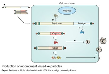
The first alphavirus vector to be constructed and the most extensively used is the recombinant VLP or ‘suicide’ particle vector (Fig. 4a). This utilises a vector backbone with the structural protein region (encoding envelope and capsid proteins) of the genome deleted and an MCS inserted. This MCS is located downstream of the 26S promoter, so that a gene cloned into the MCS is amplified similarly to the 26S RNA region in the wild-type virus. A Kozak ribosome-binding sequence and AUG initiation codon can be incorporated upstream of the MCS, and multiple stop codons downstream. This vector RNA is transcribed by SP6 or T7 polymerase from the promoter upstream of the viral sequence on the plasmid DNA, then transfected into susceptible cells in culture. To produce VLPs, RNA is transcribed from two helper plasmids that encode the envelope and capsid proteins, respectively, but lack the packaging signal located in the nonstructural region of the genome (Fig. 4b). These two helper RNAs are cotransfected into cultured cells along with the vector RNA. Replication and translation of these RNAs results in the production of ‘suicide’ VLPs (Fig. 5) that are able to infect susceptible cells, but go through only one round of multiplication because they lack the viral structural protein genes that have been supplied by the helper RNAs. In so doing, they may express at high levels a gene cloned into the vector MCS, since it is translated from RNA amplified from the 26S promoter. A second subgenomic promoter may also be inserted to allow expression of two proteins from the same vector – for example, the two subunits of interleukin (IL)-12 (Fig. 4c).
Production of recombinant virus-like particles. (a) In vitro transcribed recombinant and helper RNAs are cotransfected into BHK-21 cells by electroporation. (b) The replicase complex, translated from the recombinant RNA, amplifies all three RNA species. (c) The foreign protein is also produced from the recombinant RNA, and (d) capsid and spike proteins are produced from the helper RNAs. (e) The capsid protein packages only recombinant RNA molecules into nucleocapsids as the helper RNAs lack a packaging signal (in the nsP1 gene). (f) Assembly and budding of new particles occurs at the cell surface through an interaction between cytoplasmically preformed nucleocapsids and the viral spike proteins that have been transported to the cell membrane.

One biosafety problem with VLP generation is that recombination could occur, albeit at low probability, between vector and helper RNAs to generate wild-type virus. To reduce this possibility, two helper RNAs are used. For SFV, the capsid protein cleavage site is also mutated to prevent cleavage, thus leading to defective virus if recombination did occur (Ref. Reference Smerdou and Liljeström13). To increase further the yield of foreign protein in infected cells, a translation enhancer element from the capsid protein and a 2a protease element may be used in an enhanced expression vector (Fig. 4d)
Layered DNA–RNA vectors
A variant of the VLP system described above, which is based entirely on the expression of RNA, is the layered DNA–RNA vector (Fig. 4e). This utilises a cytomegalovirus (CMV) promoter to stimulate the transcription of a cDNA copy of the vector RNA after transfection of the DNA. This must occur in the cell nucleus, and is similar to the system used for DNA vaccines. One problem with this type of vector is the low efficiency of transfection of the DNA following administration, compared with VLP infection. However, when used as a vaccine vector, the immune response induced by this type of vector is higher than that induced by a standard DNA vector, and it has the advantage that no neutralising antibody is induced to the vector itself, since it is composed only of nucleic acid (Refs Reference Berglund14, Reference Ljungberg15).
Replication-competent vectors
Unlike the VLP vector, the replication-competent vector undergoes several rounds of multiplication when administered to the host. Originally based on the SIN genome (Ref. Reference Tamang16), it has a second 26S promoter and MCS inserted upstream of the structural protein region (Fig. 4f). Thus, this vector has two 26S promoters: one expresses cloned genes from the MCS, and the other expresses the viral structural proteins. To attenuate this type of vector, mutations or deletions may be introduced into the regions of the vector derived from the virus genome (Ref. Reference Galbraith, Sheahan and Atkins17); one such SFV vector has mutations introduced into the nuclear localisation signal in the nsP2 gene that renders it noncytopathic (Refs Reference Casales18, Reference Lundstrom19). A variation of this type of replication-competent vector, based on the avirulent A7[Reference Chikkanna-Gowda74] strain of SFV, has the second 26S promoter and MCS inserted into the 3' untranslated region of the genome, downstream of the viral structural proteins and first 26S promoter. The problem with this vector is its instability (Ref. Reference Vähä-Koskela20).
Comparison with other vectors
Viral vectors have mostly used DNA viruses (adeno, adeno-associated, herpes, pox) or those that replicate via a DNA intermediate (retroviruses). However, recently some interest has been shown in RNA virus alternatives to these standard viruses, such as measles virus and reovirus (Ref. Reference Vähä-Koskela, Heikkilä and Hinkkanen21). Alphavirus vectors have advantages over DNA systems in that transient high-level expression of cloned genes occurs, multiplication is purely cytoplasmic (so there is little danger of insertion of DNA into the host genome and consequent insertional mutagenesis), and dispersal of the vector usually occurs only to the local lymph node following intramuscular injection so is not systemic (Ref. Reference Morris-Downes22). Levels of expression are higher than those produced with most other systems, including adenovirus, and induction of neutralising antibodies by the input virus is less of a problem since lower titres are produced (Ref. Reference Riezebos-Brilman23).
The limitations to the use of alphavirus vectors are the possibility of reversion to wild-type virus for VLPs (discussed above) and transient expression. The transient but high-level expression of cloned genes by alphavirus vectors means that they cannot be used for the long-term correction of genetic function. Because of this, the main applications that have been investigated are those that require transient high-level expression: these are mainly recombinant vaccine construction, therapy of CNS disease and cancer therapy.
Applications of alphavirus vectors
The first and the most extensively studied application of alphavirus vectors was as potential vaccine vectors. For this application, recombinant VLPs or the layered DNA–RNA vector were the vectors of choice. Later, alphavirus vectors of all types were studied as potential CNS vectors and cancer therapy agents.
Vaccine vectors
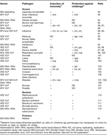
SIN, VEE and SFV vectors have all been utilised to construct prototype vaccines for animal testing. Both recombinant VLPs and the layered DNA–RNA system have been tested, but the problem with the latter system is, although it has higher biosafety as a result of its inability to generate wild-type virus and its lack of virion proteins, it induces a less efficient immune response to transgene-encoded proteins than recombinant VLPs (Refs Reference Berglund14, Reference Ljungberg15). Naked RNA vectors can also induce an immune response, but are also less efficient than VLPs, and RNA is inherently unstable (Ref. Reference Fleeton24). Alphavirus vaccines express immunogenic proteins that protect against challenge or stimulate immunity (see below and Table 1). Usually a T helper (Th) 1 response is induced following inoculation. The strategies used include ‘prime–boost’ with the same vector construct, and prime–boost with the same antigen expressed by an alphavirus vector and a different vector (not necessarily in this order). The latter strategy has the advantage that it circumvents any immunity that may be induced to the vector itself following priming. Several reviews have been published (Refs Reference Atkins, Sheahan and Liljeström11, Reference Riezebos-Brilman25, Reference Lundstrom26) that summarise the early work that was carried out on vaccine construction following initial description of the vectors in the 1990s (Refs Reference Strauss and Strauss1, Reference Pushko10, Reference Bredenbeek27). It was during this period that the efficacy of the vectors in inducing immune responses to a variety of antigens was established. More recently, prototype vaccines have been constructed against several infectious diseases (both human and veterinary), and against tumour antigens.
Experimental alphavirus-based vaccines

aTested in mice unless otherwise specified: ca, cats; ch, chickens; gp, guinea pigs; ma, macaques; mi, mice; ho, horses; pi, pigs; ra, rabbits; sh, sheep.
Abbreviations: DNA–RNA, layered cDNA vector transcribed to RNA; ND, not done; replicating, replication-competent vector with second 26S promoter; SFV, Semliki Forest virus; SIN, Sindbis virus; VEE, Venezuelan equine encephalitis virus; VLP, recombinant virus-like particle. See text for full explanation.
The development of an efficient, safe and inexpensive vaccine for human immunodeficiency virus (HIV) is one of the most intractable current medical problems. Alphavirus recombinant VLP vectors based on SIN, VEE, SFV and VEE–SIN chimaeras (Ref. Reference Perri28) have been used to study induction of immune responses to HIV proteins in mice, and simian immunodeficiency virus (SIV) proteins in macaques. Both immunisation with the alphavirus vectors alone and prime–boost in combination with other vectors have been studied.
In mice, vaginal immunisation with SIN VLPs expressing the HIV Gag protein induced both local mucosal cell-mediated responses and protection against vaginal challenge with a vaccinia vector expressing Gag. This was proof of principle that alphavirus VLPs could induce protective anti-HIV immunity at mucosal surfaces, the portal of entry of HIV in humans (Ref. Reference Vajdy29). It has also been shown that neutralising antibody responses can be induced in mice by immunisation with the HIV gp160 envelope protein expressed by VEE VLPs, and that these are similar in potency and crossreactivity to those found in the chronically infected, long-term nonprogressive donor of the virus (Ref. Reference Dong30). For SFV recombinant VLPs, priming with SFV VLPs followed by boosting with vaccinia MVA (modified vaccinia Ankara) expressing an HIV clade A antigen induced efficient and long-term anti-HIV T cell responses in mice (Ref. Reference Hanke31). However, it is also clear that homologous immunisation and boosting with SFV VLPs induces efficient anti-HIV T cell responses (Ref. Reference Sundback32), and also enhances antibody responses to envelope glycoproteins (Refs Reference Forsell33, Reference Forsell34).
SIV infection of macaques has been used as a primate model of HIV infection for testing alphavirus vectors. The study by Davis et al. (Ref. Reference Davis35) using VEE VLPs and small numbers of macaques exemplifies several early studies in this area. Using four animals, it was shown that subcutaneous injection followed by intravenous boost with VLPs expressing SIV structural proteins protected against challenge and reduced viral load following challenge, although it did not prevent infection. Both cell-mediated and antibody immunity were induced by vaccination. For SFV, it was found that a prime–boost regime that involved initial vaccination with SFV VLPs expressing SIV antigens, followed by a boost by a MVA (pox) virus vector expressing the same antigens, was more effective at inducing an immune response than the use of either vector alone. However, the prime–boost regime did not protect the animals against rectal challenge (Ref. Reference Nilsson36). For VEE VLPs expressing a cocktail of SIV antigens used to vaccinate macaques, virus loads were reduced after rectal challenge, but this reduction was not significant (Ref. Reference Johnston37). A similar reduction in viral load (in this case significant) has been obtained after immunisation of macaques with a VEE–SIN chimaeric vector expressing SIV antigens, but boosted with viral protein (Ref. Reference Xu38).
As well as the work described above for HIV, alphavirus vector systems have been investigated as vaccine vectors in several animals (mostly mice) for a large number of pathogens, both human and veterinary. The pathogens have usually been viral, but vaccines for bacteria and protozoa (malaria) have also been investigated. Such studies, too numerous to be described in detail here, are summarised in Table 1.
Central nervous system vectors
Since alphaviruses are naturally neurotropic viruses, their use to treat CNS diseases has been investigated – although so far only in mouse models and with SFV. Many studies have shown that virulent wild-type SFV infects and destroys neurons in the CNS, thus killing the mouse, but that avirulent strains are less cytopathic for neurons in adult mice (Ref. Reference Atkins, Sheahan and Liljeström11). Studies have been carried out with recombinant VLPs, and with replication-competent vectors expressing the enhanced green fluorescent protein (EGFP) reporter gene. Using hippocampal slices, it was shown that a replication-competent vector derived from the A7[Reference Chikkanna-Gowda74] avirulent strain of SFV, and expressing EGFP, infects neurons less efficiently than glial cells (Ref. Reference Ehrengruber39). However, stereotaxic intracerebral inoculation of mice with VLPs resulted in CNS damage, so use of the vectors to treat CNS disease by this route of inoculation may not be feasible (Ref. Reference Graham40).
The progression of experimental autoimmune encephalomyelitis (EAE) in the mouse CNS has been investigated following administration of SFV VLPs expressing EGFP and cytokine genes. EAE is an experimental inflammatory disease induced in mice by injection of myelin or a myelin component plus adjuvant, and is an animal model of multiple sclerosis. The vector was given intranasally because it had already been shown for the wild-type virus that this route of administration leads directly to CNS infection via the olfactory bulb, and that the virus reaches the CNS through the limbic system, thus bypassing the blood–brain barrier (Ref. Reference Sammin41). When mice were infected intranasally with VLPs expressing EGFP, fluorescence was detected in the olfactory bulb as early as one day after administration, and persisted up to 7 days. No virus-induced damage was detected in the CNS (unlike with the wild-type virus) and virus RNA could be detected only in the nasal mucosa. For a similar vector expressing IL-10, an anti-inflammatory cytokine, clinical EAE was reduced for intranasally treated mice but exacerbated in mice treated intranasally with particles containing no cloned gene (Ref. Reference Jerusalmi42). Similar experiments have been reported for an SFV VLP vector expressing interferon β (IFN-β; Fig. 3c and d). This cytokine is used to treat chronic–relapsing multiple sclerosis, with partial success in some patients. The IFN-β-expressing VLPs resulted in higher levels of IFN-β throughout the CNS after intranasal administration. However, in this case inhibition of EAE with IFN-β-expressing particles was dependent on the number and timing of treatments. Fewer treatments, given before the effector stage of the disease (i.e. before inflammation was induced), led to improvement in clinical and pathology score. It was concluded that the timing and frequency of intranasal administration of IFN-β-expressing particles are critical to disease outcome, with treatment prior to the effector stage being most effective (Ref. Reference Quinn43). However, these experiments on EAE do indicate that, in a mouse model at least, intranasal administration of SFV VLPs may be used for noninvasive treatment with biologically active proteins in the CNS, bypassing the blood–brain barrier. Any damage to the nasal mucosa can be rapidly repaired and, since only the protein encoded by the vector enters the CNS, no damage is apparent.
A replication-competent vector based on the avirulent A7[Reference Chikkanna-Gowda74] strain of SFV has also been used to treat EAE. Such a vector expressing transforming growth factor β (Ref. Reference Vähä-Koskela44) or an inhibitor of metalloproteinase 2 (Ref. Reference Nygårdas45), and given intraperitonally, was able to cross the blood–brain barrier and inhibit EAE in mice.
Vectors for tumour therapy/prophylaxis
Alphavirus vectors have been investigated for two applications as anticancer agents. First, they have been used to stimulate antitumour immunity – either prophylactically (i.e. as anticancer vaccines given before the tumour is established) or therapeutically (i.e. to treat established tumours). Second, they have been assessed as oncolytic agents – that is, to stimulate the killing of tumour cells by apoptotic or other mechanisms, either through the action of the virus itself or through the expression of proapoptotic genes. Also, some studies have used a combination of these two approaches.
Several recent reviews have dealt with the use of alphaviruses and their vectors as anticancer agents (Refs Reference Riezebos-Brilman25, Reference Kelly, Fleeton and Atkins46, Reference Atkins47, Reference Yamanaka48, Reference Lundstrom49), as well as their oncolytic capacity in comparison with other vectors (Ref. Reference Vähä-Koskela, Heikkilä and Hinkkanen21). Much of this earlier work was concerned with the induction of antitumour immune responses (Ref. Reference Riezebos-Brilman25).
Papillomavirus-associated carcinoma
Use of alphavirus vectors in the therapy and/or prophylaxis of papillomavirus-associated carcinoma has been examined in mouse models. Vaccination of mice with VEE VLPs expressing the papillomavirus oncogenic antigen E7 resulted in elimination of some E7-bearing tumours and prevention of growth of all tumours (Ref. Reference Velders50). Similar results were obtained using an SFV VLP vector expressing a fusion protein of the papillomavirus E6 and E7 oncogenic proteins (Ref. Reference Daemen51). Linkage of calreticulin to the papillomavirus E7 antigen, and expression by a SIN VLP, has been shown to produce a strong antitumour effect against E7-expressing tumours. Calreticulin associates with transporters involved in antigen presentation, and enhances the presentation of E7 through the major histocompatibility class I pathway. It may also have an effect on angiogenesis, thus inhibiting the formation of metastases (Ref. Reference Cheng52). Expression by a VEE VLP of E6 and E7 proteins that had been mutated to disrupt their oncogenic potential also inhibited tumour growth (Ref. Reference Cassetti53). Furthermore, an SFV VLP expressing E6 and E7 was shown to induce E6/E7-specific T cell activity in immunotolerant transgenic mice (Ref. Reference Riezebos-Brilman54).
Breast cancer
For the breast-tumour-associated self-antigen NEU (ERBB2), immune tolerance was overcome by expression of the unaltered antigen from a VEE VLP, with the protection of 50% of challenged rats and the induction of T cell immunity (Ref. Reference Laust55). For transgenic mice expressing NEU, vaccination with VEE VLPs expressing this antigen completely inhibited tumour cell challenge, either in the mammary glands or elsewhere. It was suggested that this construct could form the basis of a vaccine for this tumour (Ref. Reference Wang56).
Melanoma
Two recent studies have utilised alphavirus vectors in the treatment of melanoma in mouse models of melanoma, an almost invariably fatal metastatic cancer. In one regime, mice were vaccinated with a SIN DNA–RNA layered vector encoding MUC-18 (MCAM), an adhesion molecule expressed on invasive and metastatic melanoma but not on normal melanocytes. This vaccine induced T cell immunity to MUC-18 and resistance to challenge with MUC-18-expressing tumour cells, thus indicating it could be used in the immunotherapy of melanoma (Ref. Reference Leslie57). The second study utilised an oncolytic virus approach for the treatment of human melanoma cells growing as xenographs in severe combined immunodeficiency syndrome (SCID) mice. Growth of the tumours was inhibited following both peripheral and intratumoural injection. However, some tumour cells showed regrowth, and damage was sustained by other tissues, so this treatment was only partially successful (Ref. Reference Vähä-Koskela58).
Glioma
Glioblastoma multiforme is a relatively common fast-growing human brain tumour that is usually fatal within a year of diagnosis, even when treated by resection, drugs and radiotherapy, which slow progression of the disease. Therefore oncolytic virus vectors, including alphavirus vectors, have been considered as a potential treatment for this disease. In one study, nine different oncolytic viruses, which included SIN as a representative alphavirus, were compared with regard to their ability to infect and kill human glioblastoma cells. Of these viruses, SIN and vesicular stomatitis virus showed the greatest oncolytic activity (Ref. Reference Wollmann, Tattersall and van den Pol59). A SIN VLP vector expressing a hyperfusogenic mutant of the gibbon ape leukaemia virus envelope glycoprotein (GALV.fus) has been shown to selectively kill human glioblastoma cells, both in cell culture and in a nude mouse xenograph model (Ref. Reference Zhang, Frolov and Russell60). An SFV VLP vector expressing an antiangiogenesis gene (angiostatin) and two proapoptotic genes (p53 and PTEN) showed more efficient apoptotic activity in glioma cell culture and antitumour activity in a nude mouse xenograph model than these genes expressed separately (Ref. Reference Lee61). A recent study has used the wild-type avirulent A7[Reference Chikkanna-Gowda74] strain of SFV and its derived replication-competent vector expressing a reporter gene to treat implanted rat gliomas (Ref. Reference Maatta62). Although one intracranial injection of either virus inhibited tumour growth, complete regression did not occur, even following multiple injections, and the animals did not survive. However, this preliminary study indicates that further manipulation of the virus and modification of the treatment regime could have more beneficial results.
Prostate cancer
Recently, three studies using VEE VLPs have indicated the potential of alphavirus vectors for the treatment of prostate cancer. Vaccination against the six-transmembrane epithelial antigen of the prostate (STEAP), an antigen expressed in normal prostate tissue but overexpressed in tumours, inhibited tumour growth in both therapeutic and prophylactic mouse models, without inducing autoimmunity (Ref. Reference Garcia-Hernandez Mde63). In a second study, similar results were obtained in prostate-cancer-prone transgenic adenocarcinoma mouse prostate (TRAMP) mice using a vaccine against prostate stem cell antigen, which is overexpressed in prostate cancer and metastases. Again, no autoimmunity was induced (Ref. Reference Garcia-Hernandez64). Another candidate for vaccination against prostate cancer is prostate-specific membrane antigen (Ref. Reference Durso65).
Tumour treatment strategy
Most cancer deaths are caused by the inability to target and treat rapidly growing, infiltrative tumours that may also have formed metastases. Such properties may evolve following treatment, or more rarely such tumours may be intractable to treatment when they arise. For this reason alphavirus and other oncolytic vectors have been investigated as a last resort for the treatment of rapidly growing, otherwise untreatable tumours. All work in this area for alphavirus vectors has so far been carried out using animal models.
It has been shown that SIN virus and its derived vector can target model tumors in mice when given peripherally, and that this is due to the increased concentration of the virus receptor in tumour tissue (Refs Reference Tseng66, Reference Hurtado67, Reference Tseng68, Reference Tseng69). This does not appear to occur for SFV, which does not specifically target tumour tissue if given peripherally. However, if given intratumourally, the vector remains within the tumour (Ref. Reference Rodriguez-Madoz, Ptrieto and Smerdou70).
Recombinant SFV VLPs have been tested by direct injection of subcutaneous human lung carcinoma tumours in nude mice, derived from the p53-deficient H358a cell line. Even with a vector expressing a reporter gene only, complete regression of some tumors occurred and the growth of others was substantially inhibited (Ref. Reference Murphy71). However, this treatment was less successful on the fast-growing rat AT3 prostate carcinoma cells in nude mice, although inhibition of tumor growth did occur (Ref. Reference Murphy, Sheahan and Atkins72). For several tumour models in immunocompetent mice, vectors expressing T cell epitopes and cytokines such as IL-12 and IL-18 were effective in inhibiting the growth of fast-growing tumours (Refs Reference Smyth73, Reference Chikkanna-Gowda74, Reference Chikkanna-Gowda75, Reference Rodriguez-Madoz, Prieto and Smerdou76). Also, vaccination with SFV VLPs expressing the envelope proteins only, followed by treatment with the wild-type virus, inhibited tumour growth (Ref. 73; Fig. 3e and f). Inhibition of lung metastases for the 4T1 breast carcinoma model by an enhanced IL-12-expressing vector was significant, since it could result from inhibition of angiogenesis (Ref. Reference Chikkanna-Gowda75). This was followed by a demonstration that peripheral immunisation with an SFV vector expressing FLK-1 (equivalent to human vascular endothelial growth factor receptor 2) could inhibit tumour growth in these models by inhibition of angiogenesis or an antivascular effect (Ref. Reference Lyons77). This was effective both prophylactically (i.e. before the tumour was established) and therapeutically (i.e. after the tumour was established), although the latter was less effective.
As indicated above, the replication-competent vector based on the avirulent A7[Reference Chikkanna-Gowda74] SFV strain has also been utilised for tumour treatment. It has been used to treat several model tumours by viral oncolysis (Refs Reference Vähä-Koskela58, Reference Maatta62). It has also been shown that direct intratumoural injection of the virulent wild-type virus is very effective at inducing tumour regression in vaccinated mice (Ref. 73; it kills a proportion of unvaccinated mice), so an attenuated replication-competent vector should have the same effect. This is because the viral envelope proteins expressed by replicating virus have strong T cell epitopes, inducing an intense inflammatory response that can be augmented by cytokine expression by the vector.
Clinical and commercial potential of alphavirus vectors
Alphavirus VLP replicon vectors were first described in 1991 (Ref. Reference Strauss and Strauss1), so it is surprising that only one preclinical study on an alphavirus-based influenza vaccine has been reported (Ref. Reference Hubby78). In terms of potential vaccines, numerous animal studies have been carried out, as described above and in Table 1, that indicate the efficacy and safety of such vaccines. However, the slowness of commercial and clinical exploitation of alphavirus-based vaccines may not be confined to this area, since it appears to be a characteristic of potential new recombinant vaccines in general. For example, two new vaccines that have reached or are about to reach the marketing phase are those for human papillomavirus (Ref. Reference Villa79) and Ross River virus (Ref. Reference Kistner80); both of these are based on viral protein only that is not delivered by vectors.
Although clinical trials on alphavirus-derived vaccines are now being initiated, their use as CNS or anticancer agents has not yet reached the clinical stage. The protocol for the use of a liposome-encapsulated SFV VLP vector expressing IL-12 to treat glioblastoma multiforme has been described (Ref. Reference Ren81), but the results of such a clinical trial have never been published. Although the use of alphavirus vectors as oncolytic anticancer agents is a relatively new application, they would possibly supplement and/or add to existing treatments, and possibly could be developed for tumours that are at present untreatable. One possibility is to combine the use of alphavirus vectors as CNS vectors with their oncolytic action in the treatment of glioblastoma multiforme. Further development of vector-based treatments, and clinical trials in this area, are therefore badly needed.
Conclusions and summary
Alphavirus vectors have several advantages over more conventional vectors for some applications. These include the improbability of genome integration and/or insertional mutagenesis (the layered DNA–RNA vector may be an exception here), the high levels of expression, and the simplicity and ease of manipulation of the genome. However, because of their transient expression, they cannot be used for the permanent replacement of gene function. In some situations, such as vaccine construction, this may be an advantage. Animal experiments have demonstrated their probable biosafety. However, two major problems remain in their clinical application: the development of antivector immunity on multiple administration; and the lack of reproducible targeting strategies, particularly in tumour treatment (Ref. Reference Rodriguez-Madoz, Ptrieto and Smerdou70). However, alphaviruses represent a viable alternative to existing and more commonly used strategies for these applications, and should be further investigated and exploited.
Acknowledgements and funding
We thank Dorothy Mooney and Alex Whelan for practical help with the work carried out in our laboratories. Many students and postdoctoral researchers worked with us over the years and their efforts are gratefully acknowledged. We also thank the referees for their constructive comments. Our work has been funded by Science Foundation Ireland, the Wellcome Trust, the Health Research Board, the Multiple Sclerosis Society of Ireland, the European Union, Enterprise Ireland and Cancer Research Ireland.