Hypoplastic left-heart syndrome and critical aortic stenosis with severely restrictive or intact atrial septum are associated with high prenatal and postnatal mortality. The 1-month survival rate is ∼33%.Reference Rychik, Rome, Collins, DeCampli and Spray1–Reference Lowenthal, Kipps, Brook, Meadows, Azakie and Moon-Grady5 Long-term outcome after univentricular palliation procedures – that is, bidirectional Glenn or stage 2 palliation, total cavopulmonary connection or stage 3 palliation – remains poor compared with those without a restrictive oval foramen.Reference Lowenthal, Kipps, Brook, Meadows, Azakie and Moon-Grady5–Reference Jaeggi, Ryan, Seed and Chaturvedi7 Restriction of the interatrial communication occurs in ∼20%, with complete closure in 6–10% of the foetuses with hypoplastic left-heart syndrome.Reference Rychik, Rome, Collins, DeCampli and Spray1, Reference Lowenthal, Kipps, Brook, Meadows, Azakie and Moon-Grady5
In severe left-heart obstruction, unobstructed left-to-right shunting across the foramen ovale is vital (Fig 1a). If the left-to-right shunt is restricted or absent, blood delivered from the pulmonary veins cannot exit the left atrium (Fig 1b). This results – if there is no other shunt – in pulmonary venous hypertension. Severe changes in the pulmonary vascular system and abnormal lung lymphatic development are known to occur in the foetus; however, data on the exact timing and irreversibility of these changes are unknown.Reference Rychik, Rome, Collins, DeCampli and Spray1–Reference Glatz, Tabbutt and Gaynor3, Reference Taketazu, Barrea, Smallhorn, Wilson and Hornberger8, Reference Graziano, Heidelberger, Ensing, Gomez and Ludomirsky9 After birth, a substantial increase in the pulmonary venous return to the left atrium leads to further rises in pulmonary venous pressure, pulmonary oedema, and pulmonary hypertension (Fig 1c). Neonates born without an adequate interatrial communication and atrial mixing present with severe hypoxaemia, and 50–65% of them die, despite immediate surgical or catheter-based intervention.Reference Rychik, Rome, Collins, DeCampli and Spray1–Reference Glatz, Tabbutt and Gaynor3, Reference Marshall, Levine and Morash10
(a) Foetal circulation in hypoplastic left-heart syndrome: obligate left-to-right shunt across the oval foramen (red arrow): blood returning from the pulmonary vasculature via the pulmonary veins must cross the atrial septum from left to right. (b) Foetus with hypoplastic left-heart syndrome and intact atrial septum: obstruction to left atrial egress leads to left atrial and pulmonary venous congestion and secondary changes in the pulmonary vasculature. (c) Neonate with hypoplastic left-heart syndrome and intact atrial septum: after birth, the lungs expand with an increase in pulmonary blood flow and pulmonary venous return: increase in left atrial and pulmonary congestion and pressure. The lack of interatrial mixing results in marked hypoxaemia.

Recent studies have shown that mortality remains high, despite early and accurate prenatal diagnosis and optimised perinatal management.Reference Glatz, Tabbutt and Gaynor3, Reference Lowenthal, Kipps, Brook, Meadows, Azakie and Moon-Grady5 In a group of 73 foetuses with either classic hypoplastic left-heart syndrome or critical aortic stenosis with poor left ventricular function, the outcome of foetuses with a restrictive oval foramen was worse: four of five foetuses with severe interatrial restriction died after perinatal intervention, and only three foetuses with an additional decompressing vein survived long term. Furthermore, the 2-year survival for the group with severe obstruction was only 29% compared with 83% in the non-restrictive group. Foetuses with moderate restriction survived the neonatal period but showed significant and late interstage mortality (40 versus 9.3%, p=0.04). On an intent-to-treat basis, the 2-year survival of babies born with any degree of atrial restriction was significantly lower (42.9%) than in the non-restrictive group (83%, p<0.0001). Correlation of survival with severity of restriction has been confirmed by others.Reference Vlahos, Lock, McElhinney and van der Velde2, Reference Lowenthal, Kipps, Brook, Meadows, Azakie and Moon-Grady5, Reference Vida, Bacha and Larrazabal6, Reference Marshall, Levine and Morash10
If adequate prenatal diagnosis and immediate postnatal therapy do not improve neonatal and long-term outcomes, foetal cardiac intervention may offer the potential to improve postnatal outcome.
The rationale for performing in utero treatment is to relieve left atrial and pulmonary hypertension, to prevent the anatomic changes within the pulmonary vascular system that are associated with long-lasting hypertension, and to ensure neonatal survival by creating an interatrial communication.
Foetal atrial balloon septostomy, laser perforation, and stenting of the foetal interatrial septum are the current options for foetal therapy.
Sonographic definition of severely restrictive interatrial septum
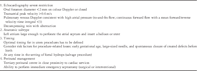
Prenatal detection of restrictive atrial septum is possible in hypoplastic left-heart syndrome, using a combination of two-dimensional Doppler echocardiography (Table 1). In the restrictive oval foramen, the diameter is <2 mm on colour Doppler echocardiography, and the transatrial peak velocity is increased.Reference Glatz, Tabbutt and Gaynor3, Reference Taketazu, Barrea, Smallhorn, Wilson and Hornberger8 In the closed structure, decompressing veins may develop as alternative pathways for the pulmonary venous drainage, that is, levoatrial cardinal veins or decompressing pathways to the coronary or hepatic veins. The presence and patency of these abnormal venous connections is an important predictor of neonatal survival.Reference Rychik, Rome, Collins, DeCampli and Spray1, Reference Lowenthal, Kipps, Brook, Meadows, Azakie and Moon-Grady5
Case selection for foetal atrial septostomy/stent implantation in hypoplastic left-heart syndrome with restrictive or intact atrial septum.
Reports suggest that severity of the restriction can be assessed most accurately by the pulmonary venous Doppler signal.Reference Lowenthal, Kipps, Brook, Meadows, Azakie and Moon-Grady5, Reference Jaeggi, Ryan, Seed and Chaturvedi7, Reference Taketazu, Barrea, Smallhorn, Wilson and Hornberger8, Reference Chintala, Tian, Du, Donaghue and Rychik11, Reference Divanović, Hor, Cnota, Hirsch, Kinsel-Ziter and Michelfelder12
The best predictors of the need for emergency atrial septostomy after delivery include a pulmonary venous Doppler waveform with a short to-and-fro flow pattern and a decrease in the combined forward-to-reverse flow velocity-time integral of <3.Reference Jaeggi, Ryan, Seed and Chaturvedi7, Reference Divanović, Hor, Cnota, Hirsch, Kinsel-Ziter and Michelfelder12 However, it is not certain whether these are also the best predictors for performing foetal interventions. Figure 2 depicts a typical case for foetal intervention.
A typical case for foetal intervention: foetus with mitral stenosis, aortic atresia, and intact atrial septum. There is moderate mitral regurgitation, the interatrial septum is bowing to the right. High atrial pressure because of the severe restriction is depicted by the typical pulmonary venous Doppler pattern with to-and-fro flow in the right upper pulmonary vein. Gestational age: 30+3 weeks.
Foetal atrial balloon septostomy (Figs 3 and 4)
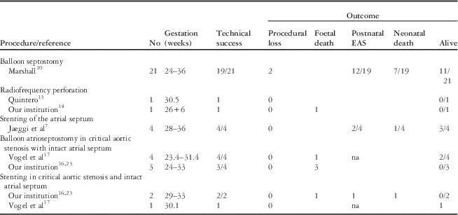
The largest series of foetal atrial septostomies to date has been published by the Boston group.Reference Marshall, Levine and Morash10 Of the 21 procedures attempted between 24 and 34 gestational weeks, 90% were technically successful. There was an intervention-related foetal loss of 10%. Following direct puncture of the atrial septum, a balloon was dilated several times across the atrial septum. An adequate defect was judged to be more than 3 mm and correlated with higher postnatal oxygen saturation (p<0.001), but was achieved in only 30% of the cases. Recoil of the septal tissue following dilatation contributed to procedure failure and this is the experience of other centres. Overall survival in this foetal series was 52%, and despite fetal intervention 12 neonates needed urgent postnatal atrial decompression. Survival was marginally better in those with larger defects – six of seven neonates – compared with 5 of 12 neonates with small defects. There was also a trend to improved post-surgical survival of neonates with restrictive oval foramen who underwent a foetal septostomy (69 versus 23%), but numbers are too small to assess statistical significance.Reference Vida, Bacha and Larrazabal6
Technique of foetal atrial septostomy: the foetal intervention is performed under light maternal sedation and with local anaesthesia. After positioning the foetus in the adequate supine position, lying with the right atrium superiorly (a), foetal anaesthesia is administered. An ultrasound-guided percutaneous puncture of the foetal atrial septum is performed with a long needle (18 G), which is advanced through the maternal abdominal wall, uterine wall, amnion, the foetal chest, and right atrium. The needle is used to pierce the atrial septum and to reach the left atrium (b). A floppy 0.014′′ coronary wire can be passed across the needle into the pulmonary vein, and over the wire, a coronary balloon catheter can be advanced into the left atrium (c). During retraction of the needle tip into the right atrium, a balloon is positioned into the atrial septum and inflated several times to create an appropriate communication (d). Then the balloon is deflated, retracted into the needle, and the needle and wire are withdrawn (e).
Atrial balloon septostomy in a foetus with an intact atrial septum (33+3 weeks of gestation). (a) Balloon inflated in the atrial septum (4.5 mm coronary balloon). (b) Defect created within the atrial septum (*). (c) Doppler flow across the created defect with left-to-right shunt. IAS=intact atrial septum; LA=left atrium; LV=left ventricle; RA=right atrium; RV=right ventricle.
Laser atrial septostomy
Laser atrial septostomy has been used to create a permanent atrial communication in a single case with hypoplastic left-heart syndrome, intact atrial septum, and an additional decompressing vein.Reference Quintero, Huhta, Suh, Chmait, Romero and Angel13 This was a technical failure owing to immediate spontaneous closure of the small defects created by the laser and highlights the limitation of these procedures to alter outcome.Reference Jaeggi, Ryan, Seed and Chaturvedi7, Reference Kohl, Müller, Tchatcheva, Achenbach and Gembruch14
Foetal stenting of atrial septum (Figs 5 and 6)
Stenting has been explored to create a larger and persistently patent atrial communication, and may be promising.Reference Jaeggi, Ryan, Seed and Chaturvedi7, Reference Marshall, Levine and Morash10, Reference Kohl, Müller, Tchatcheva, Achenbach and Gembruch14 After an initial report of a single case in 2005, successful atrial stenting has recently been reported in four foetuses with hypoplastic left-heart syndrome and restrictive atrial septum who were operated between 28 and 36 gestational weeks (Table 2).Reference Jaeggi, Ryan, Seed and Chaturvedi7 Coronary stents were deployed percutaneously into the foetal atrial septum (diameter between 2.75 and 3.0 mm) and dilated up to 3.2 mm. Of the four foetuses, two showed in-stent stenosis; however, all foetuses survived, with two of them requiring emergency atrial septostomy after delivery. All foetuses underwent Norwood stage 1 surgery, and three of them are alive. The limitations of this technique include partial lumen occlusion by rapid proliferation of endocardial cells, which has also been shown in animal studies,Reference Schmidt, Jaeggi and Ryan15 and acute stent thrombosis within hours after successful foetal stent implantation.Reference Herberg16
Technique of foetal atrial stenting. Similar to foetal atrial septostomy, a coronary stent mounted on a balloon is positioned in the atrial septum and inflated (a). After maximal inflation, the balloon is deflated and retracted, the stent remains in position (b).
Atrial stenting in a foetus with an intact atrial septum (27+1 weeks of gestation): (a) The stent is placed within the atrial septum. (b) On colour Doppler (*) and pulsed-wave Doppler (c, upper image), there is continuous left-to-right shunt across the stent. (c, lower image) PW Doppler of a pulmonary vein after foetal intervention: improvement in the Doppler profile. IAS=intact atrial septum; LA=left atrium; LV=left ventricle; PW=pulsed wave; RA=right atrium.
Details, procedural, and postnatal outcomes of foetal atrial septostomy and atrial stenting.
EAS=emergency atrioseptostomy; na=not applicable.
The published experiences of cardiac interventions in intact atrial septum are summarised in Table 2. This does not represent the total experience, as other centres have performed on a few cases, but these are unreported.
Prenatal interventions in foetal aortic stenosis and intact atrial septum
In foetuses with critical aortic stenosis, poor left ventricular function and severe mitral regurgitation, the oval foramen can be highly restrictive or closed. Typically, the left atrium is severely dilated, the right heart is compressed, and the foetus often develops severe hydrops associated with 90% foetal and perinatal mortality.Reference Rychik, Rome, Collins, DeCampli and Spray1, Reference Vogel, McElhinney and Wilkins-Haug17 Foetal aortic valve dilatation with improvement of forward flow, reduction in left ventricular afterload, improvement of function, and decrease in mitral valve regurgitation could theoretically improve the haemodynamics.Reference Vogel, McElhinney and Wilkins-Haug17–Reference Arzt, Wertaschnigg, Veit, Klement, Gitter and Tulzer19 Tulzer and colleagues reported two cases, in which the sealed oval foramen reopened after successful foetal aortic ballooning and improvement in foetal function.Reference Tulzer18, Reference Gembruch, Geipel, Herberg and Berg20 In cases of intact septum that do not reverse following successful foetal aortic valvuloplasty, creation of an atrial communication has been performed as salvage rescue therapy.Reference Marshall, Levine and Morash10, Reference Herberg16, Reference Vogel, McElhinney and Wilkins-Haug17, Reference Rogers, Peterson, Gaynor, Rome, Weinberg and Rychik21
Technical considerations
There are significant risks associated with foetal atrial septostomy or stenting. Bradycardia and pericardial effusion have been reported in 7 of the 21 cases after intrauterine balloon septostomy.Reference Marshall, Levine and Morash10
Timing of foetal cardiac intervention
Physiologically, the foetus should benefit from the earliest possible decompression of left atrial hypertension to avoid persistent changes to the pulmonary vasculature. However, there are technical limitations in creating a large and persistent atrial defect in small foetuses, including an increased risk of procedural losses in early gestation by the use of relatively large needles and the natural history of closure of atrial communications in this disease. Reference Marshall, Levine and Morash10, Reference McElhinney, Tworetzky and Lock22
Perinatal management
Foetuses with restrictive oval foramen or intact atrial septum and severe left-heart obstruction are at high risk for perinatal demise. Severe left atrial hypertension and inadequate left atrial egress may critically limit pulmonary blood flow and oxygenation in the perinatal period, despite successful prenatal intervention. In an interdisciplinary approach, delivery should be performed in a tertiary centre with proximity to the catheter lab and operating room, providing the option of immediate postnatal intervention and surgery.
Conclusion
Hypoplastic left-heart syndrome and critical aortic stenosis is associated with a high perinatal and postnatal morbidity and mortality when there is a severely restricted or intact atrial septum. Prenatal diagnosis and optimised perinatal planning with emergent intervention has not improved outcomes impressively. The in utero creation of a persistent and adequately sized atrial septal defect reduces the need for immediate postnatal intervention and seems to improve short- and medium-term outcomes. Furthermore, larger studies are needed to assess the perinatal and postnatal survival benefit of in utero cardiac interventions in this group of critically ill foetuses.
Acknowledgement
None.