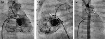
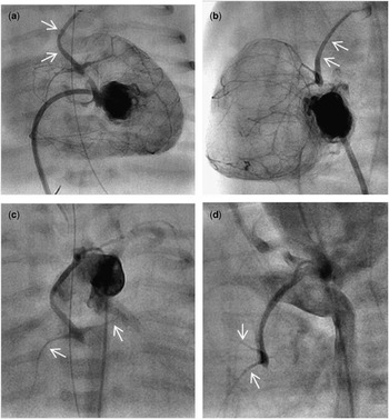
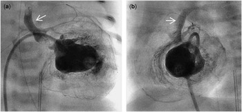
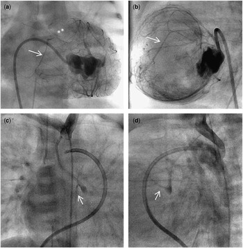
In infants with hypoplastic left heart syndrome, left ventricular to coronary connections occur in >50% of patients when mitral stenosis and aortic atresia are present.Reference Sathanandam, Cui and Nguyen 1 Infants with a hypoplastic left heart and significant left ventricular to coronary connections may have adverse outcomes after intervention. We therefore currently perform angiography in the left ventricle and ascending aorta before performing a hybrid procedure. We present brief clinical summaries and images in four infants with hypoplastic left heart syndrome and significant left ventricular to coronary connections who were referred for heart transplantation. Patient 1: Pulmonary artery bands were placed on day 3 after birth; on day 7, balloon atrial septostomy and subsequent angiography suggested atresia of the proximal right coronary artery and possible left ventricle-dependent coronary circulation. Repeat catheterisation was carried out on day 15 (Fig 1). Heart transplantation was performed at 3 weeks of age, and the patient is alive at 3.5 years of age. Patient 2: Catheterisation was carried out on day 2 after birth, which showed extensive left ventricular to coronary connections and very hypoplastic proximal coronary arteries (Fig 2). Pulmonary artery bands were placed on day 7 and heart transplantation was carried out at 3 months of age; the patient is alive at 3 years of age. Patient 3: Catheterisation was carried out on day 2 after birth and it showed extensive left ventricular to coronary connections (Fig 3). Pulmonary artery bands were placed on day 10 after birth; balloon atrial septostomy and ductal stenting were performed on day 21; atrial stenting was performed at 3 months of age, followed by Norwood procedure and cavopulmonary shunt and takedown at 5 and 11 months of age, respectively. Poor right ventricular function led to heart transplantation at 17 months of age, but the patient died of rejection at 28 months of age. Patient 4: Catheterisation on day 4 after birth showed atresia of the proximal left coronary artery and possible left ventricle-dependent coronary circulation (Fig 4). Pulmonary artery bands were placed on day 7 and the patient was referred for heart transplantation.
( a ) Anteroposterior (AP) view and ( b ) lateral view of the left ventriculogram, showing retrograde filling of both the right and left coronary arteries. The arrows in ( a ) and ( b ) show atresia of the proximal right coronary artery; the left coronary artery is in continuity with the aortic root. ( c ) AP view of a transverse aortogram, showing filling of a diminutive ascending aorta with the arrow indicating opacification of only the left coronary artery.

( a ) Anteroposterior (AP) view ( b ) lateral view of a left ventriculogram, with arrows in ( a ) and ( b ) showing the ascending aorta that is filled by left ventricular (LV) to coronary connections. ( c ) AP view and ( d ) lateral view of transverse aortogram, showing the diminutive ascending aorta, with arrows in ( c ) and ( d ) showing diminutive coronary arteries.

( a ) Anteroposterior (AP) view and ( b ) lateral view of a left ventriculogram, showing flow into both right and left coronary arteries with the arrows indicating contrast filling a good sized ascending aorta.

( a ) Anteroposterior (AP) view and ( b ) lateral view of a left ventriculogram, showing extensive left ventricular (LV) to coronary connections with the arrows in ( a ) and ( b ) indicating filling of the proximal right coronary artery, which eventually filled the aortic root; the asterisks indicate atresia of the proximal left coronary artery. ( c ) AP view and ( d ) lateral view of the aortogram, showing a diminutive ascending aorta, with arrows in ( c ) and ( d ) showing filling of only the right coronary artery.

Acknowledgements
The authors thank Dr. Humberto Restrepo for his editorial assistance.
Financial Support
This research received no specific grant from any funding agency, commercial, or not-for-profit sectors.
Conflict of Interest
None.