Introduction
Reproductive failure in high-yielding dairy cattle is a multifaceted problem. The pathogenesis of their sub-fertility is complex. The interactions between negative energy balance (NEB), early post partum and the hypothalamus–pituitary–ovary–uterus axis have been studied thoroughly (Ducker et al., Reference Ducker, Morant, Fisher and Haggett1985; Lucy, Reference Lucy2001; Butler, Reference Butler2003). Energy partitioning favoring the mammary tissue makes it possible to produce large volumes of milk, putting the remainder of the body in a status of severe NEB. Disturbed endocrine signaling during NEB leads to a retarded resumption of ovarian cyclicity post partum that has been recognized as a major factor contributing to the reproductive failure often exhibited by high-yielding dairy cattle (Opsomer et al., Reference Opsomer, Coryn, Deluyker and de Kruif1998). However, attention has recently been shifting towards the ubiquitously reported disappointingly low conception rates (Royal et al., Reference Royal, Mann and Flint2000; Lucy, Reference Lucy2001; Bousquet et al., Reference Bousquet, Bouchard and DuTremblay2004) and the remarkably high incidence of early embryonic mortality (Dunne et al., Reference Dunne, Diskin, Boland, O’Farrell and Sreenan1999; Mann and Lamming, Reference Mann and Lamming2001; Bilodeau-Goeseels and Kastelic, Reference Bilodeau-Goeseels and Kastelic2003) in these cattle. Therefore, it is critical to further investigate the quality of the oocyte and embryo, and to elucidate the direct effects of their micro-environments in order to adequately resolve the problem of sub-fertility (O’Callaghan and Boland, Reference O’Callaghan and Boland1999).
Recent studies have confirmed that the quality of the female gamete and the embryo is under pressure (Kruip et al., 1995; Kendrick et al., Reference Kendrick, Bailey, Garst, Pryor, Ahmadzadeh, Akers, Eyestone, Pearson and Gwasdauskas1999; Gwazdauskas et al., Reference Gwazdauskas, Kendrick, Pryor and Bailey2000; Wiltbank et al., Reference Wiltbank, Sartori, Sangsritavong, Lopez, Haughian, Fricke and Gumen2001; Walters et al., Reference Walters, Bailey, Pearson and Gwazdauskas2002). Oocytes retrieved from high genetic merit cows, irrespective of their milk production value, resulted in significantly lower blastocyst yields in vitro (Snijders et al., Reference Snijders, Dillon, O’Callaghan and Boland2000). This suggests possible adverse effects on fertility of the enforced genetic selection towards milk production.
In addition to oocyte quality, embryo quality also seems to be reduced in high-producing dairy cows compared to their non-lactating counterparts (Wiltbank et al., Reference Wiltbank, Sartori, Sangsritavong, Lopez, Haughian, Fricke and Gumen2001), and what is more, a high proportion of non-viable embryos were produced by lactating cows (Sartori et al., Reference Sartori, Sartor-Bergfelt, Mertens, Guenther, Parrish and Wiltbank2002). More studies are needed to obtain an informative and complete picture of average embryo quality in high-yielding dairy cows. Moreover, in the literature, many suggestions are made and much speculation offered on the causes of the reduction in quality of oocytes and embryos in dairy cattle; examples of these are provided by the following questions:
• Is oocyte growth and maturation adversely affected before ovulation due to biochemical or endocrine alterations in the intra-follicular environment (O’Callaghan and Boland, Reference O’Callaghan and Boland1999; Lozano et al., Reference Lozano, Lonergan, Boland and O’Callaghan2003)?
• Has the micro-environment of the oviduct or uterus been changed due to dietary and metabolic changes in the modern dairy cow, thus creating a hostile environment for the early embryo (Elrod and Butler, Reference Elrod and Butler1993; McEvoy et al., Reference McEvoy, Robinson, Aitken, Findlay, Palmer and Robertson1995; Kenny et al., Reference Kenny, Humpherson, Leese, Morris, Tomos, Diskin and Sreenan2002)?
• Is something defective in the genetic structure of modern dairy cow oocytes due to consecutive years of rigorous genetic selection towards milk yield? Or in other words, is there a genetic correlation between merit for milk production and oocyte or embryo quality (Snijders et al., Reference Snijders, Dillon, O’Callaghan and Boland2000; Hayhurst et al., 2007)?
In contrast with the extensive knowledge of disturbed endocrine signaling and ovarian function, clear evidence concerning the deficiencies in oocyte and/or embryo quality, and the consequent impact on the reproductive performance in high-producing dairy cows is severely lacking. In the present article, we will review a number of possible mechanisms linking NEB to oocyte quality. Furthermore, in the event an embryo is formed after fertilization, we will investigate whether the quality of early life is impaired. Accordingly, oocyte and embryo quality were investigated and compared in lactating high-yielding dairy cows, non-lactating dairy heifers and beef cows.
Follicular fluid, the missing link between blood and gamete
An NEB is characterized by a number of typical endocrine and biochemical changes in the blood of modern dairy cows (Herdt, Reference Herdt2000). Some studies have already inferred these metabolic changes in serum affect oocyte quality, but the physiological connection between blood and oocyte is still not well established, i.e. the follicle and the follicular fluid (FF) (Hashimoto et al., Reference Hashimoto, Minami, Yamada and Imai2000; De Wit et al., Reference De Wit, Cesar and Kruip2001; Jorritsma et al., Reference Jorritsma, Cesar, Hermans, Kruitwagen, Vos and Kruip2004).
The follicle is an avascular ‘compartment’ filled with FF in which the oocyte undergoes the fine-tuning process of oocyte growth, prematuration and final maturation (Bagavandoss et al., Reference Bagavandoss, Midgley and Wicha1983; Gosden et al., Reference Gosden, Hunter, Telfer, Torrance and Brown1988). The physiological properties of FF have been reviewed by Gosden et al. (Reference Gosden, Hunter, Telfer, Torrance and Brown1988). During the process of follicular growth, the physiochemical properties of the blood–follicle barrier transform thoroughly, suggesting that the oocyte’s environment undergoes compositional changes (Edwards, Reference Edwards1974; Wise, Reference Wise1987; Gosden et al., Reference Gosden, Hunter, Telfer, Torrance and Brown1988). In addition, the active transport mechanisms through the follicular wall may also alter during follicular growth. Argov et al. (Reference Argov, Arav and Sklan2004), for example, recently demonstrated that while lipoproteins are predominantly internalized by endocytosis in small follicles, this is not the case in large follicles, in which circulating lipoproteins contribute their cholesterol esters by selective uptake and without internalization of the lipoprotein as such. Hence, to further investigate the biochemical milieu of the oocyte prior to ovulation, the composition of FF originating in three differently sized follicles was determined and compared to the composition of serum from 30 dairy cows shortly post mortem (Leroy et al., Reference Leroy, Vanholder, Delanghe, Opsomer, Van Soom, Bols and de Kruif2004a). The data confirmed that the FF’s composition changes as the follicle grows. Another important finding was that the FF’s composition is correlated with that of the serum. These correlations are ‘static’, however, and for the confirmation of correlations in this particularly ‘dynamic’ system, the composition of FF of high-yielding dairy cows was established during the early post partum period by means of repeated trans-vaginal follicle puncture and FF aspiration.
In a subsequent study, Leroy et al. (Reference Leroy, Vanholder, Delanghe, Opsomer, Van Soom, Bols, Dewulf and de Kruif2004b) concentrated on compositional changes over time in the FF of high-yielding dairy cows early post partum; here the data were compared with the extensively reported biochemical alterations in blood during NEB (see above). NEB typically causes some obvious changes in serum, such as high non-esterified fatty acids (NEFA) and β-hydroxybutyrate (β-OHB) concentrations, or low glucose concentrations (Baird, Reference Baird1982; Chilliard et al., Reference Chilliard, Bocquier and Doreau1998; Duffield, Reference Duffield2000; Herdt, Reference Herdt2000). Urea concentrations can also be elevated, due to an increased amino acid metabolism for gluconeogenesis, or the intake of a protein-rich diet (Butler, Reference Butler1998; Sinclair et al., Reference Sinclair, Sinclair and Robinson2000). Britt (Reference Britt1992) hypothesized that these features of the NEB can directly affect the follicle and its enclosed oocyte, leading to the ovulation of an inferior oocyte. This hypothesis is interesting because it is generally accepted that oocytes are highly vulnerable to any disruption in their environment (O’Callaghan and Boland, Reference O’Callaghan and Boland1999), a point of view that has been more or less confirmed by others (Armstrong et al., Reference Armstrong, McEvoy, Baxter, Robinson, Hogg, Woad, Webb and Sinclair2001; Boland et al., Reference Boland, Lonergan and O’Callaghan2001). The data of Leroy et al. (Reference Leroy, Vanholder, Delanghe, Opsomer, Van Soom, Bols, Dewulf and de Kruif2004b) provided evidence to, at least partially, answer the question raised by Britt (1994): ‘How does NEB directly influence the micro-environment that is most intimately linked with the oocyte?’
An adapted ovum pick-up technique (Bols et al., Reference Bols, Vandenheede, Van Soom and de Kruif1995) was used at six different points in time during the early post partum period to collect FF from the dominant follicle of high-producing dairy cows. Given that follicular size is related to FF composition (Leroy et al., Reference Leroy, Vanholder, Delanghe, Opsomer, Van Soom, Bols and de Kruif2004a), similar-sized follicles were aspirated throughout the entire study. Due to the reduced accessibility of the ovaries during the puerperium period, FF was only collected from day 14 post partum onwards. Good correlations were found between serum and FF composition for glucose, β-OHB, urea and total cholesterol. Based on the results of repeated measurements (dynamic correlations) (Leroy et al., Reference Leroy, Vanholder, Delanghe, Opsomer, Van Soom, Bols, Dewulf and de Kruif2004b), we can state that typical post partum serum fluctuations are more or less reflected in the FF of the dominant follicle. For urea and β-OHB, no concentration differences between serum and FF could be detected. It is important to mention, though, that the follicle appears to be able to maintain higher glucose and lower NEFA concentrations than does serum. In other words, it can be suggested that the oocyte is isolated, possibly even protected, from excessively low glucose or high NEFA concentrations in the blood.
Despite the follicle’s buffering capacities, glucose concentrations do decrease, and NEFA concentrations significantly rise, in FF during NEB. Also Jorritsma et al. (Reference Jorritsma, Groot, Vos, Kruip, Wensing and Noordhuizen2003) and Comin et al. (Reference Comin, Gerin, Cappa, Marchi, Renaville, Motta, Fazzini and Prandi2002) described an increase in NEFA concentrations in FF due to an acute dietary restriction. However, in these studies no concentration gradients between serum and FF were mentioned, there is now enough evidence to conclude that the growing and maturing oocyte is directly exposed to the typical biochemical changes that occur in high-yielding dairy cows early post partum. It has furthermore been established that high urea concentrations, also found in FF (Hammon et al., Reference Hammon, Holyoak and Dhiman2005), can be toxic to oocytes during maturation (Ocon and Hansen, Reference Ocon and Hansen2003; Iwata et al., Reference Iwata, Inoue, Kimura, Kuge, Kuwayama and Monji2006), probably through an inhibition of the polymerization of tubulin into microtubules (De Wit et al., Reference De Wit, Cesar and Kruip2001). The same is true for the lowered glucose concentrations observed. Adequate glucose supplies are necessary to support normal cumulus expansion and nuclear maturation (Krisher and Bavister, Reference Krisher and Bavister1998; Sutton-McDowall et al., Reference Sutton-McDowall, Gilchrist and Thompson2004). Similarly, high NEFA and β-OHB concentrations are probably harmful to the oocyte’s developmental competence, but this has, to our knowledge, never been substantiated.
Negative energy balance and its direct consequences for oocyte quality: an in vitro model
Initially, it was important to pay attention to the NEFA fraction of the FF, as not only the absolute NEFA concentration but also the NEFA composition is physiologically important. Samples were analyzed by means of a combined thin layer and gas chromatography (Folch et al., Reference Folch, Lees and Sloane Stanley1957). The results surprisingly revealed that both the NEFA concentration (see above) and the NEFA composition significantly differed between serum and FF. Differences in albumin content (on which NEFA are predominantly bound) between the two compartments could not be found, and thus did not offer any clues for explaining the observed differences in NEFA concentration and composition. Disconcertingly, the described dynamic interchange of NEFA between serum and FF (Moallem et al., Reference Moallem, Folman, Bor, Arav and Sklan1999) was also not totally in agreement with these findings. The study of Chung et al. (Reference Chung, Tallis, Cho, Segrest and Henkin1995), however, offered a possibly useful clarification. In the presence of high NEFA levels, a substantial portion of the NEFA in serum is partitioned into low-density lipoproteins (LDL). The saturated fatty acids in particular are bound on LDL, while the unsaturated ones are preferentially bound on albumin (Chung et al., Reference Chung, Tallis, Cho, Segrest and Henkin1995). Because LDL are absent in FF, these findings could account for the differences in concentration and composition of NEFA in FF compared to serum early post partum (Wehrman et al., Reference Wehrman, Welsh and Williams1991).
Chromatographic analyses revealed that the three most abundant free fatty acids present in FF during NEB were oleic, palmitic and stearic acid. The NEB-associated FF concentrations of these three free fatty acids were applied to an in vitro serum-free maturation model to evaluate their effect on oocyte quality (Leroy et al., Reference Leroy, Vanholder, Mateusen, Christophe, Opsomer, de Kruif, Genicot and Van Soom2005a). In these tests, oleic acid had no discernable effect on the oocyte’s developmental capacity. However, exposing oocytes to palmitic and stearic acid, at concentrations comparable to those assessed in vivo, resulted in reduced maturation rates, thus leading to unsatisfactory fertilization and cleavage rates. Also, cumulus expansion was adversely affected. In these cumulus cells, a significantly higher rate of apoptosis and even necrosis could be detected after 24 h of exposure to elevated stearic or palmitic acid concentrations. Very recently, it was shown that this fatty acid exposure during maturation has carry-over effects on embryo quality in terms of open pulled straw freezeability (Shehab-El-Deen et al., in press). This also suggests that the fatty acid composition of the oocyte, and thus the embryo, is altered, leading to reduced freezing-warming survival rates. Rooke et al. (Reference Rooke, Ewen, Reis and McEvoy2006) demonstrated that changes in the FF fatty acid composition are also reflected in the fatty acid content and profile of the cumulus-oocyte complex.
Similar toxic effects of high NEFA concentrations on bovine or human granulosa cells in vitro have been shown in other studies (Mu et al., Reference Mu, Yanase, Nishi, Tanaka, Saito, Jin, Mukasa, Okabe, Nomura, Goto and Nawata2001; Jorritsma et al., Reference Jorritsma, Cesar, Hermans, Kruitwagen, Vos and Kruip2004; Vanholder et al., Reference Vanholder, Leroy, Van Soom, Opsomer, Maes, Coryn and de Kruif2005). Optimal granulosa and cumulus cell functioning are indispensable for oocyte maturation because these cells are responsible for endocrine and paracrine signaling (Bilodeau-Goeseels and Panich, Reference Bilodeau-Goeseels and Panich2002; Tanghe et al., Reference Tanghe, Van Soom, Nauwynck, Coryn and de Kruif2002). Therefore, it is most likely that the toxic effect of NEFA on oocyte quality is partly an indirect effect. In contrast with the results of Leroy et al. (Reference Leroy, Vanholder, Mateusen, Christophe, Opsomer, de Kruif, Genicot and Van Soom2005a), Jorritsma et al. (Reference Jorritsma, Cesar, Hermans, Kruitwagen, Vos and Kruip2004) did find detrimental effects of oleic acid. In their study, however, oleic acid was bound on albumin and was added in supra-physiological concentrations to an undefined in vitro maturation medium (contained fetal calf serum, an undefined source of fatty acids). Moreover, it is unclear whether these adverse effects were caused by the addition of BSA itself or by oleic acid. Homa and Brown (Reference Homa and Brown1992) showed that albumin-bound linolenic acid in in vitro maturation medium inhibited germinal vesicle breakdown in denuded oocytes.
Similar toxic effects of NEFA have also been described for Leydig cells, muscle cells and pancreatic β-cells, and it is the induction of apoptosis and/or insulin resistance as well as changes in membrane properties that have been specifically suggested as potential mechanisms for explaining the observed toxic effects (Shimabukuro et al., Reference Shimabukuro, Zhou, Levi and Unger1998; Maedler et al., Reference Maedler, Spinas, Dyntar, Moritz, Kaiser and Donath2001; Hirabara et al., Reference Hirabara, de Oliveira Carvalho, Mendonça, Piltcher Haber, Fernandes and Curi2003; Lu et al., Reference Lu, Mu, Wang, Li, Lu, Li, Pan, Yanase and Nawata2003; Jorritsma et al., Reference Jorritsma, Cesar, Hermans, Kruitwagen, Vos and Kruip2004).
These indications of NEFA toxicity at the oocyte and follicle level not only are important in relation to the sub-fertility issue in modern dairy cows, but may also provide a valuable model for human research. Obesity and diabetes are characterized by increased concentrations of NEFA due to high adipose sensitivity for lipolytic triggers (Herdt, Reference Herdt2000; Cnop et al., Reference Cnop, Hannaert, Hoorens, Eizirik and Pipeleers2001). Our data suggest that the frequently reported fertility disorders in obese or diabetic women (Pasquali et al., Reference Pasquali, Pelusi, Genghini, Cacciari and Gambineri2003) may be not only due to the toxic effects of NEFA on granulosa cells, which mainly lead to amenorrhea (Mu et al., Reference Mu, Yanase, Nishi, Tanaka, Saito, Jin, Mukasa, Okabe, Nomura, Goto and Nawata2001), but could also originate from direct detrimental effects on the cumulus-oocyte complex. The latter could explain the disappointing IVF or intracytoplasmic sperm injection (ICSI) results, and the higher risk for early pregnancy loss in obese women, as has been documented by Fedorcsak et al. (Reference Fedorcsak, Storeng, Dale, Tanbo and Abyholm2000 and Reference Fedorcsak, Dale, Storeng, Ertzeid, Bjercke, Oldereid, Omland, Abyholm and Tanbo2004) and Pasquali et al. (Reference Pasquali, Pelusi, Genghini, Cacciari and Gambineri2003). Ongoing research on humans should confirm or refute the validity of applying this bovine model in human studies.
Not only high NEFA but also elevated ketone concentrations are a distinctive characteristic of NEB (Sato et al., Reference Sato, Matsumoto and Hanasaka1999). High ketone concentrations generally go together with hypoglycemia (Herdt, Reference Herdt2000). Therefore, in a second in vitro maturation model (Leroy et al., Reference Leroy, Vanholder, Opsomer, Van Soom and de Kruif2006), the effects of combined high β-OHB and low glucose concentrations, ascertained in measurements of FF of dairy cows during NEB, were investigated. The primary conclusion of this study was that the in vitro model imitating sub-clinical ketosis had no effect on the oocyte’s developmental capacity in vitro. Clinical ketosis, however, turned out to be detrimental to oocyte quality in vitro; this was presumed to be due to the effect of depleted glucose, rather than high β-OHB concentrations. Thus, the toxicity of β-OHB, as has been described for polymorphonuclear cells and macrophages (Hoeben et al., Reference Hoeben, Heyneman and Burvenich1997; Sartorelli et al., Reference Sartorelli, Paltrinieri and Comazzi2000), could not be confirmed for cumulus-oocyte complexes. Conversely, it can be assumed, with a high degree of certainty, that inadequate glucose supplies compromise oocyte developmental competence, and this is a conclusion that is in agreement with other studies (Krisher and Bavister, Reference Krisher and Bavister1998; Cetica et al., Reference Cetica, Pintos, Dalvit and Beconi2002; Sutton-McDowall et al., Reference Sutton-McDowall, Gilchrist and Thompson2004).
When interpreting these in vitro results and explaining them in terms of sub-fertility in high-producing dairy cows, some prudence is warranted. In studies described above, it was hypothesized that elevated NEFA or β-OHB concentrations, together with low glucose concentrations, contributed to reducing fertility in high-yielding dairy cows by exerting detrimental effects on oocyte developmental competence. These findings more or less resemble the hypothesis of Britt (1994), who stated that a follicle grown during the period of NEB early post partum could be affected by unfavorable metabolic changes, and may contain a developmentally incompetent oocyte; following a growing and maturation phase of several weeks, this inferior oocyte is ovulated at the time of the initial insemination (Lucy, Reference Lucy2003). This hypothesis has been partially confirmed in recent in vivo studies (Gwazdauskas et al., Reference Gwazdauskas, Kendrick, Pryor and Bailey2000; Snijders et al., Reference Snijders, Dillon, O’Callaghan and Boland2000; Sartori et al., Reference Sartori, Sartor-Bergfelt, Mertens, Guenther, Parrish and Wiltbank2002). It is important, however, to mention that the combined in vitro and in vivo model used in the studies of Leroy et al. (Reference Leroy, Vanholder, Mateusen, Christophe, Opsomer, de Kruif, Genicot and Van Soom2005a and Reference Leroy, Vanholder, Opsomer, Van Soom and de Kruif2006) described above was not entirely appropriate for investigating the carry-over effect on oocyte quality as hypothesized by Britt (1994). The results only documented the FF composition in the dominant follicle during the NEB mimicked in vitro. Quiescent follicles, however, provide a much poorer isolation from the extrafollicular environment and blood serum for the oocytes embedded within them. As a consequence, such oocytes are probably exposed to even higher NEFA concentrations (Zamboni, Reference Zamboni1974). Another possibility is that oocytes of primordial follicles are completely insensitive to all these metabolic disruptions. Moreover, the cumulus-oocyte complexes were exposed to elevated NEFA or β-OHB and low glucose concentrations for only 24 h, whereas in vivo the oocytes are exposed to these concentrations for several days or even weeks. In the ideal model, primordial follicles should be cultivated in high NEFA conditions for several weeks. However, maintaining such long-term cultures of primordial follicles presents major obstacles, and growing bovine primordial follicles up to the preovulatory stage has so far proven to be unachievable (Gutierrez et al., Reference Gutierrez, Ralph, Telfer, Wilmut and Webb2000); although, successes have been obtained in mice with prolonged primordial and preantral follicle culture leading to in vitro ovulation and, after fertilization, the birth of pups (Cortvrindt and Smitz, Reference Cortvrindt and Smitz2001). Current research uses this murine model to investigate the consequences of long-term exposure to elevated NEFA on follicular growth and subsequent oocyte development. Nevertheless, it is believed that the data from the present model revealed for the first time the possible toxic effects of high FF NEFA and low glucose concentrations on the developmental competence of bovine oocytes in vitro.
Back to the field: a closer look at embryo quality
From the data presented above, it can be concluded that the oocyte is vulnerable to at least some of the metabolic alterations associated with NEB. Clearly, obvious adverse effects on oocyte quality were observed in our in vitro model. Logically, the next step would be to investigate the consequences of these effects on embryo quality. It has been suggested by Rizos et al. (Reference Rizos, Ward, Duffy, Boland and Lonergan2002) that the conditions prior to fertilization are the determinants for embryo yield, while the embryo’s culture environment is crucial for embryo quality. On the basis of that theory, the toxic effects of high NEFA and low glucose concentrations during oocyte maturation that have been demonstrated in high-producing dairy cows (Leroy et al., Reference Leroy, Vanholder, Mateusen, Christophe, Opsomer, de Kruif, Genicot and Van Soom2005a and Reference Leroy, Vanholder, Opsomer, Van Soom and de Kruif2006) should primarily lead to low fertilization (i.e. conception) rates. However, based on the reports of Royal et al. (Reference Royal, Mann and Flint2000) and Bousquet et al. (Reference Bousquet, Bouchard and DuTremblay2004), this does seem to be the case. Also, the environment of both the oocyte and the embryo is said to be crucial to ultimate fertility. As suggested by Kenny et al. (Reference Kenny, Humpherson, Leese, Morris, Tomos, Diskin and Sreenan2002), Elrod and Butler (Reference Elrod and Butler1993) and McEvoy et al. (Reference McEvoy, Robinson, Aitken, Findlay, Palmer and Robertson1995), high-energy and/or high-protein diets can alter the micro-environment of the embryo in the oviduct and uterus (for review see Leroy et al., 2008). Such changes are expected to be detrimental to embryo quality (Rizos et al., Reference Rizos, Ward, Duffy, Boland and Lonergan2002), which has been shown experimentally in heifers by Wrenzycki et al. (Reference Wrenzycki, De Sousa, Overström, Duby, Herrmann, Watson, Niemann, O’Callaghan and Boland2000). However, it has never been demonstrated whether or not this is also the case in high-producing dairy cows. Accordingly, a field trial was arranged to compile data on the embryo quality of high-producing dairy cows in comparison with non-lactating dairy heifers and (non-lactating) beef cows. Through this field trial it was possible to investigate simultaneously the effects of milk production and breed (or genetic background) (Leroy et al., Reference Leroy, Opsomer, De Vliegher, Vanholder, Goossens, Geldhof, Bols, de Kruif and Van Soom2005b). Briefly, embryos from lactating dairy cows clearly displayed inferior quality as assessed by morphological evaluation, compared to dairy heifers or beef cows. Furthermore, it was possible to demonstrate by means of a multivariable regression model that the state of producing milk v. not producing milk was highly correlated with embryo quality. It is also important to mention that no differences were found in fertilization rates or in the number of transferable embryos per embryo collection among these animals.
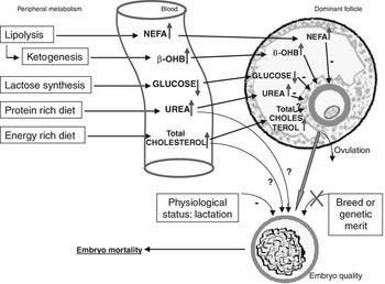
Because the embryos of the lactating dairy cows were on average collected around day 230 post partum, it is very unlikely that a carry-over effect of NEB, as has been hypothesized by Britt (Reference Britt1992), is responsible for their poor quality. This could have been a likely explanation when embryo collection was performed on average around 2 to 3 months after calving, as was done by Sartori et al. (Reference Sartori, Sartor-Bergfelt, Mertens, Guenther, Parrish and Wiltbank2002); he also found an obvious difference in embryo quality between lactating dairy cows and maiden heifers, suggesting that genetic merit for milk production does not account for the reduction in embryo quality observed in lactating dairy cows. In contrast with our results, Sartori et al. (Reference Sartori, Sartor-Bergfelt, Mertens, Guenther, Parrish and Wiltbank2002) reported not only inferior embryo quality but also a lower fertilization rate, illustrated by a higher proportion of unfertilized oocytes present in the uterine flushing of lactating dairy cows. Sartori et al. (Reference Sartori, Sartor-Bergfelt, Mertens, Guenther, Parrish and Wiltbank2002) may have described the adverse influences of a carry-over effect of NEB on oocyte quality (reduced fertilization rates), combined with the possible negative effects of lactation, management or diet on the micro-environment of the oviduct or uterus (reduced embryo quality), similar to what we found in our field trial. By way of summary, all suggested mechanisms that could potentially diminish embryo quality are diagrammatically represented in Figure 1. Further research should reveal the exact mechanism through which embryo quality is adversely affected in lactating dairy cows.
Diagrammatic representation of possible mechanisms by which embryo quality can be impaired in high-yielding dairy cows.

One of the major morphological characteristics evaluated in the study of Leroy et al. (Reference Leroy, Opsomer, De Vliegher, Vanholder, Goossens, Geldhof, Bols, de Kruif and Van Soom2005b) was embryo color. By means of a new lipid evaluation technique (Leroy et al., Reference Leroy, Genicot, Donnay and Van Soom2005c) it could be demonstrated that embryo color is correlated with lipid content, as has previously been suggested by others (Sata et al., Reference Sata, Tsujii, Abe, Yamashita and Hoshi1999; Abe and Hoshi, Reference Abe and Hoshi2003). Lactating dairy cow embryos were generally dark and contained as much lipid as in vitro produced embryos that are known to accumulate excessive amounts of lipids (Abe et al., Reference Abe, Yamashita, Itoh, Satoh and Hoshi1999). This has never been shown before. A high lipid content has undeniably been linked with impaired embryo quality (Reis et al., Reference Reis, Rooke, McCallum, Ewen, Staines, Lomax and McEvoy2003; Rizos et al., Reference Rizos, Gutierrez-Adan, Perez-Garnelo, De La Fuente, Boland and Lonergan2003). The underlying mechanism(s) linking milk production or nutrition with embryo color and/or lipid content is unknown, and further research is necessary. A broader overview on factors influencing oocyte and embryo quality as well as corpus luteum quality was presented in earlier work (Leroy et al., 2008).
Perspectives for future research and some food for thought
As has been mentioned above, future research should enlighten us more about the interactions between the blood, the micro-environment in the oviduct or uterus and embryo metabolism. Furthermore, several studies have indicated that NEB is also associated with depressed immunity during the first weeks post partum, thus leading to an increased susceptibility to infectious diseases such as mastitis and metritis (Hoeben et al., Reference Hoeben, Monfardini, Opsomer, Burvenich, Dosogne, De Kruif and Beckers2000; Lacetera et al., Reference Lacetera, Scalia, Bernabucci, Ronchi, Pirazzi and Nardone2005). Bearing this in mind, it becomes important to consider not only the direct link between NEB and fertility but also that reproductive functions are affected indirectly by an increased incidence of infectious diseases. Mastitis, for example, that, together with low fertility, is the principal reason for the culling of dairy cows, has been shown to be directly linked to the retarded onset of ovarian activity post partum (Loeffler et al., Reference Loeffler, de Vries and Schukken1999; Rajala-Schultz and Gröhn, Reference Rajala-Schultz and Gröhn2001; Huszenicza et al., Reference Huszenicza, Janosi, Kulcsar, Korodi, Reiczigel, Katai, Peters and De Rensis2005). Whether infectious diseases can affect the oocyte and/or the embryo in a direct way has been poorly studied, and certainly requires further investigation (Hansen et al., Reference Hansen, Soto and Natzke2004). Furthermore, environmental pollution has also been associated with direct harmful effects on oocyte quality through the generation of endocrine disrupters (Brevini et al., Reference Brevini, Cillo, Antonini and Gandolfi2005), and this merits further study.
Is there still a need for high-producing dairy cows? Yield maximization per animal is indeed preferable from both an economic and environmental point of view. However, only outstanding herd management can guarantee the animal’s welfare under such pressures. But even with excellent management, the demands on these animals remain high as evidenced by the fact that they are rapidly culled for reasons such as reduced fertility, metabolic disorders and infectious diseases. Now, it is becoming clear that even the oocyte and the embryo may directly suffer from high levels of productivity.
Conclusions
It can be concluded that the typical biochemical serum changes observed in dairy cattle during NEB early post partum are well reflected in the FF of the dominant follicle, thus exposing the granulosa cells and the maturing oocyte. In vitro maturation models revealed that NEB associated with elevated NEFA and lowered glucose concentrations are indeed toxic to the oocyte, resulting in both diminished oocyte maturation and embryo developmental competence.
Even after the period of NEB, and when the carry-over effects of the NEB were no longer present, high-yielding dairy cows produced statistically significantly inferior embryos in comparison with dairy heifers and beef cows. With a newly developed lipid evaluation technique, it was possible to demonstrate that high-producing dairy cow embryos contained up to 45% more lipids, compared to the embryos of non-lactating animals. These findings imply that it is not genetic merit for milk production or breed that has an adverse impact on embryo quality, but rather that a variety of factors associated with milk production as such (metabolism, nutrition, management) induce hostile conditions that prevent optimal embryo development. Further research is required to fully comprehend how a dairy cow’s milk production and nutrition influences embryo health and metabolism via an altered environment in the oviduct and uterus.
Acknowledgments
The authors thank Silke Andries for the critical reading and formatting of the manuscript.