Introduction
Gap junctions are clusters of intercellular conduits that connect the cytoplasms of two contacting cells and allow metabolic and electrical coupling. Gap junctions are formed by proteins called connexins. Six of these protein molecules form a connexon, and the docking of two connexons from two adjacent cells generates a functional gap junction channel (reviewed by Bloomfield & Völgyi, Reference Bloomfield and Völgyi2009). Homotypic gap junctions are composed of two identical connexons; heterotypic gap junctions are built from two different connexons.
In the mammalian retina, gap junctions are present in all neuronal cell classes (Vaney, Reference Vaney1991) where they lead to coupled networks. These neuronal networks are modulated by light and neuromodulators that can alter the extent of coupling when the visual environment changes (for review, see Weiler et al., Reference Weiler, Pottek, He and Vaney2000). These adaptational processes are presumably mediated by protein kinases, which can alter gap junction properties by phosphorylating either the connexins (Urschel et al., Reference Urschel, Höher, Schubert, Alev, Söhl, Wörsdörfer, Asahara, Dermietzel, Weiler and Willecke2006) or other target proteins, which can in turn change connexin characteristics.
Among the connexins in retinal neurons, Cx36 has been described in photoreceptors (Deans et al., Reference Deans, Volgyi, Goodenough, Bloomfield and Paul2002; Lee et al., Reference Lee, Han, Kim, Kim, Lee, Oh, Chung and Chun2003; Feigenspan et al., Reference Feigenspan, Janssen-Bienhold, Hormuzdi, Monyer, Degen, Söhl, Willecke, Ammermüller and Weiler2004), bipolar cells (Feigenspan et al., Reference Feigenspan, Janssen-Bienhold, Hormuzdi, Monyer, Degen, Söhl, Willecke, Ammermüller and Weiler2004; Han & Massey, Reference Han and Massey2005; Lin et al., Reference Lin, Jakobs and Masland2005), AII amacrine cells (Feigenspan et al., Reference Feigenspan, Teubner, Willecke and Weiler2001; Mills et al., Reference Mills, O’Brien, Li, O’Brien and Massey2001), and alpha ganglion cells (Schubert et al., Reference Schubert, Degen, Willecke, Hormuzdi, Monyer and Weiler2005a; Völgyi et al., Reference Völgyi, Abrams, Paul and Bloomfield2005). AII amacrine cells form homotypic gap junctions composed of Cx36 to one type of ON cone bipolar cell (Han & Massey, Reference Han and Massey2005; Lin et al., Reference Lin, Jakobs and Masland2005), heterotypic gap junctions consisting of Cx36 and Cx45 to other ON cone bipolar cell types (Maxeiner et al., Reference Maxeiner, Dedek, Janssen-Bienhold, Ammermüller, Brune, Kirsch, Pieper, Degen, Krüger, Willecke and Weiler2005; Dedek et al., Reference Dedek, Schultz, Pieper, Dirks, Maxeiner, Willecke, Weiler and Janssen-Bienhold2006), or bihomotypic gap junctions consisting of Cx36 and Cx45 (Li et al., Reference Li, Kamasawa, Ciolofan, Olson, Lu, Davidson, Yasumura, Shigemoto, Rash and Nagy2008). Cx45 is also expressed in OFF bipolar cells (Maxeiner et al., Reference Maxeiner, Dedek, Janssen-Bienhold, Ammermüller, Brune, Kirsch, Pieper, Degen, Krüger, Willecke and Weiler2005), bistratified ganglion cells (Schubert et al., Reference Schubert, Maxeiner, Kruger, Willecke and Weiler2005b), and amacrine cells (Maxeiner et al., Reference Maxeiner, Dedek, Janssen-Bienhold, Ammermüller, Brune, Kirsch, Pieper, Degen, Krüger, Willecke and Weiler2005; Pérez de Sevilla Müller et al., Reference Pérez de Sevilla Müller, Shelley and Weiler2007; Dedek et al., Reference Dedek, Breuninger, Pérez de Sevilla Müller, Maxeiner, Schultz, Janssen-Bienhold, Willecke, Euler and Weiler2009). Two other retinal connexins, Cx57 and Cx50, mediate the extensive coupling of horizontal cells (Hombach et al., Reference Hombach, Janssen-Bienhold, Söhl, Schubert, Büssow, Ott, Weiler and Willecke2004; O’Brien et al., Reference O’Brien, Li, Pan, Keung, O’Brien and Massey2006; Janssen-Bienhold et al., Reference Janssen-Bienhold, Trümpler, Hilgen, Schultz, Pérez de Sevilla Müller, Sonntag, Dedek, Dirks, Willecke and Weiler2009).
In addition to these four gap junction proteins, it seems likely that there are still other connexin genes expressed in the mammalian retina (Kihara et al., Reference Kihara, de Castro, Moriscot and Hamassaki2006). For example, Xin and Bloomfield (Reference Xin and Bloomfield1997) showed that many ganglion cell types exhibit tracer coupling. Using antibodies directed against Cx36, Hidaka et al. (Reference Hidaka, Kato and Miyachi2002, Reference Hidaka, Akahori and Kurosawa2004) showed that Cx36 mediates coupling between alpha ganglion cells of the rat retina. With the help of connexin-deficient mouse lines, other reports have shown that Cx36 mediates the coupling between alpha ganglion cells (Schubert et al., Reference Schubert, Degen, Willecke, Hormuzdi, Monyer and Weiler2005a; Völgyi et al., Reference Völgyi, Abrams, Paul and Bloomfield2005) and that Cx45 mediates the coupling between bistratified ganglion cells (Schubert et al., Reference Schubert, Maxeiner, Kruger, Willecke and Weiler2005b). Though Pan et al. (Reference Pan, Paul, Bloomfield and Völgyi2010) recently showed that Cx36 is required for gap junctional coupling of most ganglion cell subtypes in the mouse retina, for several retinal ganglion cell types, the connexin that mediates coupling is still unknown. Therefore, we studied the expression pattern of the gap junction protein Cx30.2 in the mouse retina. Cx30.2 was first shown to be expressed in cardiac myocytes (Kreuzberg et al., Reference Kreuzberg, Söhl, Kim, Verselis, Willecke and Bukauskas2005). Recently, Kreuzberg et al. (Reference Kreuzberg, Deuchars, Weiss, Schober, Sonntag, Wellershaus, Draguhn and Willecke2008) also found Cx30.2 in neurons, namely in inhibitory interneurons of the hippocampus, and suggested that most Cx30.2-expressing neurons also express Cx36. To study Cx30.2 expression in the mouse retina, we used a transgenic mouse line that expressed a lacZ reporter gene instead of the Cx30.2 coding region (Kreuzberg et al., Reference Kreuzberg, Schrickel, Ghanem, Kim, Degen, Janssen-Bienhold, Lewalter, Tiemann and Willecke2006). We found lacZ expression for Cx30.2 in six different types of ganglion cells and studied protein kinase–induced phosphorylation as a potential mechanism to modulate ganglion cell networks coupled by Cx30.2.
Materials and methods
Mouse strains and tissue preparation
Retinae were obtained from several different connexin-deficient mouse lines: Cx30.2lacZ/lacZ (Kreuzberg et al., Reference Kreuzberg, Schrickel, Ghanem, Kim, Degen, Janssen-Bienhold, Lewalter, Tiemann and Willecke2006), Cx45fl/fl:Nestin-Cre (Maxeiner et al., Reference Maxeiner, Dedek, Janssen-Bienhold, Ammermüller, Brune, Kirsch, Pieper, Degen, Krüger, Willecke and Weiler2005), Cx36−/− (Güldenagel et al., Reference Güldenagel, Ammermüller, Feigenspan, Teubner, Degen, Söhl, Willecke and Weiler2001), and Cx40−/− (Kirchhoff et al., Reference Kirchhoff, Nelles, Hagendorff, Krüger, Traub and Willecke1998). Mice were killed by cervical dislocation in accordance with the institutional guidelines for animal welfare issued by the Federal Republic of Germany. Eyes were enucleated and kept at room temperature in a Petri dish with carboxygenated Ames’ medium (pH 7.4; Sigma, Seelze, Germany). Retinae were mounted photoreceptor side down on black filter paper (Millipore Corporation, Bedford, MA).
Intracellular injections
Intracellular injections were carried out as described previously (Schubert et al., Reference Schubert, Degen, Willecke, Hormuzdi, Monyer and Weiler2005a; Pérez de Sevilla Müller et al., Reference Pérez de Sevilla Müller, Shelley and Weiler2007). Briefly, borosilicate glass electrodes (Hilgenberg, Malsfeld, Germany) were pulled with a Sutter P-97 puller (Sutter, Novato, CA). Electrode tips were filled with 1% Alexa594 or Alexa568 sodium hydrazide (Invitrogen, Karlusruhe, Germany) or Lucifer Yellow (Sigma) and 4% N-(2-aminoethyl-)-biotinamide hydrochloride (Neurobiotin; Vector, Burlingame, CA). Electrodes were backfilled with Tris buffer, pH 7.4, and typically had resistances between 100 and 140 MΩ. To visualize ganglion cell bodies in wild-type (WT) retinae, a few drops of Acridine Orange (1 μM; Sigma) were added to the bath solution. After a few minutes, retinae were washed and ganglion cells were injected. Alexa dye/Lucifer Yellow was iontophoresed with a current of −1 nA (750 ms at 1 Hz). When the dendritic morphology of the cell could be discerned, the direction of the current was reversed to +1 nA to inject Neurobiotin.
Labeling lacZ-expressing cells
To label lacZ-expressing cells in vivo, retinae from Cx30.2lacZ/lacZ and Cx30.2+/lacZ mice were incubated with fluorescein-di-beta-d-galactopyranoside (FDG; Sigma). FDG is a fluorogenic substrate for beta-galactosidase and can be used to visualize lacZ-expressing cells (Nirenberg & Cepko, Reference Nirenberg and Cepko1993). It was applied in Ames’ medium at a concentration of 1.5 mg/ml in 1.25% dimethyl sulfoxide (Sigma). After 1- to 2-min incubation, retinae were washed in the dark for 15 min in carboxygenated Ames’ medium. LacZ-positive ganglion cells, now labeled with fluorescein, were injected as described above.
Pharmacological experiments
In another set of experiments, we aimed to identify the protein kinase involved in the modulation of tracer coupling between Cx30.2-expressing RGA1 cells and displaced amacrine cells. For that purpose, retinae of WT mice were incubated with staurosporine (10 μM; Sigma) or 4-beta-phorbol-12,13-dibutyrate (PDBu, 10 μM; Sigma) prior to injection. Retinae were washed in Ames’ medium before injecting RGA1 cells for 3 min. After the last injection, the retinae remained for 20–30 min in the recording chamber, allowing diffusion of Neurobiotin.
Immunostaining in whole-mount retinae
After intracellular dye injection, retinae were fixed in 4% paraformaldehyde (PFA) for 15 min. Fixed retinae were washed for at least 30 min in 0.1 M phosphate buffer (PB), pH 7.4. Neurobiotin was visualized by incubating injected retinae overnight with streptavidin–indocarbocyanine (Cy3, 1:500; Jackson ImmunoResearch, West Grove, PA) in 0.1 M PB containing 0.3% Triton X-100 (Sigma).
Some retinae from Cx30.2lacZ/lacZ and Cx30.2+/lacZ mice were processed using the beta-galactosidase assay as described by Feigenspan et al. (Reference Feigenspan, Janssen-Bienhold, Hormuzdi, Monyer, Degen, Söhl, Willecke, Ammermüller and Weiler2004). Briefly, whole-mount retinae were washed in lacZ washing solution and incubated with the beta-galactosidase substrate X-gal for 3–4 days at 37°C. Neurons with beta-galactosidase reactivity were identified by the reaction product, which consisted of one blue dot in the cell’s nucleus.
In order to reveal the stratification level of ganglion cell processes within the inner plexiform layer (IPL), the two plexuses of cholinergic starburst amacrine cells, which mark the ON and OFF sublaminae, were immunocytochemically labeled and used as landmarks. Polyclonal anti-goat choline acetyltransferase (ChAT) antibodies (1:500; Chemicon International, Chandlers Ford, UK) were used as a specific marker for cholinergic amacrine cells.
LacZ staining in retinal cryosections
Mouse eyecups were fixed in 2% PFA for 20 min. Vertical cryosections (12–14 μm) from Cx30.2+/lacZ and Cx30.2lacZ/lacZ mice were stained for beta-galactosidase activity as described previously (Feigenspan et al., Reference Feigenspan, Janssen-Bienhold, Hormuzdi, Monyer, Degen, Söhl, Willecke, Ammermüller and Weiler2004). Briefly, slices were washed in lacZ washing solution and incubated with the beta-galactosidase substrate X-gal for 1–2 days at 37°C.
Immunostaining in retina cryosections
To label lacZ-expressing retinal neurons, we used a mouse monoclonal antibody against beta-galactosidase (1:500; Sigma). To label GABAergic neurons, a polyclonal antibody against GABA was used (rabbit, 1:2000; Sigma). Cryosections were blocked with 5% chemiblocker (Chemicon International) in 0.1 M PB + 0.5% Triton X-100 + 0.05% NaN3 at room temperature for 2 h. Sections were incubated with the primary antibodies at 4°C overnight. After three washes, the corresponding secondary antibodies (goat anti-mouse Alexa488, 1:500; Invitrogen, and goat anti-rabbit Cy5; Jackson ImmunoResearch) were applied at room temperature for 2 h. Finally, sections were washed and mounted in Vectashield. To control for unspecific binding of the secondary antibodies, controls were performed in which the primary antibody was omitted; labeling of blood vessels was observed for the secondary goat anti-mouse Alexa488 antibody.
Image analysis and statistics
Images were taken with a Leica TCS SL confocal microscope with a 40× HCX PL APO oil immersion objective (numerical aperture 1.25). Intensity and contrast of the final images were adjusted in Adobe Photoshop 7.0 (Adobe, San Jose, CA). All values are given as mean ± s.d. and were compared for statistical differences using the unpaired t-test in SigmaPlot (Systat Software Inc., San Jose, CA).
Results
Localization of Cx30.2 in the mouse retina
Recently, a novel connexin, Cx30.2, was described in the central nervous system of the mouse (Kreuzberg et al., Reference Kreuzberg, Deuchars, Weiss, Schober, Sonntag, Wellershaus, Draguhn and Willecke2008). To study whether Cx30.2 is expressed in the retina, we used a transgenic mouse line in which the coding region of the Cx30.2 gene was replaced by a lacZ reporter gene expressed under the control of Cx30.2 gene regulatory elements (Kreuzberg et al., Reference Kreuzberg, Schrickel, Ghanem, Kim, Degen, Janssen-Bienhold, Lewalter, Tiemann and Willecke2006).
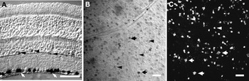
In retinal cryosections, staining for beta-galactosidase activity in Cx30.2lacZ/lacZ mice revealed lacZ-positive cells in the ganglion cell layer (GCL) (Fig. 1A) and in the inner nuclear layer (INL, arrowheads). Based on the localization of the lacZ signal and the size of the labeled cells in the INL, we hypothesized that all lacZ-positive neurons in the INL corresponded to amacrine cells and that these cells comprised at least two different types of neurons. One subtype had its nucleus close to the border between the INL and the IPL, whereas the second type had its nucleus further away from that border. Rarely, we found nuclei in the INL that were as large as the ones in the GCL. These cells might correspond to displaced ganglion cells or large amacrine cells.
Expression of Cx30.2/lacZ in retinae from Cx30.2lacZ/lacZ mice. (A) Expression pattern of the lacZ reporter gene visualized by staining for beta-galactosidase activity. LacZ staining (X-gal) was found in amacrine cells in the INL (arrowheads) and ganglion cells in the GCL (arrows). (B) Whole-mount view of the GCL. Xgal staining labeled large (arrows) and small (arrowheads) cells for beta-galactosidase activity. (C) Retinal whole mount stained with FDG and fixed in 4% paraformaldehyde shown as a maximum projection of a collapsed confocal stack (thickness: 13.6 μm). Fluorescein, indicating beta-galactosidase activity, was present in large (arrows) and small (arrowheads) cells. Occasionally, the primary dendrites of the stained neurons can be seen. Scale bars: (A) 40 μm and (B and C) 25 μm.

We studied the lacZ-positive cells in the GCL by staining whole-mount retinae from Cx30.2lacZ/lacZ mice. X-gal staining (Fig. 1B) revealed big (arrows) and small (arrowheads) lacZ-positive nuclei, suggesting that there were at least two subpopulations of Cx30.2-expressing neurons in the GCL. This was also confirmed using FDG as a fluorescent substrate for beta-galactosidase (Fig. 1C). Cells with large nuclei in the GCL are presumably retinal ganglion cells, and cells with small nuclei in the GCL most likely represent small ganglion cells or displaced amacrine cells, which are very frequent in the mouse retina (Jeon et al., Reference Jeon, Strettoi and Masland1998; Badea & Nathans, Reference Badea and Nathans2004; Lin & Masland, Reference Lin and Masland2006; Pérez de Sevilla Müller et al., Reference Pérez de Sevilla Müller, Shelley and Weiler2007).
Electroretinograms of WT and Cx30.2-deficient mice did not show any significant differences in a and b waves (L. Pérez de Sevilla Müller, unpublished observation). Considering the lacZ expression pattern described above, this was not surprising since the a and b waves are mainly generated by photoreceptor and ON bipolar cell responses, respectively, and these two cell types appeared to lack any lacZ signal in Cx30.2-deficient mice.
Immunocytological characterization of lacZ-positive cells in Cx30.2-deficient retinae
To examine the morphology of the Cx30.2-expressing cells in detail, we focused on the GCL. To analyze whether the Cx30.2-expressing cells comprised several cell types, retinae from Cx30.2lacZ/lacZ mice were stained for the calcium-binding proteins calbindin, calretinin, and parvalbumin (data not shown). LacZ-positive retinal cells were differentially immunolabeled by these markers. For example, some large lacZ-positive ganglion cells expressed parvalbumin, whereas smaller lacZ-positive ganglion cells expressed calretinin or calbindin instead, suggesting that Cx30.2 is expressed in multiple ganglion cell types in the mouse retina.
Classification of Cx30.2-expressing ganglion cells
To analyze Cx30.2-expressing ganglion cells, we injected lacZ-positive cells with dye. For this purpose, we labeled living cells with FDG (Nirenberg & Cepko, Reference Nirenberg and Cepko1993; Nirenberg & Meister, Reference Nirenberg and Meister1997). Hydrolysis of FDG by beta-galactosidase leads to an accumulation of fluorescein in the cytoplasm of the lacZ-positive cell. This approach enabled us to visualize the lacZ-positive cells, now green fluorescent, and made them easy to target with sharp dye-filled electrodes (Fig. 1C).
In order to characterize and classify the Cx30.2-expressing ganglion cells, we injected ganglion cells prelabeled with FDG (n = 62) with Neurobiotin and Alexa568 or Alexa594 hydrazide. Both the Alexa dyes and the Neurobiotin filled the cells completely, demonstrated by the tapering endings of their dendrites. A morphological catalog of the injected ganglion cells was created using the dendritic field and soma sizes, dendritic depth within the IPL, general morphology, and coupling pattern as characteristics. Dendritic depth within the IPL was determined by labeling the retina with anti-ChAT antibodies, which visually define the five strata (S1–S5) of the IPL. Cells stratifying in layers S1 and S2 of the IPL were classified as OFF cells, while those stratifying in layers S3, S4, or S5 were classified as ON cells. Cells stratifying in both ON and OFF strata were classified as ON-OFF ganglion cells. This classification was made with retinae from Cx30.2+/lacZ and Cx30.2lacZ/lacZ animals; all example images for the different cell types are from Cx30.2+/lacZ mice.
ON-OFF cells
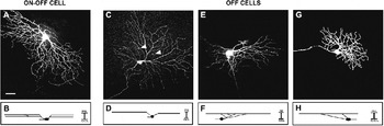
Only one type of Cx30.2-expressing ON-OFF cell was found. This bistratified ganglion cell type (Fig. 2A) had dendritic arbors with a diameter of 280 ± 60 μm (n = 6). Cell bodies had a mean diameter of 18.7 ± 0.7 μm. The dendrites were recursive and loop forming (Fig. 2A). Bistratified ganglion cells injected in Cx30.2+/lacZ mice never showed tracer coupling. The ON dendrites stratified in stratum S4 and the OFF processes in stratum S2 of the IPL (Fig. 2B), suggesting that this cell type is identical with the ON-OFF direction-selective ganglion cell RGD2 (Sun et al. Reference Sun, Li and He2002a; Weng et al., Reference Weng, Sun and He2005), M12 cells in the cluster analysis of mouse ganglion cells presented by Coombs et al. (Reference Coombs, van der List, Wang and Chalupa2006), and G17 cells (Völgyi et al., Reference Völgyi, Cheda and Bloomfield2009). Interestingly, RGD2 cells have been shown to be coupled to other bistratified ganglion cells by Cx45 (Schubert et al., Reference Schubert, Maxeiner, Kruger, Willecke and Weiler2005b; see Discussion).
Examples of the Cx30.2-expressing ON-OFF and OFF ganglion cell types. Ganglion cells were injected in retinae from Cx30.2+/lacZ mice and their stratification patterns were determined. (A) Confocal image of a bistratified ganglion cell injected with Neurobiotin, showing the dendritic branching pattern in tangential view. This cell type was similar to the RGD2 cell described by Sun et al. (Reference Sun, Li and He2002a). (B) Stratification of the bistratified ganglion cell. Dendrites stratified in strata S2 and S4 of the IPL. (C) Confocal image of the OFF type 1 ganglion cell. Coupled cells are marked with arrowheads. (D) Stratification of OFF type 1 ganglion cells. These cells stratified in stratum S1 of the IPL close to the INL. (E) Confocal image of the asymmetric OFF type 2 ganglion cell in tangential view. (F) OFF type 2 ganglion cells stratified in stratum 1 of the IPL. (G) Morphology of the OFF type 3 ganglion cell. These cells had the smallest dendritic fields of all Cx30.2-expressing ganglion cells and stratified in stratum 1 of the IPL. (H) All micrographs are maximum projections of collapsed confocal stacks (thickness: 20–29 μm). Scale bar: 40 μm.

OFF cells
Three types of OFF ganglion cells were lacZ positive. OFF type 1 cells (Fig. 2C) had large cell bodies with diameters of 14.2 ± 1.6 μm and large dendritic arbors of 370 ± 50 μm (n = 16), which stratified close to the INL in stratum S1 of the IPL (Fig. 2D). Two of the 16 injected OFF type 1 cells were coupled to amacrine cells that had their somata located in the INL; the remaining 14 cells did not show tracer coupling. The OFF type 1 cell resembles RGC2 outer ganglion cells (Sun et al., Reference Sun, Li and He2002a), G11 cells (Völgyi et al., Reference Völgyi, Cheda and Bloomfield2009), and M6 OFF ganglion cells (Coombs et al., Reference Coombs, van der List, Wang and Chalupa2006).
OFF type 2 cells (Fig. 2E) had large and asymmetric dendritic arborizations, which extended 290 ± 125 μm in diameter (n = 7). The cell bodies were round (17.9 ± 1.4 μm). Their processes stratified in stratum S1 of the IPL close to the INL (Fig. 2F). We never observed tracer coupling in these cells.
OFF type 3 cells had the smallest dendritic fields of the Cx30.2-expressing ganglion cells identified in this study, with a diameter of 240 ± 90 μm (n = 12). The dendritic arbors were round or ovoid (Fig. 2G), and dendrites stratified in stratum S1 of the IPL (Fig. 2H). The mean diameter of the round somata was 19.2 ± 1.4 μm. This ganglion cell type seems to be identical to the RGB3 (outer) found by Sun et al. (Reference Sun, Li and He2002a), G7 cells described by Völgyi et al. (Reference Völgyi, Cheda and Bloomfield2009), and cluster 6 cells (Kong et al., Reference Kong, Fish, Rockhill and Masland2005). In contrast to previous reports (Völgyi et al., Reference Völgyi, Cheda and Bloomfield2009; Pan et al., Reference Pan, Paul, Bloomfield and Völgyi2010), we never observed tracer coupling of OFF type 3 cells to other cells.
ON cells
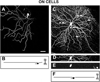
ON type 1 cells (n = 5) were defined by their small somata and medium-to-large dendritic fields. They presented round somata of 13.4 ± 1.3 μm and dendritic fields of 400 ± 80 μm in diameter (Fig. 3A). Their processes extended to stratum S5 of the IPL (Fig. 3B). The ending tips of the dendrites overlapped sparsely. This ganglion cell type had many similarities with the ganglion cell RGC1 found in the rat by Huxlin and Goodchild (Reference Huxlin and Goodchild1997) and G9 cells described in the mouse retina (Völgyi et al., Reference Völgyi, Cheda and Bloomfield2009). Consistent with previous reports (Völgyi et al., Reference Völgyi, Cheda and Bloomfield2009; Pan et al., Reference Pan, Paul, Bloomfield and Völgyi2010), we never observed coupling in these cells.
Examples of the two types of Cx30.2-expressing ON ganglion cells. Ganglion cells were injected in retinae from Cx30.2+/lacZ mice. (A) Confocal image of the ON type 1 ganglion cell (maximum projection of a confocal stack with a thickness of 25 μm). (B) These cells stratified in layer S5 of the IPL. (C) Morphology of ON type 2 cells (maximum projection of a confocal stack with a thickness of 25 μm). These cells were always coupled to displaced amacrine cells. ON type 2 cells corresponded to the RGA1 cells described by Sun et al. (Reference Sun, Li and He2002a). Coupled cells are marked with arrows. (D) Soma of the injected RGA1 cell (single scan with a thickness of 0.2 μm). (E) The injected cell was positive for FDG, indicating lacZ expression. (F) Illustration of the stratification pattern of the RGA1 cell. The dendrites of these cells ramified in stratum S5 of the IPL. Scale bar: 40 μm.

ON type 2 cells (n = 16) had large polygonal cell bodies from which three to seven primary dendrites (mean: 4.9 ± 1) left the soma to ramify in a branching radial pattern, resulting in few dendrites close to the soma (Fig. 3C). Dendrites were smooth, often overlapped at the ending tips and frequently formed hook-like terminals. The soma was always located in the center of the dendritic field. Cell somata had diameters of 18.2 ± 1.2 μm. The dendritic field size averaged 280 ± 19 μm. ON type 2 cells were always coupled to numerous small cells that had their somata in the GCL (Fig. 3C and 3E). The image in Fig. 3D corresponds to an injected cell whose soma showed the FDG signal (Fig. 3E). ON type 2 cells were monostratified with processes located in stratum S5 of the IPL, just below the cholinergic band produced by the ON starburst cells (Fig. 3F). According to the ganglion cell classification by Sun et al. (Reference Sun, Li and He2002a), the morphology of this ganglion cell type corresponded to RGA1 cells.
Tracer coupling between RGA1 and displaced amacrine cells
In the following, we focused on the RGA1 cells (ON type 2 ganglion cells), analyzing the cells’ coupling pattern and its modulation. RGA1 cells are well described in rodent retinae (Huxlin & Goodchild, Reference Huxlin and Goodchild1997; Sun et al., Reference Sun, Li and He2002a,Reference Sun, Li and Heb; Badea & Nathans, Reference Badea and Nathans2004; Kong et al., Reference Kong, Fish, Rockhill and Masland2005) and are identical to the giant cells reported by Bunt (Reference Bunt1976) in the albino rat retina. The cells were also described as M10 cells and G1 cells in the catalogs of mouse ganglion cells provided by Coombs et al. (Reference Coombs, van der List, Wang and Chalupa2006) and Völgyi et al. (Reference Völgyi, Cheda and Bloomfield2009), respectively.
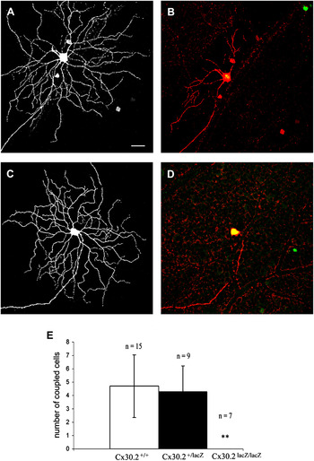
To study the coupling of RGA1 cells, we injected these cells in WT, Cx30.2+/lacZ, and Cx30.2lacZ/lacZ retinae. RGA1 cells were easy to recognize in WT retinae by the polygonal shape of their big somata. In both WT (n = 15 from nine retinae) and Cx30.2+/lacZ (n = 9 from seven retinae) mice, RGA1 cells were heterologously coupled to two to eight amacrine cells (4.7 ± 2.3 cells; Figs. 3C and 4A). The number of tracer-coupled cells was not significantly different between Cx30.2+/lacZ (4.3 ± 1.9 amacrine cells) and WT (Figs. 3C and 4; P > 0.76) mice. Tracer-coupled cells were always located within the dendritic field of the RGA1 cell and had their somata in the GCL. They had relatively round small somata with a mean diameter of 7.9 ± 2 μm. Though the cells’ dendrites could not be visualized, the small soma size suggested that these cells were most likely displaced amacrine cells. Recently, Völgyi et al. (Reference Völgyi, Cheda and Bloomfield2009) showed that RGA1 cells are coupled to two different types of displaced polyaxonal amacrine cells.
Coupling between RGA1 and displaced amacrine cells was mediated by Cx30.2. (A) Confocal image of an RGA1 cell injected in a retina from a Cx30.2+/lacZ mouse (maximum projection of a confocal stack with a thickness of 20 μm). The cell displayed coupling to three displaced amacrine cells. (B) Same injected RGA1 cell as in (A) showing the colocalization with the FDG signal (lacZ reporter gene expression). The coupled amacrine cells did not show the FDG signal and thus did not express Cx30.2. (C) An injected RGA1 cell in a Cx30.2lacZ/lacZ mouse. Coupling to displaced amacrine cells was completely absent. (D) Confocal image of the same cell as in (C) with colocalization of the FDG signal (maximum projection of a confocal stack with a thickness of 22 μm). (E) Histogram showing the mean number of coupled displaced amacrine cells in WT, Cx30.2+/lacZ, and Cx30.2lacZ/lacZ mice. Coupling patterns were not significantly different between WT and Cx30.2+/lacZ (WT, n = 15 from nine retinae; Cx30.2+/lacZ, n = 9 from seven retinae; P > 0.76) mice. In mice lacking both Cx30.2 alleles (Cx30.2lacZ/lacZ mice), coupling of RGA1 cells was completely abolished (n = 7 from four retinae). Differences to WT (P < 0.005) and Cx30.2+/lacZ (P < 0.002) mice were significant. **P values, t-test, P ≤ 0.01, scale bar: 40 μm.

An injected RGA1 cell of a Cx30.2+/lacZ mouse is shown in Fig. 4A, showing its typical tracer spread. The coupled cells (Fig. 4B) never showed the green fluorescent signal produced by FDG incubation. To exclude the possibility that the FDG signal leaked out of these cells over time, we incubated Cx30.2+/lacZ retinae with FDG and immediately looked for RGA1 cells. We never saw displaced amacrine cells positive for fluorescein in the proximity of the green-fluorescing RGA1 cells. We also injected FDG-positive cells with small somata and found them to be ON type 1 or OFF type 1 ganglion cells. Since most, if not all, displaced amacrine cells use GABA as their neurotransmitter (Kao & Sterling, Reference Kao and Sterling2006; Pérez de Sevilla Müller et al., Reference Pérez de Sevilla Müller, Shelley and Weiler2007), we stained retina sections from Cx30.2lacZ/lacZ mice using antibodies against beta-galactosidase and GABA. Only once did we find a single cell that showed both GABA- and beta-galactosidase-positive signals. However, the large majority of GABAergic cells in the GCL was negative for beta-galactosidase (Supplemental Figure 1A–1D), suggesting that nearly all Cx30.2-expressing cells in the GCL are ganglion cells. Taken together, these results suggest that Cx30.2 is not expressed by the displaced amacrine cells that are coupled to RGA1 cells. We therefore conclude that RGA1 cells most likely form heterotypic channels with displaced amacrine cells involving Cx30.2 and a yet unidentified protein.
In Cx30.2lacZ/lacZ mice, coupling of RGA1 cells (Fig. 4C and 4D; n = 7 from four retinae) was totally absent compared to WT (P < 0.005) and Cx30.2+/lacZ (P < 0.002) mice. Again, we used the FDG signal (Fig. 4D) to confirm that the injected cell (Fig. 4C) expressed the lacZ reporter gene. Thus, our results show that Cx30.2 mediates the coupling between RGA1 cells and displaced amacrine cells.
Displaced amacrine cells coupled to RGA1 cells do not express Cx36, Cx45, or Cx40
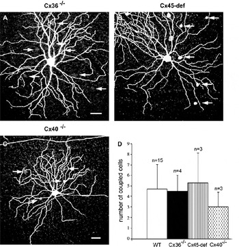
Since the displaced amacrine cells coupled to RGA1 cells did not show expression of the Cx30.2 reporter, we tested whether other known connexins are involved in this gap junction. In the mouse retina, three neuronal connexins (Cx36, Cx45, and Cx57) have been described so far. Cx57 is exclusively expressed in horizontal cells of the mouse retina (Hombach et al., Reference Hombach, Janssen-Bienhold, Söhl, Schubert, Büssow, Ott, Weiler and Willecke2004; Shelley et al., Reference Shelley, Dedek, Schubert, Feigenspan, Schultz, Hombach, Willecke and Weiler2006). Therefore, we did not test for Cx57 but for Cx36 and Cx45 (Fig. 5).
Coupling of RGA1 and displaced amacrine cells was preserved in Cx36-, Cx45-, and Cx40-deficient mice. (A) RGA1 cell injected in a Cx36-deficient mouse. Arrows point to coupled amacrine cells. (B) RGA1 cell injected in a Cx45-deficient mouse. (C) Injected RGA1 cell from a Cx40-deficient mouse. All micrographs are maximum projections of collapsed confocal stacks (thickness: 22–26 μm). (D) Histogram indicating the mean number of displaced amacrine cells coupled to the injected RGA1 cell in WT, Cx36−/−, Cx40−/−, and Cx45-deficient mice. In these transgenic mice, coupling between RGA1 and displaced amacrine cells was not significantly different from WT (WT, n = 15 from nine retinae; Cx36−/−, n = 4 from three retinae; P > 0.85; Cx45fl/fl:Nestin-Cre, n = 3 from three retinae; P > 0.65, Cx40−/−, n = 3 from three retinae; P > 0.24). Scale bar: 40 μm.

Cx36 has only been described in one type of amacrine cell: the AII amacrine cell (Feigenspan et al., Reference Feigenspan, Teubner, Willecke and Weiler2001; Mills et al., Reference Mills, O’Brien, Li, O’Brien and Massey2001). To test whether Cx36 is the connexin involved in the heterologous coupling between RGA1 and displaced amacrine cells, we injected RGA1 cells in Cx36−/− mice (Güldenagel et al., Reference Güldenagel, Ammermüller, Feigenspan, Teubner, Degen, Söhl, Willecke and Weiler2001). However, RGA1 cells were still coupled to displaced amacrine cells (Fig. 5A and 5D). There was no significant difference between the coupling in WT (4.7 ± 2.3 cells; n = 15 from nine retinae) and Cx36-deficient (4.5 ± 1.5 cells; n = 4 from three retinae, P > 0.85) mice. These results are consistent with previous findings (Schubert et al., Reference Schubert, Degen, Willecke, Hormuzdi, Monyer and Weiler2005a; Pan et al., Reference Pan, Paul, Bloomfield and Völgyi2010) and suggest that Cx36 does not participate at the junction between RGA1 and displaced amacrine cells.
In a similar way, we also tested for Cx45, which is expressed in bipolar cells (Maxeiner et al., Reference Maxeiner, Dedek, Janssen-Bienhold, Ammermüller, Brune, Kirsch, Pieper, Degen, Krüger, Willecke and Weiler2005; Dedek et al., Reference Dedek, Schultz, Pieper, Dirks, Maxeiner, Willecke, Weiler and Janssen-Bienhold2006), bistratified ganglion cells (Schubert et al., Reference Schubert, Maxeiner, Kruger, Willecke and Weiler2005b), and several types of amacrine cells, including one type of displaced amacrine cell (Maxeiner et al., Reference Maxeiner, Dedek, Janssen-Bienhold, Ammermüller, Brune, Kirsch, Pieper, Degen, Krüger, Willecke and Weiler2005; Pérez de Sevilla Müller et al., Reference Pérez de Sevilla Müller, Shelley and Weiler2007; Dedek et al., Reference Dedek, Breuninger, Pérez de Sevilla Müller, Maxeiner, Schultz, Janssen-Bienhold, Willecke, Euler and Weiler2009). To examine whether Cx45 forms heterotypic channels with Cx30.2, RGA1 cells were tracer injected in Cx45-deficient mice (Maxeiner et al., Reference Maxeiner, Dedek, Janssen-Bienhold, Ammermüller, Brune, Kirsch, Pieper, Degen, Krüger, Willecke and Weiler2005). We found that RGA1 cells were still coupled to displaced amacrine cells in Cx45-deficient mice (5.3 ± 2.8 cells, n = 3 from three retinae; Fig. 5B and 5D). Coupling in WT and Cx45-deficient mice was not significantly different (P > 0.66), indicating that these heterotypic gap junctions do not involve Cx45.
Another possible candidate connexin is Cx40 since it has been found in bovine and rat retinae (Matesic et al., Reference Matesic, Tillen and Sitaramayya2003) and recently also in the mouse retina (Kihara et al., Reference Kihara, de Castro, Moriscot and Hamassaki2006), though it is not yet clear in which retinal cell type Cx40 is expressed. Interestingly, Cx40 is strongly expressed in mouse heart, where it may form heterotypic gap junctions with Cx30.2 (Kreuzberg et al., Reference Kreuzberg, Söhl, Kim, Verselis, Willecke and Bukauskas2005). To test whether this is also the case for the coupling between displaced amacrine cells and RGA1 cells, we analyzed this coupling in Cx40-deficient mice (Kirchhoff et al., Reference Kirchhoff, Nelles, Hagendorff, Krüger, Traub and Willecke1998). We found that RGA1 cells were still coupled to displaced amacrine cells in Cx40-deficient mice (n = 3 from three retinae; Fig. 5C and 5D). The extent of coupling was similar in WT and Cx40-deficient retinae (P > 0.24), indicating that Cx40 is not involved in the gap junction between displaced amacrine and RGA1 cells. Instead, our data suggest that either the reporter gene expression in the coupled displaced cells is very low or an unidentified gap junction protein participates in the coupling between RGA1 and displaced amacrine cells.
Modulation of the tracer coupling between RGA1 and displaced amacrine cells
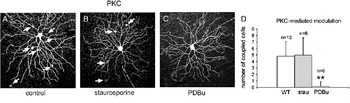
Gap junction coupling of retinal neurons is influenced by different ambient light levels and by neuromodulators (for review, see Weiler et al., Reference Weiler, Pottek, He and Vaney2000), which alter the conductance of gap junction channels most likely by inducing protein kinase–mediated phosphorylation (Xia & Mills, Reference Xia and Mills2004; Urschel et al., Reference Urschel, Höher, Schubert, Alev, Söhl, Wörsdörfer, Asahara, Dermietzel, Weiler and Willecke2006). Since Cx30.2 contains several consensus sites for protein kinase C (PKC)-mediated phosphorylation (Web-based analysis with NetPhosK 1.0 server), we tested whether the coupling between RGA1 cells and displaced amacrine cells is modulated by protein kinases by applying PKC inhibitors and activators (Fig. 6).
Tracer coupling between RGA1 and displaced amacrine cells was modulated by PKC. (A–C) Testing for PKC-mediated phosphorylation: RGA1 cells injected under control conditions (A) in the presence of staurosporine (B) and PDBu (C). Coupled cells are marked with arrows. Staurosporine did not significantly affect tracer spread (D, control, n = 13 from eight retinae, staurosporine, n = 6 from five retinae, P > 0.77). When incubated with PDBu, the number of coupled cells was significantly reduced (D, n = 6 from three retinae, P < 9 × 10⁻⁵). **P values, t-test, P ≤ 0.01, scale bar: 40 μm. All micrographs are maximum projections of collapsed confocal stacks (thickness: 13–17 μm).

RGA1 cells were injected with Neurobiotin in WT retinae. The average number of coupled displaced amacrine cells was 4.8 ± 2.2 (n = 13 from eight retinae; Fig. 6A). To test for involvement of PKC, we incubated WT retinae in staurosporine, a cell-permeable inhibitor of PKC, prior to injection (Fig. 6B). Staurosporine incubation had no effect on the coupling patterns of RGA1 cells (Fig. 6B and 6D, n = 6 from five retinae, coupled to 4.9 ± 2.7 amacrine cells, P > 0.77). In contrast, incubation with the potent PKC activator PDBu led to a significant reduction in the number of coupled cells (Fig. 6C and 6D, n = 6 from three retinae, 0.2 ± 0.7 coupled cells, P < 9 × 10⁻⁵). Thus, our results indicate that the heterotypic coupling between RGA1 and displaced amacrine cells is mediated by Cx30.2 and, most likely, an unidentified connexin and is modulated by PKC.
Discussion
In this study, we identify Cx30.2 as a novel connexin in the mouse retina. Using Cx30.2-lacZ reporter mice, we show that Cx30.2 is expressed in six different types of ganglion cells. Among these types are the RGA1 cells (Sun et al., Reference Sun, Li and He2002a), which are coupled to displaced amacrine cells. The coupling between these two cell types is abolished in Cx30.2lacZ/lacZ mice, is regulated by PKC, and is heterotypic, most likely involving Cx30.2 and a yet unidentified connexin.
Cx30.2 as a novel retinal connexin
Since Söhl et al. (Reference Söhl, Degen, Teubner and Willecke1998) first described the expression of Cx36 in the mouse retina, several new connexins have been identified in this tissue. Cx36 and Cx45 were assigned to multiple retinal cell types, whereas Cx57 was found exclusively in horizontal cells (reviewed by Söhl et al., Reference Söhl, Maxeiner and Willecke2005; Bloomfield & Völgyi, Reference Bloomfield and Völgyi2009). In this work, we show for the first time that Cx30.2 is expressed in mouse retinal neurons, namely in several different types of amacrine and ganglion cells. This confirms Cx30.2 as a new neuronal connexin in the central nervous system (Kreuzberg et al., Reference Kreuzberg, Deuchars, Weiss, Schober, Sonntag, Wellershaus, Draguhn and Willecke2008). Cx30.2 is expressed in both inhibitory interneurons (hippocampus and retina) and projecting neurons (retina).
Cx30.2 expression in the mouse retina
Since commercially available antibodies against Cx30.2 (catalog no. 40-7400, lot 27544; Invitrogen) fail to show a differential pattern in WT and Cx30.2lacZ/lacZ retinae, we had to rely on the reporter gene expression to localize Cx30.2 in the retina. Cx30.2 expression was observed in cells in the INL and GCL of the mouse retina. Cells located in the INL corresponded presumably to amacrine cells, based on nucleus size and location of the lacZ staining with respect to the borders of the INL. Thus, Cx30.2 is a likely candidate protein for mediating the coupling of different amacrine cell classes (Xin & Bloomfield, Reference Xin and Bloomfield1997; Wright & Vaney, Reference Wright and Vaney2000, Reference Wright and Vaney2004).
However, in this work, we focused on the GCL. To identify the Cx30.2-expressing cells in this layer, we labeled the lacZ-positive neurons in Cx30.2+/lacZ and Cx30.2lacZ/lacZ mice using a fluorogenic substrate (FDG). Nirenberg and Meister (Reference Nirenberg and Meister1997) showed that 95% of all lacZ-positive cells could be labeled in this way. With this method, we labeled big subsets of neurons in each Cx30.2-deficient retina. These subsets did not vary between retinae and comprised six different types of Cx30.2-expressing ganglion cells: one type of ON-OFF, three types of OFF, and two types of ON ganglion cells.
Cx30.2-mediated tracer coupling in the mouse retina
Surprisingly, only two of the six Cx30.2-expressing ganglion cell types were tracer coupled in Cx30.2+/lacZ mice: OFF type 1 cells and ON type 2 cells. In contrast, OFF type 2 and type 3 cells, ON type 1 cells, and ON-OFF type 1 cells were never found to be coupled in Cx30.2+/lacZ mice.
OFF type 1 cells presumably correspond to G11 cells (Völgyi et al., Reference Völgyi, Cheda and Bloomfield2009), which have been shown to be homologously coupled to other G11 cells and heterologously coupled to amacrine cells by Cx36 (Pan et al., Reference Pan, Paul, Bloomfield and Völgyi2010). In Cx30.2+/lacZ mice, homologous coupling of type 1 cells was absent, whereas heterologous coupling was rare: only 2 of 16 cells showed coupling. This suggests that OFF type 1 ganglion cell types express multiple connexins, an idea that has been proposed previously (Schubert et al., Reference Schubert, Maxeiner, Kruger, Willecke and Weiler2005b; Völgyi et al., Reference Völgyi, Abrams, Paul and Bloomfield2005). Indeed, a coexpression of Cx30.2 and Cx36 in neuronal tissue has been shown by Kreuzberg et al. (Reference Kreuzberg, Deuchars, Weiss, Schober, Sonntag, Wellershaus, Draguhn and Willecke2008). The weak coupling in Cx30.2+/lacZ mice suggests that Cx36 may be downregulated when one allele of Cx30.2 is missing. Similarly, in Cx36-deficient mice, Cx30.2 may be downregulated so that the lack of Cx36 is sufficient to completely abolish gap junctional coupling of G11 cells. This might also be an explanation for the lack of coupling in OFF type 3 cells, which correspond to the G7 cells described by Völgyi et al. (Reference Völgyi, Cheda and Bloomfield2009) and have been shown to be coupled by Cx36 (Pan et al., Reference Pan, Paul, Bloomfield and Völgyi2010). Similar codependencies of connexin expression levels have been suggested in the retina (Cx36/Cx45; Li et al., Reference Li, Kamasawa, Ciolofan, Olson, Lu, Davidson, Yasumura, Shigemoto, Rash and Nagy2008) and the aortic endothelium (Cx37/Cx40; Simon & McWhorter, Reference Simon and McWhorter2003).
OFF type 2 cells did not show coupling, although Völgyi et al. (Reference Völgyi, Cheda and Bloomfield2009) reported coupling in all ganglion cells stratifying in sublamina 1 of the IPL. However, since this ganglion cell type does not correspond to any previously defined subtype, it remains possible that the expression level of Cx30.2 is so low that one allele of Cx30.2 is not sufficient to promote significant dye transfer. Also, since dye transfer between retinal neurons is largely dependent on ambient light conditions (Weiler et al., Reference Weiler, Pottek, He and Vaney2000), our injection conditions may have led to a closure of gap junction channels and a lack of dye coupling in this previously undefined cell type. A similar explanation could apply to ON type 1 cells, which correspond to the G9 cells described by Völgyi et al. (Reference Völgyi, Cheda and Bloomfield2009). Under our and Völgyi’s injection conditions, these cells are not coupled although they express Cx30.2. However, again, light conditions and a low Cx30.2 expression level may lead to a lack of coupling even though reporter gene expression is detectable. Though this has never been shown before, it seems possible that unfavorable light conditions should be able to completely abolish coupling in ganglion cells that express low amounts of Cx30.2, especially since Cx30.2 has the smallest unitary conductance of all connexins (Kreuzberg et al., Reference Kreuzberg, Söhl, Kim, Verselis, Willecke and Bukauskas2005) and its permeability to dyes is generally low (Rackauskas et al., Reference Rackauskas, Verselis and Bukauskas2007). However, another explanation that may be even more likely is that Cx30.2 may function as a hemichannel in some ganglion cell subtypes. Connexin hemichannels provided by individual cells have been shown to provide a pathway for exchange of intracellular components with extracellular solution (for review, see Spray et al., Reference Spray, Ye and Ransom2006). Indeed, Bukauskas et al. (Reference Bukauskas, Kreuzberg, Rackauskas, Bukauskiene, Bennett, Verselis and Willecke2006) showed that Cx30.2 can form hemichannels in HeLa cells. This would explain why Cx30.2 expression is not always indicative of tracer coupling in the adult mouse retina.
Among the other cell types that we characterized as Cx30.2 expressing but are not coupled in Cx30.2+/lacZ mice is the RGD2 cell (Sun et al., Reference Sun, Li and He2002a; Weng et al., Reference Weng, Sun and He2005), an ON-OFF direction-selective cell that corresponds to the G17 cells recently described by Völgyi et al. (Reference Völgyi, Cheda and Bloomfield2009). This cell was shown to be homologously coupled by Cx45 (Schubert et al., Reference Schubert, Maxeiner, Kruger, Willecke and Weiler2005b). RGD2 cells retain their coupling in Cx36-deficient mice (Schubert et al., Reference Schubert, Degen, Willecke, Hormuzdi, Monyer and Weiler2005a; Pan et al., Reference Pan, Paul, Bloomfield and Völgyi2010). This suggests that Cx30.2 and Cx45 might form heteromeric channels in RGD2 cells, as was shown previously in transfected HeLa cells (Kreuzberg et al., Reference Kreuzberg, Söhl, Kim, Verselis, Willecke and Bukauskas2005). Consistent with this idea, Schubert et al. (Reference Schubert, Maxeiner, Kruger, Willecke and Weiler2005b) reported some residual tracer coupling in RGD2 cells of Cx45-deficient mice, suggesting that Cx30.2 expression in Cx45-deficient mice was still high enough to occasionally promote tracer coupling in these cells. Why then did we not observe any coupling of RGD2 cells in Cx30.2+/lacZ mice? There are two possible explanations: first, Cx45 expression in Cx30.2+/lacZ might be downregulated, as was suggested for Cx36-deficient mice (Li et al., Reference Li, Kamasawa, Ciolofan, Olson, Lu, Davidson, Yasumura, Shigemoto, Rash and Nagy2008); second, lack of Cx30.2 in putative Cx30.2/Cx45 heteromeric channels may alter channel kinetics and its modulation by ambient light in an unpredictable way. Though both Cx30.2 (~9 pS) and Cx45 (~64 pS) have low unitary channel conductances compared with other connexins (Kreuzberg et al., Reference Kreuzberg, Söhl, Kim, Verselis, Willecke and Bukauskas2005), Cx30.2 and Cx45 differ dramatically in their sensitivity to transjunctional voltage changes. Gating of Cx30.2 is almost independent of transjunctional voltage (Kreuzberg et al., Reference Kreuzberg, Söhl, Kim, Verselis, Willecke and Bukauskas2005), whereas Cx45 shows a marked sensitivity to voltage differences between adjacent cells (Bukauskas et al., Reference Bukauskas, Angele, Verselis and Bennett2002). Thus, elimination of one Cx30.2 allele may lead to channel closure under our injection conditions due to unpredictable changes in channel kinetics.
The only ganglion cell type that we found to be coupled consistently in Cx30.2+/lacZ mice was the ON type 2 cell. These cells correspond to the RGA1 cells described by Sun et al. (Reference Sun, Li and He2002a) and G1 cells described recently by Völgyi et al. (Reference Völgyi, Cheda and Bloomfield2009). For RGA1 cells, coupling was completely absent in Cx30.2lacZ/lacZ mice, indicating that coupling was mediated by Cx30.2 (see below).
Gap junction protein of the displaced amacrine cells coupled to RGA1 cells
We focused our study on the coupling of RGA1 cells. Our data strongly suggest that the RGA1 cells are coupled to displaced amacrine cells by heterotypic gap junctions involving Cx30.2 and another connexin that has not yet been identified. This conclusion is based on the following findings. 1) Coupled displaced amacrine cells do not express the lacZ reporter gene, as shown by in vitro staining with FDG, double immunostaining for beta-galactosidase and the displaced amacrine cell neurotransmitter GABA, and injection of small FDG-positive somata, which proved to be ON type 1 and OFF type 1 ganglion cells. 2) Tracer coupling between RGA1 and displaced amacrine cells is unimpaired in Cx36−/−, Cx45-deficient, and Cx40−/− mice. Though the fluorescence was slightly less intense in coupled displaced amacrine cells in Cx36-deficient mice than in WT retinae, the number of coupled cells was not significantly different. Since our study and previous reports (Schubert et al., Reference Schubert, Degen, Willecke, Hormuzdi, Monyer and Weiler2005a; Pan et al., Reference Pan, Paul, Bloomfield and Völgyi2010) did not show an impairment of RGA1 coupling to amacrine cells in Cx36-deficient mice, RGA1 cells, in contrast to most other ganglion cell types, most likely do not express Cx36. Otherwise, Cx30.2 expression would have been downregulated (see above) in Cx36-deficient mice and thus coupling would have been weaker than in WT mice. As we found the coupling of RGA1 and displaced amacrine cells also unimpaired in Cx45-deficient and Cx40−/− mice, we conclude that the displaced amacrine cells did not express Cx36, Cx45, or Cx40. Since the only other known neuronal connexin in the mouse retina, Cx57, is expressed exclusively in horizontal cells (Hombach et al., Reference Hombach, Janssen-Bienhold, Söhl, Schubert, Büssow, Ott, Weiler and Willecke2004; Janssen-Bienhold et al., Reference Janssen-Bienhold, Trümpler, Hilgen, Schultz, Pérez de Sevilla Müller, Sonntag, Dedek, Dirks, Willecke and Weiler2009), we conclude that the displaced amacrine cells coupled to RGA1 cells most likely express a yet unidentified connexin.
However, we cannot exclude that the expression of Cx30.2 in the coupled displaced amacrine cells is below the detection threshold of our lacZ reporter, though this, given the sensitivity of the FDG approach (Nirenberg & Meister, Reference Nirenberg and Meister1997), seems unlikely.
Tracer coupling of RGA1 cells is modulated by PKC
During retinal adaptation, the networks of coupled neurons are modulated by light and by neuromodulators such as dopamine (Hampson et al., Reference Hampson, Vaney and Weiler1992; He et al., Reference He, Weiler and Vaney2000). These act by activating protein kinases, which can directly or indirectly alter gap junction properties (for review, see Moreno & Lau, Reference Moreno and Lau2007). Here, we show that the serine/threonine kinase PKC is involved in the regulation of the Cx30.2-mediated coupling between RGA1 and displaced amacrine cells. Activation of PKC by PDBu leads to a significant decrease in gap junction coupling. This decrease in coupling might be caused by direct phosphorylation of Cx30.2, which harbors several consensus sites for PKC-mediated phosphorylation (Web-based analysis with NetPhosK 1.0 server). Reduction in gap junctional permeability by direct PKC-mediated phosphorylation has been shown, for example, for Cx43 (Bao et al., Reference Bao, Altenberg and Reuss2004). Since we cannot exclude that Cx30.2 forms heteromeric channels with another connexin, the decrease in coupling might also be due to PKC-mediated phosphorylation of this other connexin. Additionally, PKC activation may influence not only the connexon on the RGA1 cell side but also the unidentified connexin in the coupled displaced amacrine cells. PKC activation might also have indirect effects that could lead to the observed decrease in coupling, for example, effects on connexin interaction partners or effects on other cell types.
PKA has been shown to mediate the phosphorylation of Cx36 and to reduce the extent of coupling in AII amacrine cells (Xia & Mills, Reference Xia and Mills2004; Urschel et al., Reference Urschel, Höher, Schubert, Alev, Söhl, Wörsdörfer, Asahara, Dermietzel, Weiler and Willecke2006) and alpha ganglion cells (L. Pérez de Sevilla Müller, unpublished observations). Our results suggest that different retinal networks are modulated by different protein kinases. This may reflect the need for a precise control of coupled neuronal networks to ensure that retinal resolution and sensitivity are adapted optimally to the visual environment with its changing light conditions.
Acknowledgments
We thank Bettina Kewitz for excellent technical assistance and Jennifer Trümpler for critical reading of this manuscript. This study was supported by the Deutsche Forschungsgemeinschaft (FOR701 to R.W. and K.D., JA 854/1-1, 2 to U.J.-B. and R.W., WI 270/22-5, 6 to K.W., and Graduate School for Neurosensory Science and Systems to R.W. and L.P.de S.M.).