Introduction
Cholesteatoma is a progressive and destructive squamous epithelium that affects the middle-ear space. As a result of its location and behaviour, it has the potential to erode structures within the lateral skull base, including the labyrinth.Reference Mustafa, Heta, Kastrati and Dreshaj1 Having achieved this, the risk of sensorineural hearing loss is much increased.Reference Rosito, Netto, Teixeira and da Costa2 Moreover, this risk arises from not only the continued action of the disease, but also from surgical intervention undertaken to treat the disease.Reference Smyth and Gormley3
The otological literature contains many articles concerned with the treatment of fistulas of the labyrinth due to cholesteatoma. A recent systematic review has summarised the available information.Reference Lim, Gangal and Gluth4 In essence, there are two principal techniques for dealing with cholesteatoma over a labyrinthine fistula: (1) complete removal of cholesteatoma matrix, including over the fistula, and repair of any ensuing labyrinthine defect; and (2) exteriorisation, in which the cholesteatoma over the fistula is left in place, whilst the cholesteatoma bed is saucerised by forming a cavity with removal of the posterior ear canal wall.Reference Lim, Gangal and Gluth4
The former technique requires separation of the cholesteatoma matrix from the delicate fistula endosteum and reinforcement of the fistula cover. This procedure is dependent on the degree to which the cholesteatoma adheres to the endosteum. It may result in fistula opening, which is associated with an increased risk of sensorineural hearing loss and even dead ear.Reference Ritter5
The latter technique requires removal of the posterior wall of the external ear canal as part of the saucerisation, so that the epithelium over the fistula becomes part of the skin lining of a mastoid cavity. In this circumstance, the patient may be subject to the adverse effects of living with a mastoid cavity.Reference Lim, Gangal and Gluth4
Fistulas of the cochlea are associated both with an enhanced risk of hearing loss if an excision technique is used and long-term intrusive post-operative symptoms if an exteriorisation procedure is undertaken.Reference Lim, Gangal and Gluth4 The problems with cochlear fistulas led the senior author to consider a different approach to the treatment of labyrinthine fistulas in general.
This study was undertaken to investigate the effect of laser treatment of cholesteatoma over cochlear and vestibular fistulas. Use of the laser has been established in the senior author's practice of cholesteatoma surgery following the demonstration of greater efficacy of complete cholesteatoma removal and improved hearing through systematic preservation of the ossicles.Reference Hamilton6, Reference Hamilton7 This article describes a modified laser technique to eliminate cholesteatoma from cochlear and vestibular fistulas without the hazardous dissection of the disease from the labyrinth endothelium. The study aimed to quantify both the effectiveness of disease removal from the fistula and the effect on inner-ear hearing function resulting from the surgery.
Materials and methods
Ears that received surgical treatment by the senior author for cholesteatoma in secondary care hospitals in Gloucestershire, UK, were considered for this study. Only ears that included laser treatment of cholesteatoma causing cochlear or vestibular fistulas were included. Consecutive patients from 2005 to 2017 were included. The records of these cases were retrieved from a database (Microsoft Access; Microsoft, Redmond, Washington, USA) kept on a hospital server. Baseline information about fistula location and size, and pre-operative ipsilateral bone conduction thresholds, were recorded. Ears with no residual hearing in the ipsilateral ear prior to surgery were excluded. All patients underwent intact canal surgery with a staged, second-look operation 12 months after the first procedure.
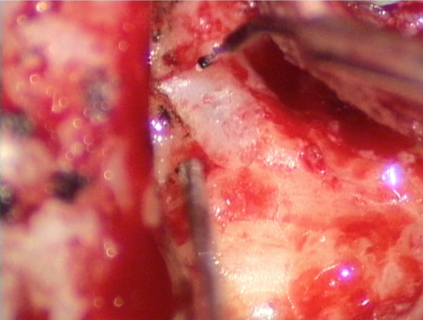
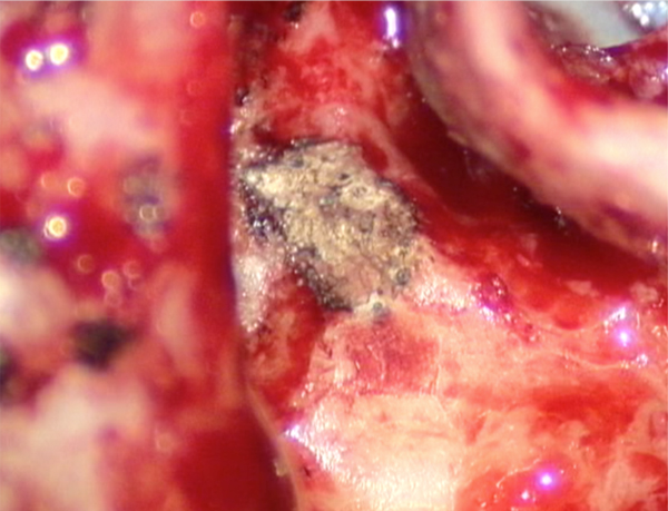
A fibre-guided potassium titanyl phosphate laser beam was used in all cases. The laser light from an optical fibre diverges and so the power density of the laser light diminishes with distance from the fibre tip. The established method for treating cholesteatoma is to approximate the target tissue to the laser fibre tip, so that the intensity of the laser light causes the target tissue to be vaporised. Clearly, this would be hazardous to the integrity of the labyrinth. Therefore, the technique used on cholesteatoma over a cochlear or vestibular fistula was to hold the fibre tip at a greater distance (1–2 cm) from the target. Hence, the power density was sufficient only to gradually heat and denature the keratin firming matrix (Figure 1). The speed of denaturing could be easily controlled by the distance between the laser fibre tip and the target, so that the integrity of the cover over the fistula was maintained (Figure 2). Once all of the cholesteatoma was deemed to have been denatured, the fistula was covered with a fascia graft, which was secured with fibrin tissue glue (Tisseel; Baxter Healthcare, Deerfield, Illinois, USA).
Cholesteatoma preserved over the lateral semicircular canal fistula. The figure shows a right ear, with the tegmen on the left side of the image and the intact canal wall in the upper right corner. The laser fibre is held a centimetre or so distant from the cholesteatoma surface and is angled away from the facial nerve.

Denatured cholesteatoma matrix after laser treatment. The same ear as Figure 1 is shown after treatment with a low energy density laser beam. Note that the soft tissue cover of the fistula remains intact, even though the heating effect of the laser has denatured the epithelial surface.

Assessment of this technique required measures of: (1) the effectiveness of disease elimination; (2) the effect on ipsilateral cochlear function; and (3) the risk of inadvertent harm. The first was measured by the presence or absence of residual cholesteatoma associated with the fistula at the second-look operation. Particular care was taken to inspect and gently palpate the area of the grafted fistula. The second was measured by comparing the pre- and post-operative bone conduction thresholds in the ipsilateral ear at three months after surgery. Bone conduction thresholds were measured according to an international standard.8 Outcomes were calculated for the entire group and the cochlear subgroup. The risk of harm was measured by the presence or absence of unintentional fistula perforation with the laser during surgery, and the presence or absence of facial palsy in the first month after surgery.
Results
Forty ears with fistulas were treated during the inclusion period. In two cases, the patients had ipsilateral dead ears prior to surgery, and so were not included in the study group. In two ears, the pre-operative audiogram could not be found. The quantitative data in this study were derived from the remaining 36 surgical procedures.
In 30 ears, the fistula affected the lateral semicircular canal. The fistula involved the cochlea in the region of the round window in four cases and the oval window in two cases. In one ear, the superior semicircular canal and oval window were eroded. The only case that involved the posterior semicircular canal also extensively involved the basal turn of the cochlea, and the patient presented with a dead ear. In all cases, the fistula was 5 mm or less in longest diameter.
Two relatively large fistulas were inadvertently perforated during diffuse laser heating during surgery during an early performance of this technique. In each case, the perforation was no larger than a pinhole. Each was closed by a minute fat graft and then covered by the same fascia technique used in all cases in this series.
No patients developed a facial palsy following surgery.
All patients underwent second-stage surgery. The cholesteatoma was completely cleared from the fistula site in all cases.
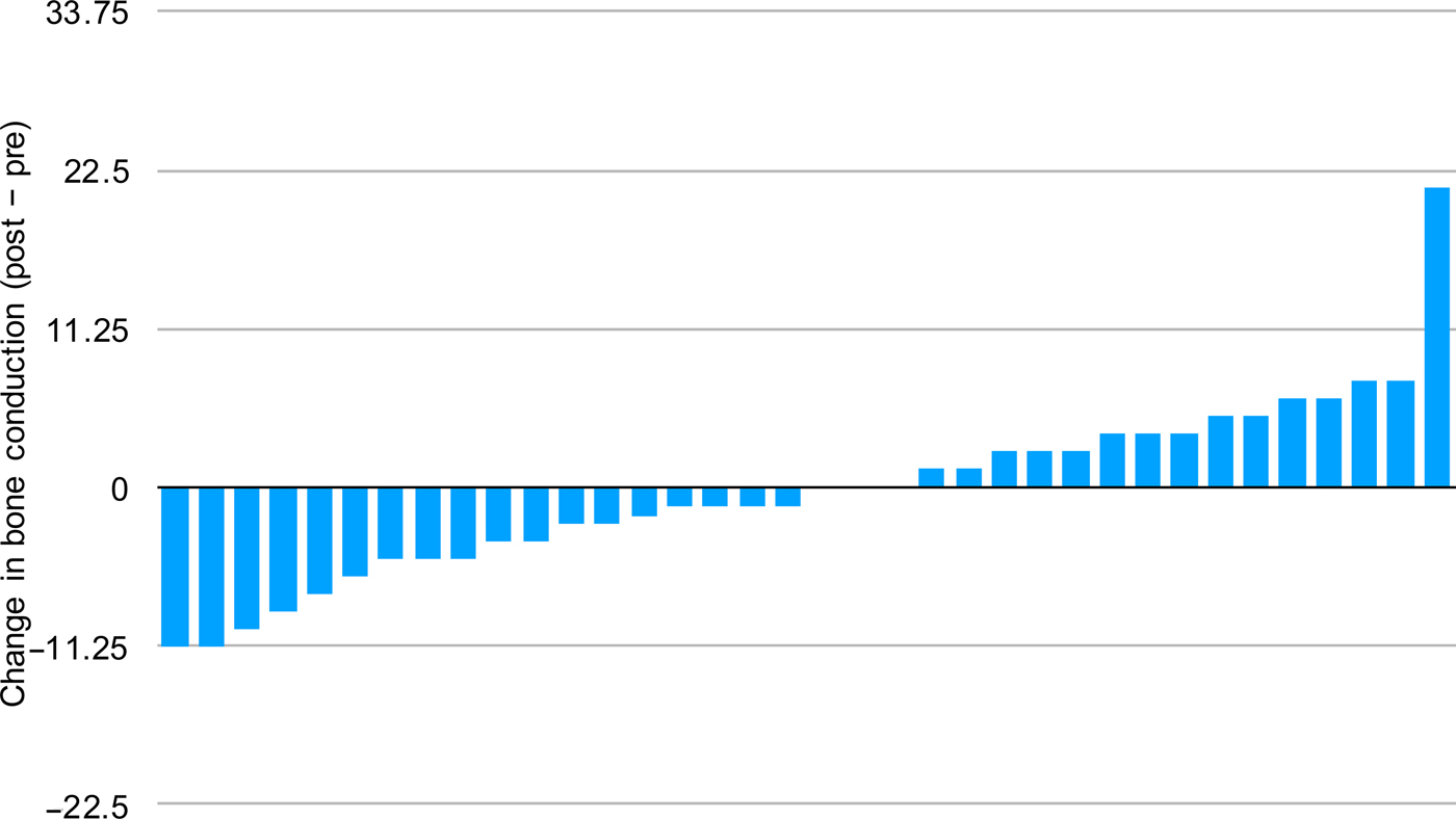
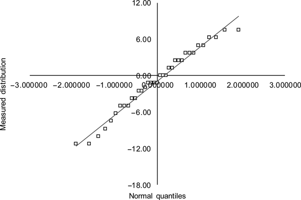
The change in bone conduction threshold as a result of the treatment is shown for each case in the entire group in Figure 3. The average change in bone conduction threshold was −0.3 dB deterioration in hearing. The changes are fairly evenly spread either side of the x-axis. Many ears appeared to have an apparent improvement in bone conduction threshold. We considered that this was unlikely to be a real phenomenon. We hypothesised that, excluding the one obvious outlier, this spread of results may be due purely to the random error associated with the difficult measurement of bone conduction thresholds. If so, the distribution of the changes in bone conduction thresholds should conform to a Gaussian distribution. This was tested using a Q-Q plot and the Shapiro–Wilk test. Both visually (Figure 4) and statistically, the distribution is confirmed to be consistent with a normal distribution (Shapiro–Wilk: z = 0.35, p = 0.36).
Change in bone conduction threshold in the operated ear. The results of the series are ordered, with the greatest deterioration in hearing to the left, and the greatest improvement in hearing to the right.

Q-Q plot of observed changes in bone conduction threshold (ordinate) against a Gaussian model based on the mean and variance of the observed sample (abscissa). The near straight line distribution provides a visual indicator that the observed distribution is also Gaussian.

The treatment of cochlear fistulas appears to be reliable; the mean change in bone conduction threshold of this subgroup was + 0.2 dB. The distribution was investigated in similar detail given the small size of the sample.
Discussion
In all surgery, the fundamental aim is to cause no harm. Hitherto, in cholesteatoma surgery, the treatment of a cochlear or vestibular fistula increased the risk of failure to achieve this aim. The surgeon faced the unenviable choice of having either to dissect the cholesteatoma matrix off the fistula endothelium or create a mastoid cavity with its attendant long-term consequences. If the fistula involved the cochlea, the surgical consequences were notably worse.Reference Lim, Gangal and Gluth4
The laser enabled us to use a novel technique that can eliminate the cholesteatoma over a fistula without having to dissect the disease from the delicate fistula endothelium. This reduces the risk of opening the fistula. The series reported in this manuscript had a median change in bone conduction threshold of −0.6 dB, a trivial change in hearing that the patient is unable to discern. The distribution of the measured changes can plausibly be accounted for by the random variation associated with the process of measuring bone conduction thresholds, rather than representing any real change in hearing.
This study did not include any fistulas over 5 mm in maximum length, and so the results of this study cannot be generalised to larger fistulas. In particular, large fistulas of the semicircular canals also have greater exposure of the transverse circumference. This means that there may be greater risk of heating and damaging the membranous labyrinth during laser denaturing of adherent cholesteatoma than with smaller fistulas. However, such large fistulas are associated with greater risk of sensorineural hearing loss with conventional cholesteatoma excision surgery, and may be vulnerable during cleaning if included in an open cavity.Reference Ikeda, Kobayashi, Kawase, Oshima and Sato9
Concern has been previously raised that long-term hearing in patients treated for cholesteatomatous fistulas may progressively deteriorate.Reference Katsura, Mishiro, Adachi, Ogino, Daimon and Sakagami10 Our study does not, at this stage, include long-term assessment of hearing after laser treatment of cochlear and vestibular fistulas.
• Conventional labyrinthine fistula surgical treatment is associated with sensorineural hearing loss, particularly for cochlear fistulas
• A new technique for labyrinthine fistula is presented: low power density laser heating of cholesteatoma matrix over the fistula
• Low power density laser energy denatures the cholesteatoma without disrupting the soft tissue fistula cover
• Low power density laser energy eliminates the cholesteatoma without loss of integrity of the fistula endosteum
• Evidence from 36 cases indicates that the technique is effective at removing cholesteatoma and maintaining cochlear function
There already exists evidence for improved outcomes of surgery for cholesteatoma when the laser is used. Use of the laser has resulted in lower rates of residual cholesteatoma in combined approach tympanoplasty.Reference Hamilton6 The laser permits higher rates of preservation of the intact chain, with associated improvement in post-operative hearing in the operated ear in both the medium and long term.Reference Hamilton7 We now have confirmed that, using a low energy density beam, the laser successfully eliminates cholesteatoma associated with labyrinthine fistulas, both cochlear and vestibular, with minimal effect on cochlear function.
Competing interests
None declared