




No CrossRef data available.
Article contents
Parametric manipulation of working memory load in traumatic brain injury: Behavioral and neural correlates
Published online by Cambridge University Press: 01 September 2004
Abstract
Traumatic brain injury (TBI) is often associated with enduring impairments in high-level cognitive functioning, including working memory (WM). We examined WM function in predominantly chronic patients with mild, moderate and severe TBI and healthy comparison subjects behaviorally and, in a small subset of moderate-to-severe TBI patients, with event-related functional magnetic resonance imaging (fMRI), using a visual n-back task that parametrically varied WM load. TBI patients showed severity-dependent and load-related WM deficits in performance accuracy, but not reaction time. Performance of mild TBI patients did not differ from controls; patients with moderate and severe TBI were impaired, relative to controls and mild TBI patients, but only at higher WM-load levels. fMRI results show that TBI patients exhibit altered patterns of activation in a number of WM-related brain regions, including the dorsolateral prefrontal cortex and Broca's area. Examination of the pattern of behavioral responding and the temporal course of activations suggests that WM deficits in moderate-to-severe TBI are due to associative or strategic aspects of WM, and not impairments in active maintenance of stimulus representations. Overall, results demonstrate that individuals with moderate-to-severe TBI exhibit WM deficits that are associated with dysfunction within a distributed network of brain regions that support verbally mediated WM. (JINS, 2004, 10, 724–741.)
Information
- Type
- Research Article
- Information
- Journal of the International Neuropsychological Society , Volume 10 , Issue 5 , September 2004 , pp. 724 - 741
- Copyright
- 2004 The International Neuropsychological Society
INTRODUCTION
Patients with even mild traumatic brain injury (TBI) often suffer from a number of enduring cognitive impairments, most notably in attention (e.g., McKinlay et al., 1981; Ponsford et al., 1995), processing speed (Ponsford & Kinsella, 1992; Ferraro, 1996; van Zomeren & Brouwer, 1987), memory (Levin et al., 1990) and negotiating multiple simultaneous task demands (i.e., dual-task performance; Cicerone, 1996; Leclercq et al., 2000; McDowell et al., 1997; Park et al., 1999). Deficits in working memory (WM) function in TBI are frequently mentioned in the literature. To date, however, only a small number of studies have explicitly examined WM function in TBI patients, behaviorally (e.g., Bublak et al., 2000; McDowell et al., 1997) or neurally (Christodoulou et al., 2001; McAllister et al., 1999, 2001), and none have involved parametric manipulation of WM load across a range of difficulties and across a range of TBI severity.
WM is a set of cognitive processes involved in actively maintaining and manipulating information in mind in order to guide contextually appropriate behavior (e.g., Baddeley, 1986; Goldman-Rakic, 1987). Thus, WM facilitates behavioral guidance through internal representations, rather than immediate external stimulation, thereby freeing the organism from stimulus-bound and reflexive responding. As such, proper WM functioning is critical to high-level cognitive activities, such as problem solving, planning, language and guidance of contextually appropriate behavior. Given the critical role of the prefrontal cortex (PFC) in WM (Cohen et al., 1997; Goldman-Rakic, 1987; Perlstein et al., 2003a), and the susceptibility of the PFC to insult in TBI (Adams et al., 1980), it is important to obtain a detailed understanding of PFC-mediated WM function in patients with a range of TBI severity.
Several previous studies have explicitly examined WM function in patients with TBI using tasks specifically designed to interrogate WM. McDowell et al. (1997; see also Leclercq et al., 2000) examined WM function in moderate-to-severe TBI using a dual-task paradigm, revealing selectively impaired performance in TBI patients under dual-task conditions. Christodoulou et al. (2001) employed a modified Paced Auditory Serial Addition Task (PASAT), a task that requires maintenance and manipulation components of WM, and found that chronic moderate-to-severe TBI patients were significantly impaired relative to healthy controls (see also Gronwall, 1986). Park et al. (1999) found similar impairments in patients with severe TBI. McAllister et al. (1999, 2001) employed an auditory n-back task, with load levels of zero through 2-back (1999) and extending to 3-back (2001), but did not observe significant differences in performance at any load level between controls and patients with acute mild TBI. Finally, Bublak et al. (2000) demonstrated impaired WM functioning in severe TBI patients using an action-sequencing task that was heavily dependent upon maintaining and manipulating information in WM.
Thus, while limited evidence points to the existence of WM impairments in patients with TBI, findings are mixed and conclusive evidence has not been demonstrated using tasks specifically designed to tap WM function across a range of WM “loads” and across a range of TBI severity. Beyond the studies cited above, most suggestions that TBI patients suffer impaired WM processes comes either from studies using experimental paradigms with a dual-task component (Leclercq et al., 2000; McDowell et al., 1997, 1998; Park et al., 1999) or from studies employing neuropsychological instruments with some WM demand (e.g., Wisconsin Card Sort Task, WCST; Greve et al., 2002; Wiegner & Donders, 1999). The introduction of a dual-task component certainly taps the “central executive” component of WM described by Baddeley (1986) and the supervisory attentional system described by Norman and Shallice (1986; Shallice & Burgess, 1996), both presumably mediated by the PFC (D'Esposito et al., 1995; Dreher & Grafman, 2003; Szameitat et al., 2002). Dual-task performance, however, requires operations on multiple domains of information, including task switching and allocation and coordination of “processing resources” (Pashler, 1994). Observed TBI-related deficits on dual-task paradigms could potentially result from limited resource pools or difficulties coordinating dual-task demands, and not necessarily from the maintenance or manipulation of representations within WM. Further, some traditional neuropsychological tasks (e.g., WCST) which have frequently been employed in studies of TBI clearly tap WM, however, they engage other cognitive processes in addition to those typically considered central to WM (e.g., learning, reinforcement) or they may be open to alternative task-performance strategies. The use of such tasks has been an important step in identifying some of the specific cognitive processes that may be impaired in patients with TBI and the brain regions most vulnerable to disruption in such patients, but their complexity makes it difficult to disentangle WM from other cognitive processes—the so-called task impurity problem (Burgess, 1997; Miyake et al., 2000; Phillips, 1997; Stuss & Alexander, 2000; Stuss & Levine, 2002)—thereby making it difficult to determine the presence of TBI-related WM deficits, as well as links between WM deficits, manifest symptomatology, and altered patterns of WM-related brain activity in patients with TBI.
The present study builds on the limited research into WM function in TBI and begins to address the limitations described above, first, by parametrically manipulating WM load and, second, by exploiting the advantages of “event-related” functional magnetic resonance imaging (fMRI) in a small subset of participants to examine the neural bases of WM dysfunction in TBI. By varying WM load in a graded fashion, it becomes possible to examine behavioral performance and selectively identify brain circuitry supporting WM in healthy subjects in a dose–response fashion (e.g., Braver et al., 1997; Cohen et al., 1994, 1997; Perlstein et al., 2001). Thus, we can evaluate dysfunction within this circuitry in TBI patients by assessing neural activity at multiple levels of WM-demand and behavioral performance. More specifically, we used a verbal sequential letter memory task—the n-back task (Braver et al., 1997; Cohen et al., 1994, 1997; Perlstein et al., 2001)—to interrogate WM functioning across a range of WM “loads” in patients with mild, moderate and severe TBI. Depending upon the load level, the n-back task requires monitoring and coding of incoming information, maintaining the appropriate number of items in a “buffer,” temporally tagging, sequencing and updating the information held in the buffer, and replacing no-longer relevant information with newer, more relevant information (Jonides & Smith, 1997). Various permutations of the n-back task have been shown to systematically engage a widespread network of regions involved in WM, particularly regions of the prefrontal, anterior cingulate and parietal cortices. For example, Cohen et al. (1994, 1997), Braver et al. (1997), and Jonides and Smith (1997) demonstrated using nearly identical versions of the n-back task used in the present research that increased WM load is associated with poorer performance and increased activation of the dorsolateral and inferior frontal (i.e., Broca's area) regions of the PFC, as well as the anterior cingulate and parietal cortices. Additionally, patients with putative dorsolateral prefrontal cortex (dlPFC) dysfunction (e.g., schizophrenia patients) show reliable impairments in task performance with concomitant alterations in dlPFC activation while performing the n-back task (Perlstein et al., 2001, 2003b). More recently, an auditory version of the n-back task has been shown to differentiate brain activity (but not behavioral performance) in patients with acute mild TBI from healthy controls during WM (McAllister et al., 1999, 2001).
The use of an event-related fMRI acquisition method confers several additional advantages over the more commonly used “blocked-design” acquisition method used by McAllister et al. (1999, 2001) and Christodoulou et al. (2001). Most important with respect to the current fMRI study is that event-related acquisition allows one to track the temporal dynamics of the hemodynamic response during the course of trials. The critical gain here is two fold: First, we can obtain event-related activity associated with stimulus encoding and manual response-related processes without a requirement for introducing a separate set of task conditions. That is, we can examine encoding and response-related activity in the context of the task that is being performed, thus, providing an important “internal activation standard” (Weinberger & Berman, 1996). Second, we can determine if activity differences between groups are reflected not only in the magnitude of load-related activation, but also in the time course of activation. That is, some group differences may not simply be reflected in the relative magnitude of task-related activation, but also in the temporal dynamics of activation (Perlstein et al., 2003b). Moreover, by examining the temporal course of the hemodynamic response during the course of a trial, we may be further positioned to make inferences regarding the potential component processes supported by particular brain regions (e.g., Cohen et al., 1997; Courtney et al., 1997) and deficient in patients with TBI. For example, using an identical task and fMRI acquisition design, Cohen et al. (1997) exploited the temporal resolution of fMRI to examine the dynamics of regional activation. Their results demonstrated that WM-load-sensitive areas dissociated into two types: (1) Those involved in the active maintenance of task-relevant representations, such as the dlPFC, and which exhibited sustained activity; and (2) those involved in more time-limited processes (e.g., updating WM contents, sequencing or assigning temporal order, comparison processes), such as Broca's area and posterior parietal cortex, which exhibited an interaction between load and time, wherein activation was greater and more prolonged as load increased.
Thus, the primary aims of the present research were to (1) examine WM performance in healthy subjects and TBI patients using a task that systematically manipulates WM load; (2) determine if TBI severity is related to the degree of WM impairment; and (3) in a small subset of TBI patients, determine the neural correlates of WM impairment using event-related fMRI. We predicted that TBI patients would exhibit deficits in WM selectively at higher levels of WM load and that greater TBI severity would be associated with greater WM impairment. We also predicted that TBI patients would show reduced activation of prefrontal cortical regions believed to support associative or executive WM functions.
METHODS
Research Participants
Experimental participants were recruited from the community through local advertisements, and included 26 healthy participants and patients with mild (n = 16), moderate (n = 8) and severe (n = 18) TBI. Patient participants were also recruited through the Florida Brain Injury Association, the Brain and Spinal Cord Injury Program of Florida and local Brain Injury Association Support Groups. Seven of the control and seven moderate-to-severe TBI participants also underwent fMRI scanning. All participants provided written informed consent according to procedures established by the Health Science Center Institutional Review Board at the University of Florida.
All participants in the TBI groups sustained a TBI as defined by the American Congress of Rehabilitation Medicine (ACRM; 1993). None of the TBI participants were actively engaged in legal action. TBI severity was determined retrospectively from comprehensive patient and significant-other interview and, when available, medical record review, related to acute neurological indices, including duration of loss of consciousness (LOC), duration of post-traumatic amnesia (PTA), and/or initial Glasgow Coma Scale (GCS) score (Teasdale & Jennett, 1974). Mild TBI was operationalized as a GCS score between 13–15, LOC < 30 minutes, and/or PTA < 24 hours (American Congress of Rehabilitative Medicine, 1993). Moderate TBI was defined as a GCS score between 9 and 12, LOC between 30 min and 6 hr, and/or PTA between 1 and 7 days (Bigler, 1990; Bond, 1986; Lezak, 1995). Severe TBI was defined as a GCS score < 9, LOC > 6 hr, and/or PTA > 6 hr (Bigler, 1990; Bond, 1986; Lezak, 1995). When multiple indices (LOC, PTA, GCS) were available, all were required to fall within the limits specified. Potential participants were excluded from study for the following reasons: history of schizophrenia or bipolar disorder, attention deficit hyperactivity disorder, learning disability, alcohol or substance abuse within 6 months prior to testing, other acquired brain disorders (e.g., epilepsy, stroke), inpatient psychiatric treatment predating brain injury, clinically significant depression or anxiety predating brain injury within two years prior to injury. Patients with language comprehension deficits, impairments of hand or finger mobility, or uncorrected visual impairments were also excluded from the study. Finally, potential participants with TBI were excluded from the study if insufficient data were available for making severity classification. All participants were paid for their participation.
Demographic characteristics of the study participants are provided in Table 1. The majority of TBI patients (85.7%) were chronic; that is 36 of the 42 TBI patients were at least 12 months post injury. For both the behavioral and fMRI studies, the groups were well matched for education and parental education (all ps > .23). While the control and TBI groups did not differ in age in the subset of participants in the fMRI study [F(1,12) ≤ 2.91, ps > .11], they did significantly differ in age in the behavioral study [F(3,64) = 5.70, p > .002]: the severe TBI group was significantly older than both the control and mild TBI groups (ps < .02). Consequently, group-related performance differences were verified on a subsample of participants that was well matched for age. The control and TBI groups differed significantly on the number of errors on the North American Adult Reading Test [NAART; Blair & Spreen, 1989; F(3,62) = 4.51, p < .007]. Data for 2 control subjects were not available. Patients in the severe TBI group made significantly more errors compared to all other groups (ps < .03). Regarding depressive and anxiety symptomatology, as measured by the Beck Depression Inventory (BDI; Beck et al., 1961) and State-Trait Anxiety Inventory (STAI; Spielberger et al., 1970), the groups did not significantly differ on total BDI or Trait Anxiety scores (ps > .10). However, the groups did significantly differ on their ratings of State Anxiety [F(3,39) = 3.84, p < .02]. Bonferroni-corrected multiple comparisons revealed that severe TBI patients reported significantly more anxiety than mild TBI patients (p < .008).
Mean (SE) demographic characteristics of experimental participants

Cognitive Tasks
Subjects performed a visual sequential-letter memory task—the “n-back” task previously used by the authors (Cohen et al., 1997; Perlstein et al., 2001, 2003b) and others (Smith & Jonides, 1998)—that parametrically-varied WM load from zero to 3 items. In the zero-back condition, the target was any letter that matched a pre-specified letter (e.g., X). In the 1-back condition, a target was any letter that was identical to the one immediately preceding it (i.e., one trial back). In the 2- and 3-back conditions, a target was any letter that was identical to the one present two and three trials back, respectively. Stimulus encoding and response demands are constant across conditions; only requirements to maintain and update increasingly greater amounts of information at higher loads differ.
The n-back task was developed on the PsyScope platform (Cohen et al., 1993) and comprised pseudo-random sequences of single consonants centrally presented on a visual display (500-ms duration). Subjects responded with a dominant-hand button press to each stimulus, pressing one button to targets (p = .33) and another to non-targets. In each trial block, a number of stimuli were non-target repeats that were included as foils (e.g., 1-back repeats in the 2-back task). For the behavioral study, the stimulus onset asynchrony (SOA) was 4 s and conditions were run in blocks of 18 stimuli (72 s), with six blocks for each load condition. For the scanning study, the SOA was 10 s (to allow for acquiring multiple volumes during the course of a trial) and conditions were run in blocks of 14 stimuli (140 s), with five blocks for each load. Order of task conditions was randomized within and across subjects, and subjects were given visual instructions regarding the task condition to be performed at the start of each trial block. Prior to performing the task, subjects were pre-practiced to ensure that they understood the task instructions and were capable of performing the task.
Analysis of Task Performance
To test the a priori hypotheses that TBI patients would perform more poorly than comparison subjects at higher load levels, mixed-design analyses of variance (ANOVAs) and tests of linear and quadratic trends over load were conducted on error rates and RTs, with the between-subjects factor of severity (control, mild, moderate, severe) and the within-subject factor of WM load (zero- through 3-back). Behavioral data from the fMRI study were similarly analyzed, but the between-subjects factor was group (controls, TBI patients). For ANOVAs where there were more than two levels of a within-subject factor, the Huynh-Feldt epsilon adjustment (Huynh & Feldt, 1976) was used; uncorrected degrees of freedom and corrected p-values are reported. Planned and follow-up contrasts were also employed and, where appropriate, used the Bonferroni adjustment for multiple comparisons (Keppel, 1982).
Functional Neuroimaging
Image acquisition
Scanning took place in a conventional 3T GE Signa whole-body scanner using a standard RF head coil. Functional images were acquired in the axial plane using a 2-interleave T2*-weighted spiral-scan pulse sequence (repetition time = 1250 ms/spiral, echo time = 18 ms, flip angle = 65°, field of view = 24 cm) (Noll et al., 1995) and were composed of isotropic voxels (3.75 mm3) acquired at 23 contiguous locations parallel to the anterior commissure–posterior commissure (AC–PC) line. Scan acquisition was time-locked to each stimulus onset, and each scan yielded four image volumes for each 10-s trial, providing four hemodynamic response points during the course of a trial. The first three trials of each block were discarded to allow for loading of WM at the outset of the task. Prior to functional scanning, T1-weighted structural images were acquired in the same planes as the functional images for anatomical localization and coregistration of images across subjects for group-wise analyses.
Image reduction and analysis
Following reconstruction, images were movement corrected using a six-parameter automated image registration algorithm (AIR; Woods et al., 1992), subject to block-wise linear detrending and normalization to a common mean signal intensity. Each subject's structural images were then co-registered to a common reference (one of the control subject's structural images) using 12-parameter AIR and smoothed using a three-dimensional Gaussian filter (8-mm FWHM) to accommodate between-subject differences in brain anatomy. Functional scans were excluded from subsequent analyses if any of their movement parameters for a given subject exceeded the 99.5% quantile for movement parameters across the two groups.1
To examine the possibility that movement artifacts impaired the detection of cortical activation in patients, we analyzed the six estimated movement parameters (pitch, roll, yaw, x, y and z) for the absolute value of scan-to-scan movement. The estimated movement parameters were subject to separate Group × Load ANOVAs, which yielded no significant differences for any of the parameters as a function of group, load or their interaction (Fs < 2.00, ps > .19). The absence of group-related movement differences suggests that the group-related activation differences cannot be attributed to differential movement in the scanner. Further evidence that movement does not contribute to the observed group-related effects is the finding of comparable task-related effects in other areas.
Imaging data were analyzed using two complementary approaches—between-group and within-group—based on voxel-wise statistical tests and follow-up contrasts on signal intensity in identified regions using between-group tests of linear and quadratic trends. Voxel-wise statistical maps were generated for each pattern of interest and then thresholded for significance using a cluster-size algorithm that protects against an inflation of the false-positive rate with multiple comparisons (Forman et al., 1995). For the between-groups analyses a cluster-size threshold of 8 voxels and a per-voxel alpha of .01 was chosen, corresponding to a corrected image-wise false-positive rate of .01. A more liberal alpha of .025 was used for the individual-group analyses in order to maximize the likelihood of obtaining suprathreshold activity in TBI patients. Image preprocessing and voxel-wise analyses were conducted using Neuroimaging Software (NIS; http://kraepelin.wpic.pitt.edu/nis/). Anatomic localization of suprathreshold activity was determined by overlaying activation maps onto the reference structural image and transformation into standard reporting coordinates (Talairach & Tournoux, 1988) using AFNI software (Cox, 1996).
The between-group analyses used voxel-wise mixed-model 2 (group) × 4 (load) ANOVAs with subject serving as the random effect. As we were interested in regions showing load-related activity that systematically increased with increased WM load, only regions showing increasing activity in the load main effect were considered as load sensitive. A second voxel-wise analysis, collapsed across groups, identified regions showing transient signal increases over time—greater during Scans 2 and 3 than Scans 1 and 4. This analysis, used to identify transient increases associated with stimulus- and response-locked events, enables examination of possible group differences in brain activity associated with stimulus-encoding and response processes and to assess for the presence of an internal activation standard. The within-group analyses employed voxel-wise monotonicity tests (Braver & Sheets, 1993), with subject as the random effect, to identify regions showing monotonic increases in activity as a function of WM load, separately for each group.2
Such focused contrasts are generally considered to be more powerful statistical tests than ANOVAs when a specific theoretical hypothesis is being examined, and have been productively used in our previous studies (Cohen et al., 1997; Perlstein et al., 2001, 2003b).
For all regions identified in the between-groups and individual-group analyses described above, the average signal intensity across all voxels in significant clusters was subject to tests of linear and quadratic trends over load for each group separately to determine if only one or both groups showed significant WM load effects.
RESULTS
N-Back Task Performance— Behavioral Study
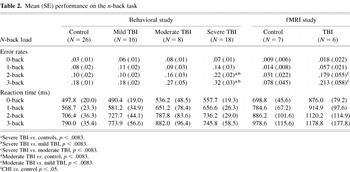
As expected, increased WM load was associated with greater errors, and with more errors at higher load levels in TBI patients compared to controls (Table 2 and Figure 1A). Additionally, greater TBI severity was associated with greater error rates, particularly at higher load levels. These observations were statistically confirmed by significant linear [F(1,64) = 164.84, p < .0001] and quadratic [F(1,64) = 5.48, p < .025] trends over load and a significant interaction of severity with the linear trend over load [F(3,64) = 6.09, p < .001]. There was also a significant main effect of severity [F(3,64) = 7.48, p < .0002] reflecting an increasing linear trend for greater error rates overall with increasing TBI severity. Follow-up group-wise contrasts using Bonferroni-corrected comparisons at each load level (critical p < .0083) revealed that the moderate and severe groups differed significantly from the control and mild TBI groups only at the 2- and 3-back levels of WM load. Correct-trial RTs similarly increased with increasing load [linear trend over load: F(1,64) = 131.24, p < .0001; cubic trend over load: F(1,64) = 4.99, p < .025], but did not significantly differ as a function of severity, either as a main effect or interaction (ps > .17).
Mean (SE) performance on the n-back task


(A) Mean error rates and reaction times for the zero- through 3-back loads on the n-back working memory task for TBI patients and healthy comparison subjects. (B) Mean error rates and reaction times as a function of trial type on the n-back task for TBI patients and healthy comparison subjects. Bars represent ± 1 SE.
Correlations between errors and RTs assessed the presence of speed-accuracy trade-offs and were conducted for all groups separately and combined, collapsed across load. There were no significant correlations for any comparison [rs ≤ −.21, ps > .30]. Thus, speed–accuracy trade-offs likely do not play a role in the pattern of findings described above.
Finally, the four groups did not differ in the number of responses overall in that they showed an equal proportion of non-responses across load levels (p > .10) suggesting that inattention or lack of behavioral engagement likely does not account for the group-related performance differences. Mean proportion of non-responses were: controls: .25 ± .08; mild TBI: .19 ± .06; moderate TBI: 1.8 ± 1.00; and severe TBI: 1.6 ± .63.
Analysis of Trial Type
We examined patterns of behavioral responding across different trial types to potentially illuminate component-process deficits, as discussed by Perlstein et al. (2001, 2003b). Specifically, within the 1- through 3-back levels of the task, there are three different trial types: targets, nontargets, and foils. Foils are nontarget repeats within the response set (e.g., 1-back match on the 2-back task, 2-back match on the 1-back task, etc); nonfoil trials are nontarget, nonrepeat trials. We compared error rates and RTs for the three trial types to determine if foils were associated with interference—greater errors and longer RTs to foils than nonfoils—to determine if all groups showed the same pattern. Such a pattern would be consistent with the hypothesis that all groups adequately maintain trace representations of stimulus identity, and that the observed WM deficit in moderate-to-severe TBI reflects impaired associative decision processes in WM, such as updating or temporal sequencing.
TBI patients showed a pattern of errors and RTs that paralleled the pattern shown by controls; that is, more errors on foil and target trials compared to nontarget, nonfoil trials (Figure 1B). Analysis of trend over trial type revealed a significant linear [F(1,64) = 4.12, p < .05] and quadratic [F(1,64) = 181.90, p < .0001] components, reflecting more errors to foil than target trials and more errors to foil and target compared to nontarget trials, respectively. Main effects of severity [F(3,64) = 7.15, p < .001] reflected increasing errors overall with increasing severity, and an interaction of severity with the quadratic trend over trial type [F(3,64) = 3.42, p < .055] reflected greater foil and target errors in the moderate and severe TBI groups compared to mild TBI and control groups. Regarding RT, all groups showed longer RTs to foil trials than to target and nonfoil, nontarget trials, indicating the foils resulted in RT interference. Trend analyses of RT yielded significant linear [F(1,64) = 64.00, p < .0001] and quadratic components [F(1,64) = 100.22, p < .0001]. There were no significant effects involving severity. The linear trend reflects the longer RTs to foil than target trials, and the quadratic effect reflect the longer RTs to foil and target compared to nontarget trials. There were no significant main effects or interactions involving group. Thus, the overall pattern of response as a function of trial type in both controls and TBI patients is consistent with the hypothesis that TBI patients adequately maintain trace representations of stimuli in WM, but are impaired in a more “executive” or temporal sequencing or tagging operation.
N-Back Task Performance— fMRI Study
Behavioral data for the subset of participants who participated in both the behavioral and fMRI sessions largely paralleled the pattern of findings described above (Table 2). Behavioral data for only six of the seven TBI patients were available due to technical difficulties acquiring one participant's behavioral data. For error rates, there was a significant linear trend over load [F(1,11) = 21.63, p < .001], a significant main effect of group [F(1,11) = 7.48, p < .02], and a significant Group × Load interaction [F(3,33) = 4.27, p < .025, Huynh-Feldt corrected], reflecting a significant interaction of group with the linear trend over load [F(1,11) = 5.80, p < .035]. Follow-up group-wise comparisons at each load level revealed that the two groups differed significantly (ps < .036) only at the 2- and 3-back load levels, with a trend toward significance at the 1-back level (p < .072). Correct-trial RT data also paralleled the pattern observed in the behavioral study. There was a significant linear trend over load [F(1,11) = 19.61 p < .0001]. A trend toward longer RTs in the TBI than control subjects did not reach statistical significance [F(1,11) = 1.86, p = .20], nor did the group interaction with the linear trend over load [F(1,11) = 0.14, p > .70].
Cross-Study Comparison of N-Back Performance
We next examined the accuracy data for the subset of participants who completed both the behavioral and fMRI sessions using a 2 (group) × 4 (load) × 2 (session) ANOVA. Data for only 6 participants from each group were available for this comparison. Analyses yielded significant main effects of group [F(1,10) = 10.52, p < .01], session [F(1,10) = 6.14, p < .05], and load [F(3,30) = 29.05, p < .0001], as well as a significant Group × Load interaction [F(3,30) = 3.87, p < .025]. The session effect reflected greater error rates during the behavioral (.102 ± .013) than fMRI (.073 ± .015) session, as might be predicted based on the more rapid stimulation rate and limited time for temporal sequencing of stimuli in WM. The other effects paralleled those described above, with increased errors as a function of increasing WM load, and greater errors in TBI patients compared to controls at higher levels of WM load. Thus, while TBI patients performed more poorly at the faster stimulation rate, the rate of stimulus presentation did not alter the pattern of group-related load effects.
Age and NAART Scores as Confounding Variables3 We chose not to conduct covariance analyses
using age or NAART score in light of discussions regarding its
appropriateness to control for differences between intact
groups (Adams et al., 1985; Miller & Chapman, 2001; Strauss, 2001).
We chose not to conduct covariance analyses using age or NAART score in light of discussions regarding its appropriateness to control for differences between intact groups (Adams et al., 1985; Miller & Chapman, 2001; Strauss, 2001).
Differences in age and NAART scores between groups in the behavioral study represent confounding variables. In our analysis, age correlated significantly with the variables that significantly differentiated the groups [error rates on the 2- and 3-back load levels: r(66) ≥ .33, p < .007]. Thus, we re-analyzed n-back data for a subset of age-matched subjects after excluding the youngest mild TBI participant and three oldest severe TBI participants. Exclusion of these participants eliminated the age differences between groups, and the matching on education remained. Results of these analyses yielded a pattern of statistically significant effects that was unchanged from the pattern described above. NAART scores similarly correlated with n-back error rates on the zero-, 2- and 3-back loads [r(64) ≥ −.29, p < .02]. Reanalysis of the error-rate data after exclusion of the three severe TBI patients who contributed most to this difference yielded an identical pattern of statistically significant effects. Thus, it is unlikely that age and NAART differences accounted for the WM deficit observed in moderate-to-severe TBI patients.
Functional Neuroimaging Data
Between-groups analysis
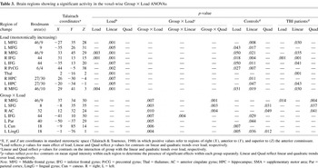
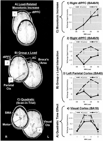
Voxel-wise Group × Load ANOVAs (Table 3) revealed significant monotonically increasing effects of WM load in a network of regions shown previously to be engaged by verbally-mediated WM (Figure 2A), primarily including superior and inferior regions of the PFC bilaterally, as illustrated in the signal intensity plots of Figure 2. The main effect of group revealed that activity in a region of the posterior parietal cortex (Brodmann Area, BA 7; Talairach coordinates: x = −18, y = −72, z = 42; p < .008) was greater in patients than controls, but this region did not differ as a function of WM load. More importantly, a number of regions showed significant Group × Load interaction (Figure 2B), including the right dlPFC (BA 46/9), left Broca's area (BA 44) and parietal cortex (BA 40), and the anterior cingulate gyrus (BA 32). These regions showed lesser magnitude increases with increased WM load in patients compared to controls, or non-linear load-related changes in fMRI signal intensity in TBI patients (Figure 2).
Brain regions showing a significant activity in the voxel-wise Group × Load ANOVAs


Functional magnetic resonance (fMRI) images showing representative regions for the grouped data that exhibited (A) main effects of working memory load, (B) Group × Load interactions and (C) scan-within-trial effects. Figures reflect overlays of thresholded group-wise statistical images onto the reference image transformed to standard Talairach space. Plots to the right reflect the mean percent change in signal intensity across all suprathreshold voxels within the specified region (signified by the numbered box) for TBI patients and healthy comparison subjects as the percent change in signal intensity from the zero-back load (A and B), and scan 1 (C). Scan-in-trial on the absissa in C reflects increments of 2.5 s, reflecting the duration of the repetition time (TR) or duration to acquire a volume of functional images (i.e., 33 slices). The onset of scan 1 was time-locked to the stimulus onset of each trial, and acquisition of the four scans spanned the duration of the 10-s stimulus onset asynchrony. Bars represent ± 1 SE.
Examination of regions exhibiting transient responses associated with stimulus encoding- and button press response-related processes (Figure 2C) revealed significant activity in regions of the supplementary motor area (SMA; BA 6), bilateral motor cortex (BA 3 and 4) and thalamus, and visual cortex (BA 18). Follow-up contrasts on activity in these regions showed that the two groups did not differ (Table 4), demonstrating that the TBI patients, while showing a number of regions that fail to activate properly as a function of WM load, do activate regions associated with visual encoding and dominant-hand motor responses.
Brain regions showing a significant activity in the voxel-wise scan-in-trial-related effects

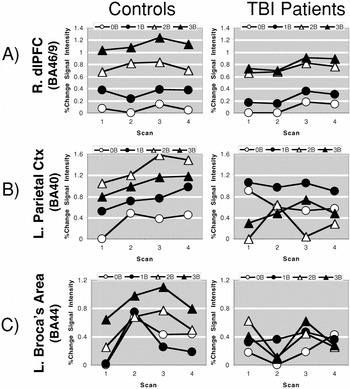
Finally, the event-related design of the fMRI acquisition enables us to examine the temporal course of the hemodynamic response during the course of trials. Consequently, we examined several additional patterns of interest, beyond load- and group-related effects and interactions. Specifically, since active maintenance and manipulation in WM can be manifest as greater intensity or more prolonged hemodynamic response as a function of increased WM load (see, e.g., Cohen et al., 1997; Perlstein et al., 2003b), we also examined the temporal course of activity (i.e., time-in-trial) in significant task-related clusters described above. Several findings converge with those reported by Cohen et al. (1997). First, activity in the region of the right dlPFC, which did not show differential activity as a function of group, increased monotonically with increasing WM load, as described above. Moreover, this activity was sustained over the course of trials in both groups (Figure 3A). In contrast, as shown in Figures 2B and 2C, illustrating signal intensity changes in the left Broca's area and the left parietal region that showed a Group × Load interaction, respectively, controls showed load-related activity that was sustained over the course of trials at higher levels of load, while returning toward baseline at lower load levels. TBI patients, in contrast, showed activity in both regions that was more transient in nature, and which did not track increasing load with increasing levels of activation.

Plots showing the percent change in signal intensity from the lowest value across load and scan-in-trial conditions as a function of working memory load and scan-within-trial for the TBI patients and healthy comparison subjects. The onset of Scan 1 was time-locked to the stimulus onset of each trial, and acquisition of the four scans spanned the duration of the 10-s stimulus onset asynchrony.
Within-group analysis
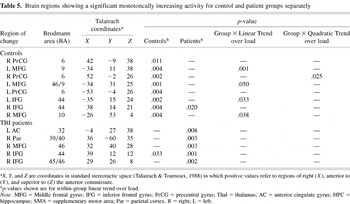
Voxel-wise tests of monotonically-increasing activity assessed the nature of load-related activity for the two groups separately. Results (Table 5; Figure 4) indicate a clear prefrontal laterality effect: Controls show monotonically-increasing activity in the left dlPFC, TBI patients show increasing activity in the right dlPFC. Furthermore, controls activated bilateral inferior frontal gyri (IFG), while TBI patients activated the IFG only on the right side. More generally, patients showed fewer regions of suprathreshold activation than controls.
Brain regions showing a significant monotoncally increasing activity for control and patient groups separately


Functional magnetic resonance images for grouped data showing representative regions that exhibited monotonically increasing activity as a function of increased working memory load separately for the TBI patients and healthy comparison subjects. Z-value indicates relative position to the anterior commissure-posterior commissure line in standardized Talairach space. dlPFC = dorsolateral prefrontal cortex; IFG = inferior frontal gyrus.
DISCUSSION
The pattern of findings that emerges from the behavioral study is that individuals who have sustained a moderate-to-severe TBI exhibit a load-related impairment in WM relative to demographically matched, neurologically-normal comparison and mild TBI subjects. This impairment, reflected in performance accuracy on the n-back task, was greater at higher levels of WM load, and more severe TBI was associated with greater impairment on both versions of the task. The small subset of moderate-to-severe TBI participants who also underwent fMRI scanning showed a high degree of cross-session consistency on task performance. These findings clearly suggest that chronic moderate-to-severe TBI is associated with impaired WM functioning in a dose–response or load-dependent fashion, similar to other patient groups with putative PFC dysfunction (e.g., schizophrenia; Perlstein et al., 2001, 2003b).
The present findings of WM impairment in patients with moderate-to-severe TBI extend previous findings suggesting the presence of WM deficits in TBI patients. Much of this previous work has used dual-task paradigms to assess WM function, demonstrating disproportionately greater dual-task performance decrements in TBI patients compared to healthy comparison subjects, particularly when the dependent variable was reaction time (e.g., McDowell et al., 1997). However, as noted in the Introduction, dual-task paradigms require a task-switching component that extends beyond active maintenance and manipulation of stimulus representations in WM and, therefore, tap into an additional set of component processes. Additionally, many of the dual-task paradigms that have been employed have also been associated with group-related performance differences on the tasks when performed individually (e.g., Leclercq et al., 2000; McDowell et al., 1997). Such differences in “baseline” performance complicate the interpretation of findings from the dual task paradigms and make it difficult to discriminate generalized from process-specific impairments.4
The issue of “baseline” performance difference and the use of difference scores in the presence of these differences have been discussed at length by Chapman and colleagues (Chapman & Chapman, 1989; Miller & Chapman, 2001).
Results of the fMRI study, which compared performance of a small subset of moderate-to-severe TBI patients to healthy comparison subjects on the n-back task, largely replicate findings from previous studies using a similar paradigm in healthy subjects (Braver et al., 1997; Cohen et al., 1997; Perlstein et al., 2001, 2003b). These studies have demonstrated load-related increases in activity in a number of brain regions that support WM processes (e.g., dlPFC, Broca's area and parietal cortexes). More central to the aims of the current research, however, was the finding that moderate-to-severe TBI patients show altered load-related activity in each of these WM-related regions. This finding contrasts with findings from our previous studies of patients with putative PFC dysfunction who evidence WM deficits assessed by the n-back task and concomitantly altered brain activity in a very localized fashion (e.g., schizophrenia patients; Perlstein et al., 2001, 2003b). Thus, as might be expected, patients with moderate-to-severe TBI show WM-related alterations in brain activity that are distributed rather than confined to a single focus.
The present fMRI results are, in some respects, comparable to those reported by McAllister et al. (1999, 2001) and Christodoulou et al. (2001) in that they show altered activation in a distributed “network” of WM-related brain regions in patients who have experienced TBI. However, our results differ from these previous findings in several important respects. First, we observed altered activity in patients in the presence of task-related performance differences, in contrast to the studies by McAllister et al. Second, the McAllister et al. studies, which employed an auditory version of the n-back task in patients with acute mild TBI, demonstrated greater increases in TBI compared to control subjects from the 1- to 2-back conditions in the right dlPFC and parietal regions, in contrast to the present finding of generally lesser magnitude load-related increases in patients compared to controls in all differentially-affected regions. The reasons for these differences are uncertain; however, in the McAllister et al. studies, patients and controls did not significantly differ in task performance at any load level, and their patients were individuals with acute mild TBI. On the other hand, Christodoulou et al. (2001), who examined brain activation concomitants of WM function in chronic patients with moderate-to-severe TBI using a modified version of the PASAT, observed that TBI patients performed more poorly than controls. These authors showed that while TBI patients generally activated similar regions during task performance relative to controls, they also displayed a more regionally dispersed and right-lateralized pattern of activation relative to control. Our results, at least with respect to the analyses of the control and TBI groups separately, showed that the two groups activated a rather different set of load-related regions, and that TBI patients showed greater activation of right PFC and controls showed greater activation of left PFC.
What cognitive mechanism(s) may account for the observed WM impairment in moderate-to-severe TBI? It is unlikely that impairment of a single cognitive mechanism can account for the observed WM dysfunction given the heterogeneity of brain injury in this patient group. However, the current findings suggest some possibilities when considered in light of theories of WM and component processes required for n-back task performance, including active maintenance of stimulus representations, coding of sequential order, updating, etc. The detailed breakdown of task performance as a function of trial type (i.e., foils, nonfoils, and targets) suggests a potential deficit in associative processes—coding or maintaining sequential order information—rather than processing speed or simple active maintenance of representations within WM. Additional support for this interpretation comes from the observation that the group differences in behavioral performance emerged in the 2- and 3-back load levels, the load levels that require maintaining the target set of n-back stimulus representations and coding and maintaining temporal position in the sequence. In contrast, neither the zero nor 1-back levels require sequencing operations, since only a single letter must be kept in mind at any given time. Results of the fMRI study are consistent with this interpretation. Specifically, a region of the dlPFC that exhibited sustained, load-sensitive and presumably active maintenance processes, did not differ between the groups; both the control and TBI groups showed sustained activity that increased with increasing load, and that was not affected by time in trial. However, activity in Broca's area and parietal cortex was more transient in TBI patients, and did not show systematic load-related increases in activity.
An alternative but not mutually exclusive interpretation of the observed impairment in moderate-to-severe TBI patients is that differences in behavioral performance and brain activation reflect, in part, generalized, rather than specific deficits. Importantly, the four groups did not differ in the overall rate of nonresponding on the n-back task, or on error rates at the zero and 1-back load levels, suggesting that moderate and severe TBI patients were at least minimally engaged in the task and sustained sufficient attention and motivation in conditions where minimal effort was required. This finding, however, does not rule out the possibility that the greater error rates of TBI patients in the 2- and 3-back conditions may be due to a generalized effect of task difficulty. That is, TBI patients' performance may decrease relative to control participants as task difficulty increases, independent of WM-related processes. Indeed, general factors such as poor concentration, lack of effort, frustration or anxiety are more likely to result in decreased performance as task difficulty increases. For instance, a review by Humphreys and Revelle (1984) showed than anxiety increases performance for easy tasks and decreases performance for difficult tasks. In our study, severe TBI patients demonstrated greater state anxiety than mild TBI patients, and they may have felt more anxious or frustrated under the more difficult 2- and 3-load conditions. Similarly, limited attention and concentration are frequent symptoms after moderate and severe TBI (McKinlay et al., 1981; Ponsford et al., 1995). The more effortful 2- and 3-load conditions may have simply exceeded moderate and severe TBI patients' attention abilities or capacity (Callicott et al., 1999). Future research using conditions equated for task difficulty is needed to disentangle the differential contributions of generalized versus specific deficits (Chapman & Chapman, 1989; Miller & Chapman, 2001) in TBI populations.
How might the fMRI findings be interpreted? Unfortunately, the findings from the present study, and from the three previously published TBI-related fMRI studies of WM (Christodoulou et al., 2001; McAllister et al., 1999, 2001) are complex and do not give rise to completely parsimonious explanations. We discuss potential limitations and interpretational conundrums inherent in functional neuroimaging studies of TBI below. However, findings from the current fMRI study are consistent with the hypothesis that patients with moderate-to-severe TBI are impaired in the executive or strategic aspects of task performance. Specifically, analyses of the temporal response function demonstrated that controls showed activity in Broca's area that increased monotonically with increased WM load and, furthermore, that at higher load levels this activity was more sustained during the course of trials, but returned toward baseline at lower load levels. Cohen et al. (1997) suggested that this pattern of activity might reflect the invocation of verbally mediated rehearsal mechanisms that aid in actively maintaining and sequencing stimulus representations. In light of this view, the finding that TBI patients showed a pattern of Broca's area activity (and parietal activity) which did not follow a meaningful pattern both with respect to load and time-in-trial, suggests that TBI patients may be deficient in strategic aspects of task performance, such as subvocal rehearsal. Finally, our sample of moderate-to-severe TBI patients and controls showed comparable levels of activation in regions associated with visual encoding and motor response-related processes, suggesting that alterations in WM-related activation are not due to a generalized inability to activate cortex.
Despite evidence provided by our study that is consistent with the predictions outlined in the Introduction, limitations and alternative explanations require discussion. As discussed in the Introduction, one potential problem in interpreting differences in task performance between TBI patients and healthy control concerns whether the impaired performance reflects a nonspecific deficit in patients, such as reduced processing speed (Ferraro, 1996; Salthouse, 1996), generalized inattention, or lack of behavioral engagement. It is well known that TBI patients are generally slower in performing many tasks (Ferraro, 1996), and often show generalized inattention (e.g., Miller, 1970). Importantly, the TBI and control groups did not differ in the overall rate of nonresponding on the n-back task, or on error rates on the zero- and 1-back load levels, suggesting that lack of engagement in the tasks was not a factor in producing the pattern of results observed. Furthermore, the TBI and control groups did not differ in RTs, and no group showed evidence of speed-accuracy trade-offs. The finding of impaired performance on the n-back task in moderate-to-severe TBI patients is likely not due to reduced processing speed or time pressure. Such a deficit might be involved in the temporal sequencing of stimulus representations which requires time within the intertrial interval. While the behavioral study presented stimuli at a rate of 1 per 4 s, the fMRI study had a significantly longer stimulus interval (1 stimulus per 10 s), and is likely to be adequate for the sequencing operations to be performed with considerably reduced time pressure. Although the moderate-to-severe TBI patients performed more poorly than controls in the behavioral study with the more rapid stimulus delivery rate, they also performed more poorly than controls in the fMRI study with the slower stimulus rate. The absence of a significant interaction of session (i.e., stimulus rate) with group and/or load suggests that the increased difficulty associated with the increased stimulation rate between the two versions of the task suggests that the behavioral and imaging versions of the task are tapping similar cognitive phenomena.
There are also several issues of relevance regarding the patient sample in the present study. First, chronicity of TBI patients was confounded with injury severity. That is, both mild and moderate TBI patients were, on average, tested at a shorter post-injury period compared to severe TBI patients. However, re-analysis of the data following removal of the “acute” (i.e., post-injury < 1 year) patients yielded an identical pattern of statistically significant results to that described for the full patient sample. This finding suggests that the observed deficits were relatively stable and persistent in the chronic moderate-to-severely injured patients. Second, we did not have access to neuroradiological findings for the majority of our patient sample and, therefore, could not determine relations between objective neurological injury and behavioral performance. It is likely that the more severe TBI patients had focal in addition to diffuse injury, whereas the more mild TBI patients likely had more diffuse than focal injury. Thus, relationships between neurological insult, symptomatology and task performance could not be determined. Third, the issue of injury severity classification must be considered in light of the necessity to generalize findings across studies. There is considerable variability in the literature regarding severity classification, particularly regarding moderate TBI severity, and the variables employed for establishing severity criteria also differ across many studies. For the current study, for example, we did not have all three classification variables—initial GCS scores, duration of LOC, or PTA—for all patients.
Regarding potential limitations of the fMRI study, several considerations must be kept in mind. First, fMRI in TBI is subject to a number of inherent interpretational challenges. Observed differences in activation between TBI and control groups could be due to several factors that are not directly related to impairments in task performance. These include (1) possible fundamental anomalies in cerebral vasculature in patients with TBI, (2) some alteration in the relationship between neuronal activity and the blood flow response induced by the brain injury, (3) alterations in apparent blood flow or volume due to alterations in the ratio of gray to white matter resulting in cortical atrophy (partial volume effects), and/or (4) some unanticipated artifact of experimental design (Price & Friston, 1999, 2001). The existence of cortical contusions or hematoma may also play a role in giving rise to group differences, due to magnetic susceptibility effects that may give rise to inhomogeneities of signal variance. Heterogeneity of potential injury sites and the possibility of DAI also may contribute to the appearance of functional activation differences between TBI patients and controls. Finally, the present imaging results are based on a small sample size and, therefore, must be considered cautiously, particularly with respect to generalization to other TBI patient samples.
In conclusion, our findings strongly indicate that patients with moderate-to-severe chronic TBI exhibit impairments in WM. Decomposition of task-performance components suggests that this impairment may be due to more executive, associative or strategic components of WM, such as coding of temporal order and/or verbally mediated rehearsal processes, rather than processes involved in the active maintenance of stimulus representations per se. Additionally, patients showed an impaired ability to track WM load in brain activity in several load-sensitive (i.e., WM-related) regions, suggesting that TBI is associated with distributed rather than focal impairments in brain function. Whether the observed TBI-related impairment in WM reflects specific or more generalized deficit is uncertain. However, the present results suggest that generalized inattention or lack of task engagement do not account for the observed differences. Ongoing studies in our laboratory are aimed at decomposing component processes of prefrontally-mediated cognitive functions to determine what aspects of executive function may mediate TBI-related cognitive dysfunction in TBI, including studies that examine the effects of chronicity and recovery, both behaviorally and neurally, aimed at more closely linking the proposed WM deficits to symptomatic state.
ACKNOWLEDGMENTS
Supported by grants from the McKnight Brain Research Grant Program, the Brain and Spinal Cord Injury Research Trust Fund, and the National Institute of Mental Health (K01 MH01857) to W.M.P. We thank Kay Waid-Ebbs for her assistance with patient recruitment, Sarah LageMan for her assistance in fMRI data acquisition, and Dr. Jane Mathias for providing us with the self- and other-rating versions of the revised Neurobehavior Rating Scales. Portions of this research were presented at the 31st Annual Meeting of the International Neuropsychological Society and the 32nd Annual Meetings of the Society for Neuroscience.
References
REFERENCES
Adams, K.M.,
Brown, G.G., &
Grant, I.
(1985).
Analysis of covariance as a remedy for demographic mismatch of
research subject groups: Some sobering simulations.
Journal of Clinical and Experimental Neuropsychology,
7,
445–462.CrossRefGoogle Scholar
Adams, J.H.,
Scott, G.,
Parker, L.S.,
Graham, D.I., &
Doyle, D.
(1980).
The contusion index: A quantitative approach to cerebral contusions
in head injury.
Neuropathology and Applied Neurobiology,
6,
319–324.CrossRefGoogle Scholar
American Congress of Rehabilitative Medicine.
(1993).
Definition of mild traumatic brain injury.
Journal of Head Trauma Rehabilitation,
8,
86–87.Google Scholar
Baddeley, A.D.
(1986).
Working memory.
New York:
Oxford University Press.
Beck, A.T.,
Ward, C.H.,
Mendelson, M.,
Mock, J.E., &
Erbaugh, J.K.
(1961).
An inventory for measuring depression.
Archives of General Psychiatry,
4,
561–571.CrossRefGoogle Scholar
Bigler, E.D.
(1990).
Neuropathology of traumatic brain injury. In
E.D. Bigler (Ed.),
Traumatic Brain Injury (pp.
13–49).
Austin, TX:
Pro-ed.
Blair, J.R. &
Spreen, O.
(1989).
Predicting premorbid IQ: A revision of the National Adult Reading
Test.
Clinical Neuropsychologist,
3,
129–136.CrossRefGoogle Scholar
Bond, M.R.,
(1986).
Neurobehavioral sequelae of closed head injury. In
I. Grant &
K.M. Adams (Eds.),
Neuropsychological Assessment of Neuropsychological Disorders (pp.
347–373).
New York:
Oxford University Press.
Braver, T.S.,
Cohen, J.D.,
Nystrom, L.E.,
Jonides, J.,
Smith, E.E., &
Noll, D.C.
(1997).
A parametric study of prefrontal cortex involvement in human
working memory.
Neuroimage,
5,
49–62.CrossRefGoogle Scholar
Braver, S.L. &
Sheets, V.L.
(1993).
Monotonic hypothesis in multiple group designs: A Monte Carlo
study.
Psychological Bulletin,
113,
379–395.CrossRefGoogle Scholar
Bublak, P.,
Schubert, T.,
Matthes-von Cramon, G.,
and von Cramon, D.Y.
(2000).
Differential demands on working memory for guiding a simple action
sequence: Evidence from closed-head-injured subjects.
Journal of Clinical and Experimental Neuropsychology,
22,
176–189.CrossRefGoogle Scholar
Burgess, P.W.
(1997).
Theory and methodology in executive function research. In
P. Rabbitt (Ed.),
Methodology of frontal and executive function (pp.
81–116).
Hove, UK:
Psychology Press.
Callicott, J.J.,
Mattay, V.S.,
Bertolino, A.,
Finn, K.,
Coppola, R.,
Frank, J.A.,
Goldberg, T.E., &
Weinberger, D.R.
(1999).
Physiological characteristics of capacity constraints in working
memory as revealed by functional MRI.
Cerebral Cortex,
9,
20–26.CrossRefGoogle Scholar
Chapman, L.J. &
Chapman, J.P.
(1989).
Strategies for resolving the heterogeneity of schizophrenia and
their relatives using cognitive measures.
Journal of Abnormal Psychology,
98,
357–366.CrossRefGoogle Scholar
Christodoulou, C.,
DeLuca, J.,
Ricker, J.H.,
Madigan, N.K.,
Bly, B.M.,
Lange, G.,
Kalnin, A.J.,
Liu, W-C.,
Steffner, J.,
Diamond, B.J., &
Ni, A.C.
(2001).
Functional magnetic resonance imaging of working memory impairment
after traumatic brain injury.
Journal of Neurology, Neurosurgery and Psychiatry,
71,
161–168.CrossRefGoogle Scholar
Cicerone, K.D.
(1996).
Attention deficits and dual task demands after mild traumatic brain
injury.
Brain Injury,
10,
79–89.CrossRefGoogle Scholar
Cohen, J.D.,
Forman, S.D.,
Braver, T.S.,
Casey, B.J.,
Servan-Schreiber, D., &
Noll, D.C.
(1994).
Activation of the prefrontal cortex in a nonspatial working memory
task with functional MRI.
Human Brain Mapping,
1,
293–304.CrossRefGoogle Scholar
Cohen, J.D.,
Macwhinney, B.,
Flatt, M.R., &
Provost, J.
(1993).
PsyScope: A new graphic interactive environment for designing
psychology experiments.
Behavioral Research Methods, Instruments and Computers,
25,
257–271.CrossRefGoogle Scholar
Cohen, J.D.,
Perlstein, W.M.,
Braver, T.S.,
Nystrom, L.E.,
Noll, D.C.,
Jonides, J., &
Smith, E.E.
(1997).
Temporal dynamics of brain activation during a working memory task.
Nature,
386,
604–608.CrossRefGoogle Scholar
Courtney, S.M.,
Ungerleider, L.G.,
Keil, K., &
Haxby, J.V.
(1997).
Transient and sustained activity in a distributed neural system for
human working memory.
Nature,
386,
608–612.CrossRefGoogle Scholar
Cox, R.W.
(1996).
AFNI: Software for analysis and visualization of functional
magnetic resonance neuroimages.
Computational and Biomedical Research,
29,
162–173.CrossRefGoogle Scholar
D'Esposito, M.,
Detre, J.A.,
Alsop, D.C.,
Shin, R.K.,
Atlas, S., &
Grossman, M.
(1995).
The neural basis of the central executive system of working memory.
Nature,
378,
279–281.CrossRefGoogle Scholar
Dreher, J.-C. &
Grafman, J.
(2003).
Dissociating the roles of the rostral anterior cingulate and the
lateral prefrontal cortices in performing two tasks simultaneously or
successively.
Cerebral Cortex,
13,
329–339.CrossRefGoogle Scholar
Ferraro, F.R.
(1996).
Cognitive slowing in closed-head injury.
Brain and Cognition,
32,
429–440.CrossRefGoogle Scholar
Forman, S.D.,
Cohen, J.D.,
Fitzgerald, M.,
Eddy, W.F.,
Mintun, M.A., &
Noll, D.C.
(1995).
Improved assessment of significant activation in functional
magnetic resonance imaging (fMRI): Use of a cluster-size
threshold.
Magnetic Resonance in Medicine,
33,
636–647.CrossRefGoogle Scholar
Goldman-Rakic, P.
(1987).
Circuitry of primate prefrontal cortex and regulation of behavior
by representational memory (pp.
373–417). In
F. Plum &
V. Mountcastle (Eds.),
Handbook of Physiology—The Nervous System V.
Bethesda, MD:
American Physiological Society.
Greve, K.W.,
Love, J.M.,
Sherwin, E.,
Mathias, C.W.,
Ramzinski, P., &
Levy, J.
(2002).
Wisconsin Card Sorting Test in chronic severe traumatic brain
injury: Factor structure and performance subgroups.
Brain Injury,
16,
29–40.CrossRefGoogle Scholar
Gronwall, D.
(1986).
Rehabilitation programs for patients with mild head injury:
Components, problems, and evaluation.
Journal of Head Trauma and Rehabilitation,
1,
153–162.Google Scholar
Humphreys, M.S. &
Revelle, W.
(1984).
Personality, motivation, and performance: a theory of the
relationship between individual differences and information
processing.
Psychological Review,
91,
153–184.CrossRefGoogle Scholar
Huynh, H. &
Feldt, L.S.
(1976).
Estimation of the box correction for degrees of freedom from sample
data in the randomized block and split-plot designs.
Journal of Educational Statistics,
1,
69–82.CrossRefGoogle Scholar
Jonides, J. &
Smith, E.E.
(1997).
The architecture of working memory. In
M.D. Rugg (Ed.),
Cognitive neuroscience (pp.
243–276).
Cambridge, MA:
MIT Press.
Keppel, G.
(1982).
Design and analysis: A Researchers Handbook
(2nd ed.).
Englewood Cliffs, NJ:
Prentice Hall.
Leclercq, M.,
Couillet, J.,
Azouvi, P.,
Marlier, N.,
Martin, Y.,
Strypstein, E., &
Rousseaux, M.
(2000).
Dual task performance after severe diffuse traumatic brain injury
or vascular prefrontal damage.
Journal of Experimental and Clinical Neuropsychology,
22,
339–350.CrossRefGoogle Scholar
Levin, H.S.,
Gary, H.,
Eisenberg, H.,
Ruff, R.,
Barth, J.,
Kreutzer, J.,
High, W.,
Portman, S.,
Foulkes, M., &
Jane, J.
(1990).
Neurobehavioral outcome 1-year after severe head injury: Experience
of the Traumatic Coma Data Bank.
Journal of Neurosurgery,
73,
699–709.CrossRefGoogle Scholar
Lezak, M.D.
(1995).
Neuropsychological assessment
(3rd ed.).
New York:
Oxford Press.
McAllister, T.W.,
Sparling, M.B.,
Flashman, L.A.,
Guerin, S.J.,
Mamourian, A.C., &
Saykin, A.J.
(2001).
Differential working memory load effects after mild traumatic brain
injury.
Neuroimage,
14,
1004–1012.CrossRefGoogle Scholar
McAllister, T.W.,
Saykin, A.J.,
Flashman, L.A.,
Sparling, M.B.,
Johnson, S.C.,
Guerin, S.J.,
Mamourian, A.C.,
Weaver, J.B., &
Yanofsky, N.
(1999).
Brain activation during working memory 1 month after mild traumatic
brain injury: A functional MRI study.
Neurology,
53,
1300–1308.CrossRefGoogle Scholar
McDowell, S.,
Whyte, J., &
D'Esposito, M.
(1997).
Working memory impairments in traumatic brain injury: Evidence from
a dual-task paradigm.
Neuropsychologia,
35,
1341–1353.CrossRefGoogle Scholar
McDowell, S.,
Whyte, J., &
D'Esposito, M.
(1998).
Differential effect of a dopaminergic agonist on prefrontal
function in traumatic brain injury patients.
Brain,
121,
1155–1164.CrossRefGoogle Scholar
McKinlay, W.W.,
Brooks, D.N.,
Bond, M.R.,
Marinage, D.P., &
Marshall, M.M.
(1981).
The short-term outcome of severe blunt head injury as reported by
relatives of the injured persons.
Journal of Neurology, Neurosurgery, and Psychiatry,
44,
527–533.CrossRefGoogle Scholar
Miller, E.
(1970).
Simple and choice reaction-time following severe head injury.
Cortex,
6,
121–127.CrossRefGoogle Scholar
Miller, G.A. &
Chapman, J.P.
(2001).
Commentary on two articles concerning generalized and specific
cognitive deficits.
Journal of Abnormal Psychology,
110,
31–39.Google Scholar
Miyake, A.,
Friedman, N.P.,
Emerson, M.J.,
Witzki, A.H.,
Howerter, A., &
Wager, T.
(2000).
The unity and diversity of executive functions and their
contributions to complex “frontal lobe” tasks: A latent
variable analysis.
Cognitive Psychology,
41,
49–100.CrossRefGoogle Scholar
Miyake, A.,
Emerson, M.J., &
Friedman, N.P.
(2000).
Assessment of executive functions in clinical settings: Problems
and recommendations.
Seminars in Speech and Language,
21,
169–183CrossRefGoogle Scholar
Noll, D.C.,
Cohen, J.D.,
Meyer, C.H., &
Schneider, W.
(1995).
Spiral K-space MR imaging of cortical activity.
Journal of Magnetic Resonance Imaging,
5,
49–56.CrossRefGoogle Scholar
Norman, D.A. &
Shallice, T.
(1986).
Attention to action: Willed and automatic control of behavior. In
R.J. Davidson,
G.E. Schwartz, &
D. Shapiro (Eds.),
Consciousness and self-regulation: Advances in research and
theory (Vol. 4, pp.
1–18).
New York:
Plenum.
Park, N.W.,
Moscovitch, M., &
Robertson, I.H.
(1999).
Divided attention impairments after traumatic brain injury.
Neuropsychologia,
37,
1119–1133.CrossRefGoogle Scholar
Pashler, H.
(1994).
Dual-task interference in simple tasks: Data and theory.
Psychological Bulletin,
116,
220–244.CrossRefGoogle Scholar
Perlstein, W.M.,
Carter, C.S.,
Noll, D.C., &
Cohen, J.D.
(2001).
Relation of prefrontal cortex dysfunction to working memory and
symptoms in schizophrenia.
American Journal of Psychiatry,
158,
1105–1113.CrossRefGoogle Scholar
Perlstein, W.M.,
Cole, M.A.,
Larson, M.,
Kelly, K.,
Seignourel, P., &
Keil, A.
(2003a).
Steady-state visual evoked potentials reveal frontally-mediated
working memory activity in humans. Steady-state visual evoked
potentials reveal frontally-mediated working memory activity in
humans.
Neuroscience Letters,
342,
191–195.Google Scholar
Perlstein, W.M.,
Dixit, N.K.,
Carter, C.S.,
Noll, D.C., &
Cohen, J.D.
(2003b).
Prefrontal cortex dysfunction mediates deficits in working memory
and prepotent responding in schizophrenia.
Biological Psychiatry,
53,
25–38.Google Scholar
Phillips, L.H.
(1997).
Do “frontal tests” measure executive function? Issues
of assessment and evidence from fluency tests. In
P. Rabbitt (Ed.),
Methodology of frontal and executive function (pp.
191–213).
Hove, UK:
Psychology Press.
Ponsford, J.L. &
Kinsella, G.
(1992).
Attentional deficits following closed head injury.
Journal of Clinical and Experimental Neuropsychology,
14,
822–838.CrossRefGoogle Scholar
Ponsford, J.L.,
Olver, J.H., &
Curran, C.
(1995).
A profile of outcome: 2 years after traumatic brain injury.
Brain Injury,
9,
1–10.CrossRefGoogle Scholar
Price, C.J. &
Friston, K.J.
(1999).
Scanning patients with tasks they can perform.
Human Brain Mapping,
8,
102–108.3.0.CO;2-J>CrossRefGoogle Scholar
Price, C.J. &
Friston, K.J.
(2001).
Functional neuroimaging of neuropsychologically impaired
patients. In
R. Cabeza &
A. Kingstone (Eds.),
Handbook of functional neuroimaging of cognition (pp.
379–399).
Cambridge, MA:
MIT Press.
Salthouse, T.A.
(1996).
The processing-speed theory of adult age differences in cognition.
Psychological Review,
103,
403–428.CrossRefGoogle Scholar
Shallice, T. &
Burgess, P.
(1996).
The domain of supervisory processes and temporal organization of
behavior. Philosophical Transactions of the Royal Society of London
B, Biological Sciences,
351,
1405–1411.CrossRefGoogle Scholar
Smith, E.E. &
Jonides, J.
(1998).
Neuroimaging analysis of human working memory.
Proceedings of the National Academy of Sciences,
25,
12061–12068.CrossRefGoogle Scholar
Spielberger, C.D.,
Gorsuch, R.L., &
Lushene, R.E.
(1970).
Manual for the state-trait anxiety inventory.
Palo Alto, CA:
Consulting Psychologists Press.
Strauss, M.E.
(2001).
Demonstrating specific cognitive deficits: A psychometric
perspective.
Journal of Abnormal Psychology,
110,
6–14.CrossRefGoogle Scholar
Stuss, D.T. &
Alexander, M.P.
(2000).
Executive functions and the frontal lobes: A conceptual view.
Psychological Research,
63,
289–298.CrossRefGoogle Scholar
Stuss, D.T. &
Levine, B.
(2002).
Adult clinical neuropsychology: Lessons from studies of the frontal
lobes.
Annual Reviews of Psychology,
53,
401–433.CrossRefGoogle Scholar
Szameitat, A.J.,
Schubert, T.,
Müller, K., &
von Cramon, D.Y.
(2002).
Localization of executive functions in dual-task performance with
fMRI.
Journal of Cognitive Neuroscience,
14,
1184–1199.CrossRefGoogle Scholar
Talairach, J. &
Tournoux, P.
(1988).
Co-planar stereotaxic atlas of the human brain.
Stuttgart, Germany:
George Thieme Verlag.
Teasdale, G. &
Jennett, B.
(1974).
Assessment of coma and impaired consciousness: A practical scale.
Lancet,
ii,
81–84.CrossRefGoogle Scholar
Van Zomeren, A.H. &
Brouwer, W.H.
(1987).
Head injury and concepts of attention. In
H.S. Levin,
J. Grafman, &
H. Eisenberg (Eds.),
Neurobehavioral recovery from head injury, pp.
398–415.
New York:
Oxford University Press.
Weinberger, D.R. &
Berman, K.F.
(1996).
Prefrontal function in schizophrenia: Confounds and controversies.
Philosophical Transactions of the Royal Society of London B,
351,
1495–1503.CrossRefGoogle Scholar
Wiegner, S. &
Donders, J.
(1999).
Performance on the Wisconsin Card Sorting Test after traumatic
brain injury.
Assessment,
6,
179–187.CrossRefGoogle Scholar
Woods, R.P.,
Cherry, S.R., &
Maziotta, J.C.
(1992).
Rapid automation algorithm for aligning and reslicing PET images.
Journal of Computer Assisted Tomography,
16,
620–633.CrossRefGoogle Scholar

Mean (SE) demographic characteristics of experimental participants
Mean (SE) performance on the n-back task

(A) Mean error rates and reaction times for the zero- through 3-back loads on the n-back working memory task for TBI patients and healthy comparison subjects. (B) Mean error rates and reaction times as a function of trial type on the n-back task for TBI patients and healthy comparison subjects. Bars represent ± 1 SE.
Brain regions showing a significant activity in the voxel-wise Group × Load ANOVAs
Functional magnetic resonance (fMRI) images showing representative regions for the grouped data that exhibited (A) main effects of working memory load, (B) Group × Load interactions and (C) scan-within-trial effects. Figures reflect overlays of thresholded group-wise statistical images onto the reference image transformed to standard Talairach space. Plots to the right reflect the mean percent change in signal intensity across all suprathreshold voxels within the specified region (signified by the numbered box) for TBI patients and healthy comparison subjects as the percent change in signal intensity from the zero-back load (A and B), and scan 1 (C). Scan-in-trial on the absissa in C reflects increments of 2.5 s, reflecting the duration of the repetition time (TR) or duration to acquire a volume of functional images (i.e., 33 slices). The onset of scan 1 was time-locked to the stimulus onset of each trial, and acquisition of the four scans spanned the duration of the 10-s stimulus onset asynchrony. Bars represent ± 1 SE.

Brain regions showing a significant activity in the voxel-wise scan-in-trial-related effects
Plots showing the percent change in signal intensity from the lowest value across load and scan-in-trial conditions as a function of working memory load and scan-within-trial for the TBI patients and healthy comparison subjects. The onset of Scan 1 was time-locked to the stimulus onset of each trial, and acquisition of the four scans spanned the duration of the 10-s stimulus onset asynchrony.
Brain regions showing a significant monotoncally increasing activity for control and patient groups separately
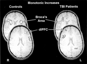
Functional magnetic resonance images for grouped data showing representative regions that exhibited monotonically increasing activity as a function of increased working memory load separately for the TBI patients and healthy comparison subjects. Z-value indicates relative position to the anterior commissure-posterior commissure line in standardized Talairach space. dlPFC = dorsolateral prefrontal cortex; IFG = inferior frontal gyrus.
 You have
Access
- 133
- Cited by