Introduction
Head and neck squamous cell carcinoma (SCC) is one of the most common types of cancer, with more than 500 000 new cases predicted annually worldwide.Reference Jemal, Murray, Ward, Samuels, Tiwari and Ghafoor1 Patients with head and neck SCC experience severe disease- and treatment-related morbidity, and have only a 50 per cent five-year survival rate. The latter statistic has not improved in more than two decades.Reference Forastiere, Koch, Trotti and Sidransky2
Head and neck SCC frequently metastasises to the regional lymph nodes, and this is the strongest predictor of disease prognosis. For most human malignancies, precise lymph node staging is necessary to establish an accurate clinical prognosis, and such staging often guides therapeutic decisions. However, currently available pre-operative clinical staging methods (including newer radiographic techniques) are suboptimal, and often misdiagnose the presence or absence of cervical nodal metastasis.Reference Forastiere, Koch, Trotti and Sidransky2, Reference Boring, Squires and Tong3 It is essential to improve our knowledge of the underlying molecular mechanisms by which tumours spread via the lymphatic vessels to the distal lymph nodes and beyond, in order to develop effective therapeutic modalities for head and neck SCC.
One of the shortcomings of previous studies of immunohistochemically assessed lymphangiogenesis has been an inability to distinguish between blood and lymphatic vessels; this is significant, as the two types of vessel play different roles in cancer progression.Reference Van Netten, Cann, Maxwell and Finegan4, Reference Van Netten, Cann and van der Westhuizen5
In contrast, the development of a polyclonal antibody recognising podoplaninReference Breiteneder-Geleff, Soleiman, Kowalski, Horvat, Amann and Kriehuber6 has enabled the current authors to stain lymphatic vessels selectively. Podoplanin (also termed D2-40) is a mucin-like transmembrane glycoprotein consisting of an extracellular domain, a single transmembrane portion, and a short cytoplasmic tail for protein kinase C and cyclic adenosine monophosphate phosphorylation.Reference Stacker, Caesar, Baldwin, Thornton, Williams and Prevo7, Reference Straume, Jackson and Akslen8 Podoplanin is highly and specifically expressed in lymphatic endothelial cells and during tumour-associated lymphangiogenesis, but is not expressed in blood endothelial cells.Reference Dadras, Paul, Bertoncini, Brown, Muzikansky and Jackson9 Podoplanin is widely used in histopathology as a specific marker for lymphatic endothelium.Reference Straume, Jackson and Akslen8 Its expression has been reported in carcinomas of the skin, lung, uterus and thyroid. A high level of podoplanin expression is significantly associated with lymph node metastasis and a poor survival rate.Reference Schacht, Dadras, Johnson, Jackson, Hong and Detmar10–Reference Giorgadza, Baloch, Pasha, Zhang and LiVolsi13 This raises the possibility that podoplanin may have biological functions in tumour cells, and may play a role in tumour-associated lymphangiogenesis and malignant transformation.
The current study used monoclonal antibody D2-40, which specifically recognises podoplanin, to determine the expression of podoplanin in patients with supraglottic laryngeal carcinoma. The study then analysed potential associations between podoplanin expression and clinicopathological factors. The potential value of lymphangiogenesis assessment in predicting disease outcome was also examined.
Materials and methods
Patients and samples
The study enrolled an initial cohort of 108 patients with supraglottic laryngeal carcinoma who were treated at the Kurume University Hospital between October 1995 and October 2004.
A total of 24 patients who did not undergo surgery, or who received radiotherapy before surgery, were excluded.
Therefore, the actual analysis was performed on 84 patients who underwent total laryngectomy either with or without radical or modified radical neck dissection. Of these 84 patients, 77 were men and seven women. Patients' ages ranged from 43 to 95 years, with a mean age of 67 years.
Histopathologically, all patients had supraglottic laryngeal carcinoma, which was well differentiated in 15 patients, moderately differentiated in 48 and poorly differentiated in 21. Patients were classified according to the 2002 tumour–node–metastasis (TNM) classification, as follows: for tumour stage, T1 = four patients, T2 = 22, T3 = 17 and T4 = 41; for node stage, N0 = 50 and N+ = 34 (N1 = 6, N2 = 27 and N3 = 1); and for metastasis stage, M0 = 71 and M1 = 13.
During follow-up examination, recurrence was detected in 11 patients, while the remaining 73 patients were free of recurrence. Overall, 84 patients had a three-year survival rate of 70.72 per cent and a five-year survival rate of 65.74 per cent.
The 84 patients' histopathological findings were initially screened at a low magnification. Cancerous and noncancerous tissues were then extracted from the original paraffin blocks for further study.
To provide a control for this study, normal supraglottic laryngeal mucosa tissue was obtained from five patients who had undergone total laryngectomy for advanced tongue cancer.
Immunohistochemical analysis
Consecutive, 3 µm thick tissue sections were placed on microscope slides, deparaffinated and dehydrated in xylol with graded alcohol. After rinsing with phosphate-buffered saline, the sections were placed in hot 10 mmol/l citrate buffer (pH 6.0) and heated in a microwave oven (twice, for 5 minutes each time) to facilitate antigen retrieval.
The Dako EnVision™ immunohistochemistry method was then utilised to stain the lymphatic microvessels with the D2-40 monoclonal antibody (antipodoplanin) (1:100, M0876; Dako, Tokyo, Japan). Using the Dako TechMate™ Horizon automated immunostainer, the slides were incubated with primary antibodies for 60 minutes. EnVision system peroxidase was used during the immunohistochemical staining process. Diaminobenzidine substrate chromogen solution was applied, followed by counterstaining with haematoxylin.
Determination of podoplanin expression
Podoplanin expression was evaluated by an independent investigator unfamiliar with the patient's history, using a digital optical microscope (VHX-200; Keyence, Osaka, Japan) at ×400 magnification. Lymphatic microvessels were counted in (1) cancer nests and stroma (i.e. intratumoural lymphatic microvessels), and (2) along the tumour–myometrial junction (i.e. peritumoural lymphatic microvessels). To determine the lymphatic microvessel count, each section was scanned at low magnifications (i.e. ×40 and ×100), and three representative areas were identified. Lymphatic microvessels in these three areas were then counted at ×200 magnification, and the mean count for the three areas was used for statistical analysis. Only lymphatic vessels containing podoplanin-expressing cells displaying cell membrane immunoreactivity were counted.
Sections of tongue SCC served as positive controls for immunohistochemical analysis. Sections incubated without the primary antibody served as negative controls.
Statistical analysis
For immunohistochemical markers, the cut-off value for the definition of subgroups was the median value.Reference Kyzas, Geleff, Batistatou, Agnantis and Stefanou14 Any correlation between immunohistochemical results and clinicopathological features was assessed using the chi-square test. Cumulative survival time was calculated using the Kaplan–Meier method, and compared using the log-rank test.
Overall survival was defined as the period from the primary surgery until the death of the patient. Death from a cause other than supraglottic laryngeal carcinoma, and survival until the end of the observation period, were considered as censoring events. Disease-free survival was defined as the period from the end of the primary therapy until the first evidence of progression of disease. Univariate analysis of overall survival and disease-free survival was performed as outlined by Kaplan and Meier.Reference Kaplan and Meier15 Multivariate analysis was based on the Cox proportional hazards regression model. A two-tailed p value of less than 0.05 was considered statistically significant.
All statistical analyses were performed using the Statistical Package for the Social Sciences version 12.0 for Windows software program (SPSS Inc, Chicago, Illinois, USA).
Results
Podoplanin expression in supraglottic laryngeal carcinoma
Immunohistochemical analysis for podoplanin was conducted in order to calculate the lymphatic microvessel density within the supraglottic laryngeal carcinoma samples (Figure 1). Podoplanin immunoreactivity was detected in the cytoplasm of lymphatic endothelial cells but not blood endothelial cells.
Photomicrographs of immunohistochemical analysis of podoplanin expression in supraglottic laryngeal carcinoma, showing (a) podoplanin-positive lymphatic vessels (arrows) (×100), (b) lymphocytes within podoplanin-positive lymphatic vessels (arrows) (×400), and (c) podoplanin-positive lymphatic vessels containing tumour cells (arrows), indicative of lymphatic invasion (×100). Note the presence of unstained blood vessels.

The majority of intratumoural lymph vessels were small and collapsed. In contrast, the peritumoural lymph vessels were often enlarged, with wide, open lumina. Lymph vessels were unevenly distributed throughout the tumour.
Relationship between lymphatic microvessel density and clinicopathological factors
For the 84 study patients, the median intratumoural lymphatic microvessel density was 0 microvessels per field (range, 0–26 vessels). As regards intratumoural vessels, 48 (57 per cent) patients were considered to have low lymphatic microvessel density, and 36 (43 per cent) to have high lymphatic microvessel density. The median peritumoural lymphatic microvessel density was four microvessels per field (range, 0–32 vessels). As regards peritumoural vessels, 46 (55 per cent) patients were considered to have low lymphatic microvessel density, and 38 (45 per cent) to have high lymphatic microvessel density.
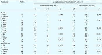
The association between lymphatic microvessel density and clinicopathological features is summarised in Table I. A high intratumoural lymphatic microvessel density was significantly associated with a more aggressive tumour N stage (p < 0.01) and M stage (p = 0.037), and a poorer prognosis (p = 0.011) at the time of diagnosis. A high peritumoural lymphatic microvessel density was significantly associated with a more aggressive tumour N stage (p = 0.004) and a poorer prognosis (p = 0.029) at the time of diagnosis.
Association of lymphatic microvessel density with clinicopathological parameters

*Assessed using podoplanin immunoreactivity. Pts = patients; mv/fld = microvessels per microscopy field; Ca dif = cancer differentiation; mod = moderate; T = tumour; N = node; M = metastasis
There was no statistically significant association between intratumoural lymphatic microvessel density and patient sex, histological differentiation, tumour T stage or recurrence. Peritumoural lymphatic microvessel density showed no significant association with sex, histological differentiation, tumour T stage, tumour M stage or recurrence.
Survival and recurrence analysis
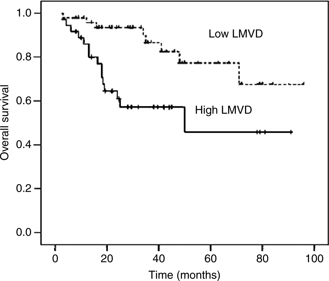
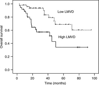
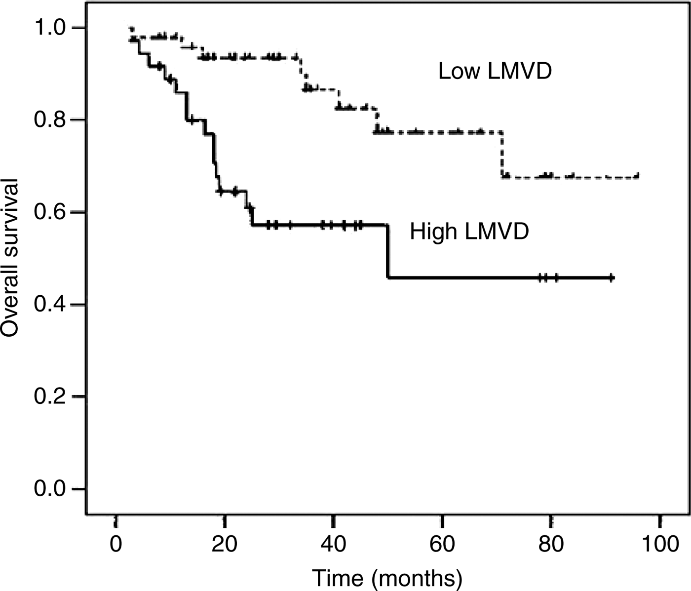
Intratumoural lymphatic microvessel density was the only significant prognostic factor for overall survival and disease-free survival in the supraglottic laryngeal carcinoma patients (log rank p = 0.005 and log rank p = 0.003, respectively) (Figures 2 and 3). The mean survival time was 34 months in patients with high intratumoural lymphatic microvessel density, and 42 months in those with low intratumoural lymphatic microvessel density.
Kaplan–Meier analysis of overall survival of patients with supraglottic laryngeal carcinoma who had high and low intratumoural lymphatic microvessel densities (LMVD). p = 0.005.
Kaplan–Meier analysis of disease-free survival of patients with supraglottic laryngeal carcinoma who had high and low intratumoural lymphatic microvessel densities (LMVD). p = 0.003.

In contrast, peritumoural lymphatic microvessel density showed no significant association with overall survival or disease-free survival.
Univariate and multivariate analyses
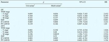
Univariate analysis showed that intratumoural lymphatic microvessel density was a significant predictor of overall survival and disease-free survival (p = 0.005 and p = 0.003, respectively) (Table II), whereas tumour TMN stage, tumour differentiation and peritumoural lymphatic microvessel density were not significant predictors of overall or disease-free survival (p > 0.05).
Patients' overall and disease-free survival*

* 84 patients with supraglottic laryngeal carcinoma. †Log rank test; ‡Cox regression. CI = confidence interval; RR = relative risk; univariate = univariate analysis; multivariate = multivariate analysis; OS = overall survival; TNM = tumour–node–metastasis; diff = tumour differentiation; IT = intratumoural; LMVD = lymphatic microvessel density; PT = peritumoural; DFS = disease-free survival
In the multivariate analysis, covariates for survival were tested using a Cox proportional hazards model (Table II). Patients with a low intratumoural lymphatic microvessel density had a significantly lower risk for disease-free survival (hazard ratio = 3.078, 95 per cent confidence interval (CI) = 1.401–6.761, p = 0.005) and for overall survival (hazard ratio = 3.221, 95 per cent CI = 1.359–7.634, p = 0.008), compared with patients with a high intratumoural lymphatic microvessel density. Moreover, multivariate analysis indicated that tumour M stage was an independent prognostic factor for disease-free survival (p = 0.0019), whereas tumour T stage was an independent predictor of overall survival (p = 0.036). Only intratumoural lymphatic microvessel density was found to be an independent predictor of both disease-free survival (p = 0.003) and overall survival (p = 0.005).
This suggests that intratumoural lymphatic microvessel density may be a stronger predictor of supraglottic laryngeal carcinoma prognosis than tumour histology.
Discussion
In patients diagnosed with supraglottic laryngeal carcinoma, the determination of lymph node status at the time of diagnosis is one of the major predictors of outcome, and is also used to guide therapeutic decisions. Despite recent advances in radiological imaging, the most reliable method of detecting lymph node metastasis remains the careful microscopic assessment of regional lymph nodes harvested from resection specimens.
However, the prognostic value of lymph node assessment is restricted by the variable yield of nodes harvested from the surgical specimens.38 It may also be adversely affected by the current recommendation to examine microscopically only one slide from each node.Reference Marcus, Arenberg, Lee, Kleer, Chepeha and Schmalbach16
Consequently, accurate assessment of the risk of lymphatic metastasis is not always possible. Therefore, a method which accurately predicted the risk of lymph node metastasis, based on characteristics of the primary tumour, would constitute a powerful prognostic tool for clinicians and pathologists.
In this study, intratumoural and peritumoural lymphatic microvessel densities were assessed within surgical specimens of supraglottic laryngeal carcinoma, using a specific lymphatic endothelial marker.Reference Breiteneder-Geleff, Soleiman, Horvat, Amann, Kowalski and Kerjaschki17, Reference Kriehuber, Breiteneder-Geleff, Groeger, Soleiman, Schoppmann and Stingl18 Previous findings have suggested that intratumoural lymphatics are nonfunctional, and that lymph node metastases occur in tumours lacking intratumoural functional lymphatics.Reference Padera, Kadambi, di Tomaso, Carreira, Brown and Boucher19 In contrast, the current study found that high densities of both intratumoural and peritumoural lymphatic microvessels were both significantly associated with the presence of lymph node metastasis. Furthermore, intratumoural lymphatic microvessel density (but not peritumoural lymphatic microvessel density) was shown to be a prognostic factor, and to be significantly correlated with overall survival and disease-free survival.
The hypothesis that intratumoural lymphatic microvessels are necessary for metastasis of tumour cells to lymph nodes is further supported by two important findings.
First, tumour cells have been observed to be present within intratumoural lymphatic microvessels. However, several studies have shown that intratumoural lymphatic vessels are often occluded by tumour cells at the time when lymph node metastases are detectable.Reference Padera, Kadambi, di Tomaso, Carreira, Brown and Boucher19, Reference Skobe, Hawighorst, Jackson, Prevo, Janes and Velasco20 Hence, tumour lymphatic vessels may have normal drainage functions in the early stages of tumour development, but these functions may be blocked in the later stages of tumour progression.Reference Cao21
Second, the present study found that lymph node metastases occurred in 25 of the 36 (69.4 per cent) patients with a high density of intratumoural lymphatic microvessels, but in only nine of the 48 (18.8 per cent) patients with a low density of intratumoural lymphatic microvessels. Furthermore, 15 of the 36 (41.7 per cent) patients with a high density of intratumoural lymphatic microvessels died of their tumour, while only eight of the 48 (16.7 per cent) patients with a low density of intratumoural lymphatic microvessels died of their tumour. Intratumoural lymphangiogenesis was significantly associated with the presence of lymph node metastasis and with poor prognosis; this indicates that intratumoural lymphangiogenesis is an active element of human lymph node metastasis.
The detection of increased lymphatic microvessel density may provide a convenient and reliable means of determining the risk of lymphatic metastasis in solid tumours. Furthermore, lymphatic microvessel density may constitute a simple target for future anti-tumour therapies.
Previous studies have reported controversial results regarding the importance of peritumoural lymphatic microvessels for the metastatic potential of laryngeal SCC. Some investigatorsReference Altman, Lausen, Sauerbrei and Schumacher22 have found a significant association between high peritumoural lymphatic microvessel density and shorter disease-free survival. In contrast, other studies have found a relationship between high peritumoural lymphatic microvessel density and more favourable outcomes.Reference Concato, Feinstein and Holford23, Reference Birner, Schindl, Obermair, Breitenecker, Kowalski and Oberhuber24
The authors of the current study did not find a significant association between peritumoural lymphatic microvessel density and overall survival or disease-free survival, although correlations were noted between higher peritumoural lymphatic microvessel density and lymph node involvement and worse prognosis.
Such contradictory results for the correlation between peritumoural lymphatic microvessels and prognosis may be due to differences in: patient selection; methods of counting lymphatic microvessels; the grade, stage and type of tumours analysed; and the local microenvironment present during the development of primary tumours.
In addition, the underlying mechanisms by which tumours induce lymphangiogenesis and lymphatic metastasis are complex, and vary significantly in tumours of different histological type and anatomical location.Reference Munoz-Guerra, Marazuela, Martin-Villar, Quintanilla and Gamallo25
• Head and neck squamous cell carcinoma frequently metastasises to the regional lymph nodes, and this is the strongest indicator of disease prognosis
• This study assessed the association of intra- or peritumoural lymphatic microvessels with clinicopathological factors and survival
• Intratumoural lymphatic microvessel density may be an independent predictor of lymphatic tumour spread
However, a main contributor to the controversy over the prognostic significance of tumour lymphatic microvessels may be the lack of appropriate lymphatic vessel markers. Previous investigators have used nonspecific or nonsensitive markers for their studies, such as Lymphatic vessel endothelial hyaluronan receptor-1 (LYVE-1). Although this is considered a specific marker for lymphatic vessels, it has also been observed in the endothelium of blood vessels.Reference Reis-Filho and Schmitt26, Reference Jain and Fenton27 The current study used podoplanin, considered to be the most specific lymphatic endothelial marker,Reference Stacker, Achen, Jussila, Baldwin and Alitalo28 and all podoplanin-positive vessels were observed to have morphological features consistent with lymphatics.
Some studies have analysed the presence of lymphangiogenesis in head and neck SCC. Maula et al. Reference Maula, Luukkaa, Grénman, Jackson, Jalkanen and Ristamäki29 studied patients with head and neck SCC, and found that intratumoural lymphatic vessels were associated with a poor disease-specific prognosis, but that a high density of peritumoural vessels was associated with favourable survival. Frech et al. Reference Frech, Hörmann, Riedel and Götte30 found that a high density of intratumoural lymphatic vessels correlated significantly with nodal metastasis in oral carcinoma patients, but showed no such association in patients with other types of head and neck SCC. The discrepancy between these two study findings may be due to differences in lymphatic vessel markers, and to the fact that these authors assessed a diverse range of tissue of varying histology arising from a variety of anatomical head and neck SCC sites.
Conclusion
These study findings establish podoplanin as a valid marker for lymphatic endothelium.
Although an increase in intratumoural lymphatic microvessel density was significantly associated with a worse prognosis in patients with supraglottic laryngeal carcinoma, the prognostic power of peritumoural lymphatic microvessel density was not strong enough to retain significance.
Intratumoural lymphatic microvessel density may have clinical utility in the evaluation of supraglottic laryngeal carcinoma, particularly for the estimation of lymph node metastatic risk. In patients with supraglottic laryngeal carcinoma, increased intratumoural lymphatic microvessel density may serve as an indicator of poor prognosis.
Acknowledgement
This work was supported by the Shanghai Pujiang Fellowship Foundation (grant number 07pj14064).