Introduction
Sjögren's syndrome is a chronic, inflammatory, autoimmune disease characterised by lymphocytic infiltration of the exocrine glands. Although keratoconjunctivitis sicca and xerostomia are prominent symptoms, systemic symptoms such as skin lesions, Raynaud's phenomenon, interstitial pneumonia, autonomic dysfunction and central nervous system dysfunction may also be present.Reference Calzada, Balaker, Ishiyama, Lopez and Ishiyama1,Reference Yerdelen, Karatas, Alkan and Tufan2 The exact aetiology is unclear; however, immune-mediated vasculopathy, vasculitis or demyelination are considered to be important underlying factors in the pathogenesis.Reference Calzada, Balaker, Ishiyama, Lopez and Ishiyama1–Reference Margaretten4
In primary Sjögren's syndrome, the reported frequency of neurological symptoms varies between 8 and 49 per cent, with an average frequency of approximately 20 per cent.Reference Margaretten4,Reference Delalande, de Seze, Fauchais, Hachulla, Stojkovic and Ferriby5 The most common peripheral neuropathies are distal sensor and sensorimotor neuropathies.Reference Margaretten4 Although cranial nerve mononeuropathy and polyneuropathy may be present, the most common neuropathy involving the craniofacial region is trigeminal neuropathy. Apart from the trigeminal nerve, involvement of VIIth, VIIIth, IIIrd, IVth and Vith cranial nerves have also been reported.Reference Margaretten4,Reference Delalande, de Seze, Fauchais, Hachulla, Stojkovic and Ferriby5 Sensorineural hearing loss, especially at high frequencies, has been reported in patients.Reference Calzada, Balaker, Ishiyama, Lopez and Ishiyama1,Reference Tucci, Quatraro and Silvestris6 In addition to findings of vestibular involvement in primary Sjögren's syndrome,Reference McCombe, Sheean, McLaughlin and Pender7,Reference Estiasari, Matsushita, Masaki, Akiyama, Yonekawa and Isobe8 some publications have reported vestibular preservation but with hearing dysfunction in primary Sjögren's syndrome patients.Reference Boki, Ioannidis, Segas, Maragkoudakis, Petrou and Adamopoulos9
Vestibular-evoked myogenic potentials testing evaluates the vestibulocollic reflex arc, measuring the potentials created by acoustic stimulation of the ear in the neck sternocleidomastoid muscle. Two types of vestibular-evoked myogenic potentials tests are used in clinical practice: cervical vestibular-evoked myogenic potentials and ocular vestibular-evoked myogenic potentials. In the cervical vestibular-evoked myogenic potentials test, the saccule, inferior vestibular nerve, lateral vestibular nucleus, medial vestibulospinal tract and spinal cord motor neuron pathway are evaluated. In the ocular vestibular-evoked myogenic potentials test, information about the superior vestibular nerve and the pathway behind is obtained. The vestibular-evoked myogenic potentials test is inexpensive and non-invasive, providing information about lesions on this entire axis in a short time.Reference Hizal, Erbek and Özlüoğlu10,Reference Rosengren, Colebatch, Young, Govender and Welgampola11
The video head impulse test is also a non-invasive test that bilaterally compares the semicircular canals and provides information about canal functions. Use of the video head impulse test as a screening test in the first step of vestibular evaluation, before the caloric test, has been proposed. The video head impulse test mainly evaluates the vestibulo-ocular reflex, and the results provide the semicircular canal vestibulo-ocular reflex gains on both sides comparatively.Reference Halmagyi, Chen, MacDougall, Weber, McGarvie and Curthoys12,Reference Alhabib and Saliba13
The present study aimed to evaluate the hearing and vestibular functions of primary Sjögren's syndrome patients to elucidate different views on vestibular involvement reported in the literature. A review of literature using the PubMed database revealed no study examining the relationships between primary Sjögren's syndrome and vestibular-evoked myogenic potentials and video head impulse test results.
Materials and methods
In the present study, 35 female volunteers with primary Sjögren's syndrome, diagnosed according to the 2002 America-Europe Consensus Group Sjögren's syndrome classification criteria,Reference Vitali, Bombardieri, Jonsson, Moutsopoulos, Alexander and Carsons14 comprised the study group and 35 healthy volunteers of a similar age comprised the control group. Local ethics committee approval (approval number: 2018/252) was obtained, as was written informed consent from all participants. This study was conducted according to the Helsinki Declaration.
The exclusion criteria were: conductive hearing loss, tympanic membrane perforation, uncontrolled hypertension, uncontrolled diabetes mellitus, a neuro-otological disease diagnosis, a history of head trauma or acoustic trauma, previous surgery in the cervical or ocular region, and age below 18 years or over 65 years.
Participants were first questioned regarding the presence of dizziness or vertigo, tinnitus, oscillopsia, hearing loss, diplopia, and dysarthria. After otoscopic examination, participants were evaluated in terms of hearing using pure tone audiometry, tympanometry and acoustic reflex tests. Participants’ vestibular systems were also evaluated, with a nystagmus examination, Romberg test, Dix–Hallpike test, and dysdiadokinesia and dysmetria tests. All participants underwent bilateral cervical and ocular vestibular-evoked myogenic potentials tests, and video head impulse tests. Likewise, the vestibular tests were applied to individuals whose hearing thresholds were better than 25 dB in a pure tone audiometry test.
Vestibular-evoked myogenic potentials test
Vestibular-evoked myogenic potentials tests of the individuals participating in the study were conducted in the audiovestibular laboratory of our hospital (using the ICS Chartr EP 200 vestibular-evoked potentials testing system; GN Otometrics, Taastrup, Denmark). Cervical and ocular vestibular-evoked myogenic potentials tests were conducted as reported by Ulusoy et al.Reference Ulusoy, Gül, Elsürer, Bozkurt, Tülek and Körez15
Video head impulse test
The video head impulse test was conducted using the video head impulse test Ulmer II system 3.1.0.8 device (Synapsys, Marseille, France) and video head impulse test Ulmer software. During preparation for the test, all individuals were first informed about the test. The distance between the camera and the individual was set to 90 cm and the height of the camera was positioned 90 cm above the ground. Each test participant was seated in a chair and asked to look at the fixed visual target on the camera. After calibration, participants were instructed to relax their head, not to tense their neck muscles, not to close their eyes, not to blink if possible, and to avoid involuntary head movements – the experimenter would carry out all necessary head movements.
For lateral semicircular canal evaluation, each participant's head was brought 30 degrees forward to the flexion position while facing the target. Then, their head was turned abruptly at an angle of approximately 15 degrees (range, 10–20 degrees) towards the side of the canal to be stimulated, in the horizontal plane.
In order to test the vertical canal pairs on the same plane for right anterior/left posterior and left anterior/right posterior tests, participants’ heads were rotated 30–45 degrees. Vertical canal pairs were measured with forward and backward pushing movements, while maintaining the head in the desired position. The average value of the five most reliable measurements of each semicircular canal was calculated. Average vestibulo-ocular reflex values were calculated for each semicircular canal on both sides.
Sample size and power analysis
An a priori power analysis was conducted using the ‘pwr’ power analysis package in R statistical software version 3.6.0 (www.r-project.org). This analysis was performed to test the difference of the N1 latency value in the ocular vestibular-evoked myogenic potentials test between the two independent groups (control and disease); a two-tailed test was used, with a medium effect size (d = 0.50) and an alpha value of 0.05. Results of the 64 participants divided equally (n = 32) was necessary to achieve a power of 0.80.
Statistical evaluation
All statistical analyses were performed using Jamovi statistical spreadsheet software version 1.2.25 (www.jamovi.org). Before the statistical analyses, data normality was checked with the Shapiro–Wilk normality test and quantile–quantile (Q–Q) plots. The homogeneity of group variances was tested with the Levene test.
The data were summarised, depending on suitability, as mean ± standard deviation or median (interquartile range). Student's t-test, Welch's t-test and Mann–Whitney U tests were used for the comparison of the patient and control groups in terms of numerical parameters. The chi-square test was used to compare hearing loss rates and to determine the rates for the inability of obtaining waves between the groups. Relationships between cervical and ocular vestibular-evoked myogenic potentials, and video head impulse test findings, were examined with Pearson and Spearman's rho correlation co-efficients. A p-value of less than 0.05 was the threshold level for statistical significance.
Results
Mean age was 50.5 ± 7.9 years for the patient group and 48.5 ± 5.4 years for the control group, with no statistically significant difference between the groups (p = 0.096).
While sensorineural hearing loss was detected in five patients (10 ears) (14.3 per cent), no hearing loss was determined in the control group. The difference between the control and patient groups in terms of hearing loss rate was statistically significant (p = 0.021). When the patients with hearing loss were excluded, the average air conduction hearing threshold of the remaining 30 patients (60 ears) was 13.92 ± 5.18 dB (range, 3–25 dB) and the mean bone conduction hearing threshold was 10.12 ± 5.71 dB (range, 0–20 dB). In the control group, the mean air conduction hearing threshold was 11.79 ± 4.36 dB (range, 3–20 dB), and the mean bone conduction hearing threshold was 8.36 ± 5 dB (range, 0–20 dB). The mean hearing thresholds of the participants in both groups were within normal limits. All participants had bilateral acoustic reflexes and type A tympanogram results.
Symptoms such as dizziness or vertigo, tinnitus, diplopia, dysarthria, and oscillopsia were not present in the participants of the present study. No vestibular pathology indicators were determined in the nystagmus, dysdiadochokinesia, dysmetria, Romberg and Dix–Hallpike test results. In order to avoid misinterpretation in the comparison of the groups’ balance test results (cervical vestibular-evoked myogenic potentials, ocular vestibular-evoked myogenic potentials and video head impulse tests), five patients with hearing loss were excluded from this test.
When comparing the groups in terms of cervical and ocular vestibular-evoked myogenic potentials, and video head impulse test measurements, the results of 30 patients (60 ears) in the patient group and 35 individuals (70 ears) in the control group were considered. Accordingly, the mean age of the patient group (n = 30) was 49.5 ± 7.5 years, which is similar to that of the control group (p = 0.417).
Cervical vestibular-evoked myogenic potential waveforms were obtained from all participants. Ocular vestibular-evoked myogenic potential waveforms were obtained from all control group ears, but not from 2 out of 60 ears in the patient group (3.3 per cent). However, the difference between the groups in this respect was not statistically significant (p = 0.157).
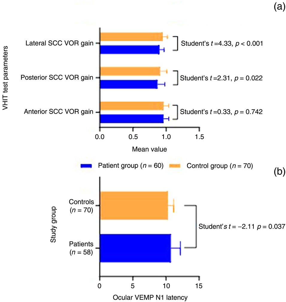
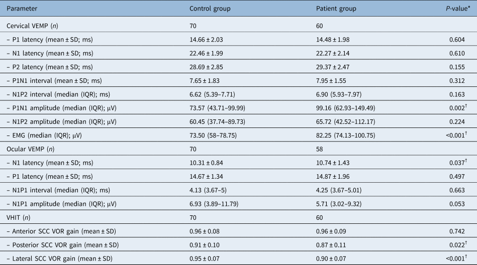
According to the ocular vestibular-evoked myogenic potentials test results, the N1 latency value was 10.74 ± 1.43 ms in the patient group and 10.31 ± 0.84 ms in the control group. The N1 latency value in the patient group was significantly longer (p = 0.037) (Figure 1 and Table 1). When the patient and control groups were compared in terms of P1 latency, N1P1 interval and N1P1 amplitude variables, no statistically significant differences could be determined (Table 1).
(a) Comparison of groups in terms of semicircular canal (SCC) vestibulo-ocular reflex (VOR) gain values in the video head impulse test (VHIT). (b) Comparison of groups according to N1 latency values in the ocular vestibular-evoked myogenic potentials (VEMP) test.

Comparison of cervical and ocular VEMPs and VHIT findings according to groups

*P-values were calculated by student's t-test or Mann–Whitney U test. †Statistically significant at the p < 0.05 level. VEMP = vestibular-evoked myogenic potentials; VHIT = video head impulse test; SD = standard deviation; IQR = interquartile range; EMG = electromyography; SCC = semicircular canal; VOR = vestibulo-ocular reflex
According to the cervical vestibular-evoked myogenic potentials test results, the median P1N1 amplitude value was 99.16 μV (range, 62.93–149.49 μV) in the patient group and 73.57 μV (range, 43.71–99.99 μV) in the control group. The higher amplitude value in the patient group was statistically significant (p = 0.002) (Table 1). There were no significant differences between the groups in terms of P1 latency, N1 latency, P2 latency, P1N1 interval, N1P2 interval and N1P2 amplitude variables (Table 1).
According to the video head impulse test results, the mean posterior canal vestibulo-ocular reflex gain value was 0.87 ± 0.11 in the patient group and 0.91 ± 0.10 in the control group. In the patient group, the posterior semicircular canal vestibulo-ocular reflex gain value was found to be significantly lower than that of the control group (p = 0.022) (Table 1). Similarly, it was observed that the lateral semicircular canal vestibulo-ocular reflex gain value of the patient group (0.90 ± 0.07) was significantly lower than that of the control group (0.95 ± 0.07) (p < 0.001) (Table 1). There was no difference between the groups in terms of the anterior semicircular canal vestibulo-ocular reflex gain value (Figure 1 and Table 1).
Discussion
The results of this study confirm audiovestibular dysfunction in primary Sjögren's syndrome patients. This is the first study (according to our search on PubMed for relevant literature) to evaluate patients with primary Sjögren's syndrome in terms of vestibular involvement, using vestibular-evoked myogenic potentials and video head impulse tests. The most important findings of the present study are the prolonged N1 latency value in individuals with primary Sjögren's syndrome, and the lower values for posterior and lateral semicircular canal vestibulo-ocular reflex gains.
Seeliger et al. reported that most patients with Sjögren's syndrome are diagnosed after the occurrence of neurological symptoms. Therefore, they recommended that all patients with polyneuropathy are screened for primary Sjögren's syndrome.Reference Seeliger, Prenzler, Gingele, Seeliger, Korner and Thiele16 Peripheral neuropathy, central nervous system involvement and cranial nerve involvement in primary Sjögren's syndrome have also been reported in numerous studies.Reference Margaretten4,Reference Delalande, de Seze, Fauchais, Hachulla, Stojkovic and Ferriby5,Reference Seeliger, Prenzler, Gingele, Seeliger, Korner and Thiele16,Reference Griffin, Cornblath, Alexander, Campbell, Low and Bird17 The involvement of immune-mediated vasculopathy, vasculitis or demyelination have been indicated in the central nervous system pathogenesis.Reference Margaretten4 Griffin et al. showed a loss of large myelinated nerve fibres, mononuclear inflammation in the cutaneous nerves, and lymphocytic infiltration in dorsal root ganglia biopsy in primary Sjögren's syndrome patients.Reference Griffin, Cornblath, Alexander, Campbell, Low and Bird17 Lymphocytosis, increased immunoglobulin G (IgG) index, increased protein level and/or oligoclonal bands in electrophoresis were determined in the cerebrospinal fluid examination of patients with primary Sjögren's syndrome and active central nervous system involvement. Lymphocytic inflammation of small vessels and ischaemic vasculopathy in the histopathological examination of brain tissue of some primary Sjögren's syndrome patients have also been reported.Reference Margaretten4 The effectiveness of magnetic resonance imaging in detecting central nervous system involvement is controversial.Reference Margaretten4
A retrospective study reported the rate of neurological involvement as 20 per cent, and described the difficulties of neurological involvement diagnosis.Reference Delalande, de Seze, Fauchais, Hachulla, Stojkovic and Ferriby5 The same study also reported focal or multifocal central nervous system involvement in 68 per cent, as well as spinal cord involvement in 34 per cent, brain involvement in 40 per cent, optic neuropathy in 16 per cent, and peripheral nerve involvement in 62 per cent (particularly trigeminal, facial and cochlear nerves). They concluded that patients with primary Sjögren's syndrome be systematically evaluated for acute or chronic myelopathy, axonal sensorimotor neuropathy, or cranial nerve involvement.Reference Delalande, de Seze, Fauchais, Hachulla, Stojkovic and Ferriby5
Although the aetiology of inner-ear involvement in primary Sjögren's syndrome has not been clearly elucidated, immune complexes may play a role in the pathogenesis by occluding small vessels and capillaries, causing ischaemia and local inflammation in the inner ear.Reference El-Minawi, El-Fouly, Abo-Eleyoun, El-Mofty, El-Azkalany and El-Refai3,Reference Tucci, Quatraro and Silvestris6,Reference Galarza-Delgado, Villegas Gonzalez, Riega Torres, Soto-Galindo, Mendoza Flores and Trevino Gonzalez18
Calzada et al. investigated the inner-ear structures of four primary Sjögren's syndrome patients histopathologically and immunohistochemically.Reference Calzada, Balaker, Ishiyama, Lopez and Ishiyama1 Severe intermediate cell loss in the stria vascularis and IgG accumulation in the basement membrane of stria vascularis blood vessels in three patients with sensorineural hearing loss were determined. In addition, spiral ganglion cell shrinkage was observed in two patients, with preserved vestibular ganglion cells. However, mild stria vascularis atrophy in the fourth patient with normal hearing was also reported.Reference Calzada, Balaker, Ishiyama, Lopez and Ishiyama1
Different rates of hearing loss have been reported in the literature, and higher rates of hearing loss have been described in studies using high-frequency audiometry. Approximately 25 per cent of primary Sjögren's syndrome patients have sensorineural hearing loss, mostly at high frequencies.Reference Tucci, Quatraro and Silvestris6,Reference Boki, Ioannidis, Segas, Maragkoudakis, Petrou and Adamopoulos9 Studies using high-frequency audiometry reported the rate of hearing loss to be 60–100 per cent.Reference Galarza-Delgado, Villegas Gonzalez, Riega Torres, Soto-Galindo, Mendoza Flores and Trevino Gonzalez18,Reference Gonzalez, Torres, Rios, Gonzalez, Mendez Saenz and Soto-Galindo19 Additionally, Galarza-Delgado et al. reported that a type A tympanogram was obtained in 98.3 per cent of the patients.Reference Galarza-Delgado, Villegas Gonzalez, Riega Torres, Soto-Galindo, Mendoza Flores and Trevino Gonzalez18 In the present study, the rate of hearing loss in primary Sjögren's syndrome patients was 14 per cent. The underlying reason for the low rate obtained in the present study is considered to be the use of the 0.5–8 kHz frequency range in the audiological examination of patients.
Studies investigating vestibular involvement in autoimmune diseases have been published. Audiovestibular dysfunction, an inability to obtain waveforms in the vestibular-evoked myogenic potentials test, and prolongation of wave latencies have also been reported in patients with autoimmune diseases such as rheumatoid arthritis and Behçet's disease.Reference Erbek, Erbek, Yilmaz, Yucel and Ozluoglu20–Reference Heydari, Hajiabolhassani, Fatahi, Movaseghi and Jalaie23
Vestibular involvement in primary Sjögren's syndrome remains a controversial issue. Galarza-Delgado et al. reported that 28.3 per cent of primary Sjögren's syndrome patients had vertigo complaints.Reference Galarza-Delgado, Villegas Gonzalez, Riega Torres, Soto-Galindo, Mendoza Flores and Trevino Gonzalez18 Other studies have shown vestibular neuropathy and brainstem involvement in primary Sjögren's syndrome.Reference Yerdelen, Karatas, Alkan and Tufan2,Reference McCombe, Sheean, McLaughlin and Pender7,Reference Estiasari, Matsushita, Masaki, Akiyama, Yonekawa and Isobe8 Conversely, one study reported no vestibular involvement in primary Sjögren's syndrome.Reference Boki, Ioannidis, Segas, Maragkoudakis, Petrou and Adamopoulos9 However, not all patients were examined; only individuals with vertigo or dizziness complaints were examined using electronystagmography. Therefore, it may be that individuals with subclinical involvement could not be detected in that study.
The PubMed review did not reveal any study examining the relationship between primary Sjögren's syndrome and vestibular-evoked myogenic potentials. However, one study examined cervical vestibular-evoked myogenic potentials in patients with secondary Sjögren's syndrome. A vestibular-evoked myogenic potentials response could not be obtained in one patient, with significant prolongation in P13 and N23 latency.Reference El-Minawi, El-Fouly, Abo-Eleyoun, El-Mofty, El-Azkalany and El-Refai3
The caloric test is the ‘gold standard’ in vestibular evaluation, especially in the qualitative and quantitative measurement of low-frequency vestibular function. However, there are some inherent disadvantages, including the necessity for more time, lateral semicircular canal evaluation only, nausea and dizziness symptoms in the patient.Reference Alhabib and Saliba13 The video head impulse test, on the other hand, shows vestibular functions at a higher frequency, and it allows rapid evaluation without causing symptoms such as nausea and vomiting, and enables comparisons of function in each semicircular canal with that of its contralateral part. In the video head impulse test, a reduction of more than 40 per cent in vestibular functions is considered as the precondition to detect abnormalities. Video head impulse test specificity is reported to be 90 per cent.Reference Alhabib and Saliba13 While cervical vestibular-evoked myogenic potentials provide information about the vestibulocollic reflex pathway, ocular vestibular-evoked myogenic potentials give more information about the vestibulo-ocular reflex arc.Reference Hizal, Erbek and Özlüoğlu10,Reference Rosengren, Colebatch, Young, Govender and Welgampola11
• Hearing loss rate was higher in primary Sjögren's syndrome patients than in healthy control subjects
• N1 latency for the ocular vestibular-evoked myogenic potentials test was significantly longer in the patient group
• Posterior and lateral semicircular canal vestibulo-ocular reflex gain values were significantly lower in the patient group
• These results indicate subclinical vestibular involvement in primary Sjögren's syndrome
• This is the first study to evaluate primary Sjögren's syndrome in terms of vestibular involvement using vestibular-evoked myogenic potentials and video head impulse tests
Our review of PubMed literature did not reveal any study examining the relationship between primary Sjögren's syndrome and results of the video head impulse test, or cervical and ocular vestibular-evoked myogenic potentials tests. In the present study, patients diagnosed with primary Sjögren's syndrome were examined in terms of vestibular involvement via a test battery comprising cervical vestibular-evoked myogenic potentials, ocular vestibular-evoked myogenic potentials and video head impulse tests, which allowed a more holistic evaluation. The present study revealed a higher N1 latency value in the ocular vestibular-evoked myogenic potentials test, and lower posterior and lateral canal gains in the video head impulse test, in patients with primary Sjögren's syndrome. In the patient group, the rate for the inability to obtain ocular vestibular-evoked myogenic potentials waveforms was 3.3 per cent, with no statistically significant between-groups difference. In addition, in the cervical vestibular-evoked myogenic potentials test, a higher P1N1 amplitude was determined in the patient group. The present study shows that primary Sjögren's syndrome is not only a disease affecting the auditory system, but also a disease affecting the vestibular system, though at a subclinical level.
Conclusion
Our results show that primary Sjögren's syndrome negatively affects the auditory and vestibular systems. The data obtained indicate that primary Sjögren's syndrome patients should be evaluated more carefully in terms of vestibular involvement. Vestibular-evoked myogenic potentials and video head impulse tests could be used effectively to determine vestibular involvement in primary Sjögren's syndrome patients.
Acknowledgement
This study was financially supported by the Selçuk University Scientific Research Projects Support Office (project number: 19401032).
Competing interests
None declared