Epicardial ventricular pacing is a common and effective means of pacing in children.Reference Walsh, McAlister, Andrews, Steeg, Eisenberg and Furman1–Reference Cohen, Vetter and Wernovsky5 Though endocardial pacing has been shown to be associated with lower rates of exit block, and greater lead longevity, than the epicardial approach, epicardial systems are generally preferable in children weighing less than 10 to 15 kilograms, those with intracardiac shunts, or those having functionally univentricular anatomy.Reference Cohen, Bush and Vetter6–Reference Hamilton, Gow, Bahaoric, Griffiths, Freedom and Williams11 Prior to the use of steroid-eluting leads, the failure rate of epicardial leads was markedly higher than that of endocardial leads.Reference Hamilton, Gow, Bahaoric, Griffiths, Freedom and Williams11, Reference Karpawich, Hakimi, Arciniegas and Cavitt12 Although the use of steroid-eluting leads has improved the longevity of epicardial pacing systems, at 5 years epicardial leads are still reported to fail in up to one-quarter of patients.Reference Noiseux, Khairy, Fournier and Vobecky4, Reference Fortescue, Berul, Cecchin, Walsh, Triedman and Alexander10, Reference Sachweh, Vazquez-Jimenz and Schondube13–Reference Horenstein, Hakimi, Walters and Karpawich21 Lead fracture, exit block, high pacing thresholds, and lead failure are common in this group, and tend to occur more frequently with epicardial than with endocardial leads.Reference Balmer, Fasnacht, Rahn, Molinari and Bauersfeld3, Reference Noiseux, Khairy, Fournier and Vobecky4, Reference Fortescue, Berul, Cecchin, Walsh, Triedman and Alexander10–Reference Karpawich, Hakimi, Arciniegas and Cavitt12, Reference Johns, Fish, Burger and Hammon19, Reference Silka and Bar-Cohen22, Reference Beaufort-Krol, Mulder, Nagelkerke, Waterbolk and Bink-Boelkens23 In pacemaker-dependent patients, this can have potentially life-threatening consequences.
We hypothesized that, for infants and children requiring epicardial VVI pacing, the placement of a second ventricular epicardial pacing lead attached to the atrial port of a dual chamber device at the time of surgical pacemaker placement would provide a safe and effective means of ventricular pacing in the setting of lead failure or fracture, and would not compromise safety. In the event of lead failure or fracture, the device could be manually re-programmed to pace from the atrial port in AAI mode, and continue to pace the ventricle, resulting in effective VVI pacing. By allowing for reprogramming of the device without the need for urgent repeated pacemaker surgery, this technique may prolong the time before a new pacemaker is needed.
Materials and methods
Having obtained approval from our Institutional Review Board, we performed a retrospective review of the charts of patients at our institution who had undergone epicardial pacing from May, 2001 through September, 2007. We reviewed our databases for paediatric cardiac surgery and paediatric cardiology to find all patients aged less than 21 years in whom we had placed epicardial pacing systems, along with those in whom 2 epicardial ventricular pacing leads had been connected to a dual-chamber device. From the charts, we obtained information on demographics, the indications for pacemaker placement, pertinent medical problems, cardiac anatomy, pre-operative echocardiographic findings, surgical data, epicardial lead data at the time of implant, time to failure of any lead or device, and most recent follow-up and pacemaker interrogation data.
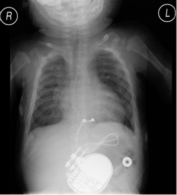
From the stance of the surgical procedure, following the administration of general endotracheal anaesthesia, a sternotomy was performed and the right ventricle was dissected free. Two unipolar or bipolar steroid eluting pacing leads were sewn to the diaphragmatic surface of the right ventricle (Fig. 1). In one patient (Table 1, Patient #1), a limited left anterior thoracotomy was performed and a portion of the left ventricular apex was identified for placement of the pacemaker leads. All leads were manufactured by Medtronic (Minneapolis, MN). The leads were CapsureEpi leads, model numbers 4965 or 4968. The leads were 15 centimetres in length in all but one patient, where a lead of 35 centimetres was used because of the size of the patient. A pacemaker pocket was created behind the rectus sheath in the left upper quadrant, and the leads brought through to the pocket. The two ventricular leads were plugged into atrial and ventricular ports of a DDD generator set at VVI. In general, the lead with the lower acute pacing threshold was placed in the ventricular port. All pacemakers were Medtronic devices. The following generators were placed: EnPulse 2DR E2DR01, Kappa 700 DR KDR721, Kappa 920 DR KDR921, Adapta DR ADDDR01, and Adapta DR ADDDRS1.
Chest X-ray after placement of the epicardial pacemaker. Note that there are two steroid-eluting, unipolar, epicardial leads placed on the diaphragmatic surface of the right ventricle, with one lead connected to the ventricular port, and the other lead connected to the atrial port of a dual-chamber pacemaker generator.

Overall data.

Using a Fisher Exact Test, we made a statistical comparison of the rates of failure of leads between the patients receiving 2 leads, and those who underwent placement of a standard epicardial pacemaker at our institution during the period of the study. A p-value of less than 0.05 was considered significant.
Results
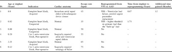
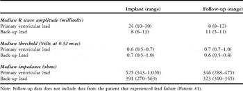
During the period of study, we placed 88 epicardial pacemakers at our institution. In 6 of these 88 patients (7%), we placed 2 ventricular leads placed as described above (Fig. 1). The decision to use this technique of pacing was based upon the preference of the surgeon. In this subset, the median age at implant was 0.92 years, with a range from 0.29 to 8 years, and the median weight was 8 kilograms, with a range from 4.2 to 31.8 kilograms (Table 1). In 2 patients, pacing was required because of symptomatic congenital atrioventricular block, 2 further patients had symptomatic acquired, high grade atrioventricular block of unclear aetiology, and the final 2 patients had postoperative atrioventricular block, this occurring in 1 following repair of a ventricular septal defect, and in the other following repair of tetralogy of Fallot. Of the other patients, 3 had normal intracardiac anatomy, and 1 had a defect of the oval fossa that was incidentally discovered at the time of presentation with heart block, and was subsequently percutaneously closed with an Amplatzer device. The patient with the atrial septal defect was 8 years of age at the time of implantation of the pacemaker, and an epicardial system being placed due to the presence of intracardiac shunting. There were no surgical complications associated with placement of the pacemakers in any of the patients. In 5 of the 6 patients (83%), we used unipolar leads, placing bipolar leads in the other. The data obtained at implantation is shown in Table 2.
Data at implant and at most recent follow-up.

Note: Follow-up data does not include data from the patient that experienced lead failure (Patient #1).
The median period of follow-up in the six patients receiving 2 ventricular pacing leads was 1.5 years, with a range from 0.3 to 4.4 years. The data obtained at the most recent follow-up is also shown in Table 2. Failure of the ventricular lead occurred 8 months after implantation in 1 patient (17%) due to high pacing thresholds. In this patient, our first in the table, pacing at maximal output failed to provide consistent ventricular capture. The impedance on the primary lead was 385 ohms, and there was no obvious fracture of the lead visible on the chest X-ray. At that time the device was re-programmed to AAI mode to pace the ventricle from the atrial port. The threshold on the back-up lead was 1.75 Volts at 0.52 msec, and the pacing impedance was 347 ohms. Pacing the ventricle from the atrial port continued for 12 months, during which time the patient underwent transcatheter closure of an atrial septal defect. The pacing system was subsequently electively changed to a transvenous dual-chamber system because there was concern that, during the interval to elective replacement, the device would revert to VVI pacing, resulting in ventricular non-capture.
In another patient, the second in our table, pacing was changed from the primary ventricular lead to the back-up ventricular lead. In this patient the threshold on the back-up lead, of 0.75 Volts at 0.52 msec, was better than that achieved using the primary lead, this being 1.5 Volts at 0.52 msec. The device was reprogrammed in an effort to extend the life of the battery, and the patient was reprogrammed back to VVI mode as the end of battery life approached to allow for safe pacing as the device approached the elective replacement interval. This patient ultimately underwent change to a dual-chamber transvenous system.
Our overall review showed that 88 patients had undergone epicardial pacing during this period. Of these systems, 11 had failed (13%) and required surgical insertion of a new pacing system. The rate of failure rate was not statistically significant for those having 1 or 2 ventricular leads. Reasons for failure in these 11 patients were high lead thresholds in 6 (55%), exit block in 2 (18%), lead fracture in 2 (18%), and infection in the other 1 (9%). Of the 11 failures, 7 occurred in patients with significant congenital cardiac disease. The median time to failure was 1.6 years, with a range from 0.01 to 5.2 years.
Discussion
Epicardial pacing systems are still routinely placed in infants and small children, and in patients with intracardiac shunts or functionally univentricular hearts, even though they are susceptible to higher than desired rates of failure due to exit block and lead fracture.Reference Balmer, Fasnacht, Rahn, Molinari and Bauersfeld3, Reference Noiseux, Khairy, Fournier and Vobecky4, Reference Hamilton, Gow, Bahaoric, Griffiths, Freedom and Williams11, Reference Karpawich, Hakimi, Arciniegas and Cavitt12, Reference Fortescue, Berul, Cecchin, Walsh, Triedman and Alexander15, Reference Johns, Fish, Burger and Hammon19, Reference Silka and Bar-Cohen22 The placement of two ventricular leads connected to a dual chamber device may offer an effective back-up system in the event of lead failure or fracture. Although the device cannot automatically change pacing modes to switch pacing from the ventricular to the atrial port, the presence of two ventricular leads attached to a dual chamber device may prevent the need for urgent surgical intervention. In those patients who would ultimately benefit from a transvenous system, this back-up lead may allow time for somatic growth. There was no demonstrable difference in the surgical approach and there was no difference in operative risk as compared to placement of a system comprising a single chamber and single epicardial lead.
In our series, 1 of the 6 patients (17%) experienced failure of the epicardial lead. Review of our overall institutional experience with epicardial pacing during this period showed that 88 epicardial devices had been placed, with 11 systems (13%) failing and requiring surgical insertion of a new pacing system. Thus, use of our new approach inserting 2 leads, when comparing with traditional epicardial pacing, does not seem to produce any increased risk of lead failure.
The technique, nonetheless, has several limitations. First, though there is not a large difference in the size between the single and dual-chamber devices, for example 12.1 cubic centimetres versus 9.7 cubic centimetres for one particular Medtronic model, this small difference in size could be problematic in infants, and promote skin erosion and infection. Second, the overall cost of the procedure will be greater, as the placement of a second ventricular pacing lead and a dual chamber device are more expensive than the standard alternative. Third, as noted above, the device cannot automatically change its programmes for pacing from the ventricular to the atrial port in the event of lead failure. Though this approach allows for re-programming without the need for surgery, in a pacemaker-dependent patient it is possible that fracture of a lead could still acutely be of great risk. Fourth, at the elective replacement interval many pacemakers will revert to VVI mode and, if the primary ventricular lead has failed or fractured, those patients that are pacing the ventricle from the atrial port may experience failed pacing when the device reverts to VVI mode.
One additional factor to consider is the magnet mode for these patients. When a magnet is placed over the generator in Medtronic devices, the pacemaker will pace asynchronously in the mode in which the device is programmed. In our subset of patients, for devices that are programmed VVI, the device will change to VOO in magnet mode. For patients who are reprogrammed to AAI, the device will change to AOO. There are other pacemaker systems that, in magnet mode, will revert all dual chamber devices to DOO, regardless of the programmed parameters. In patients with ventricular leads connected to both the atrial and ventricular ports, because both leads are on the ventricle, both leads will pace the ventricle asynchronously. This could, theoretically, carry the risk of pacing during ventricular repolarization, and producing R on T phenomenons. As mentioned above, this will not occur with present Medtronic devices.
Those patients that are pacemaker dependent, and could suffer life-threatening consequences in the event of lead failure, are a special group to consider. Though not attempted in such patients, one option might be to pace them in a DDD mode with a very short atrioventricular interval, resulting in the second ventricular pacing stimulus occurring during a refractory period, which would allow for uninterrupted ventricular capture in the event of lead failure. This approach would, however, lead to more rapid depletion of the battery.
In conclusion, placement of a dual chamber pacemaker with a back-up ventricular epicardial pacing lead connected to the atrial port can provide a safe and effective back-up for children requiring epicardial pacing, potentially obviating the need for urgent replacement of the pacemaker. Further study of this technique, including a larger, prospective, randomized study, may better determine both the utility of the approach, and the population of patients that may benefit the most from the placement of a back-up epicardial pacing lead.
Financial Supports, Grants, Commercial Support
There were no grants or other means of financial support for this investigation. The authors have no disclosures.