
Introduction
Glioblastoma (GBM) is one of the most aggressive types of cancer with a patient average life expectancy of 12–15 months.[Reference Wen and Kesari1] It mostly affects the brain, but it can also occur in other parts of the central nervous systems such as the brainstem, cerebellum, and spinal cord. To date, GBM has no cure. The current standard treatment consists of surgical resection followed by radiation and chemotherapy with temozolomide drug. Its coexistence with normal tissue, invasive nature and heterogeneity makes it impossible to make a total resection, hence, leaving malignant material behind. To avoid the recurrence of tumors, new treatment strategies are needed to specifically treat the cancerous cells and simultaneously spare healthy ones. Photodynamic therapy (PDT) is a light assisted noninvasive treatment. It is currently used in the treatment of various types of cancers, including the brain, gynecological, skin, and gastrointestinal.[Reference Dolmans, Fukumura and Jain2,Reference Dougherty, Gomer, Henderson, Jori, Kessel, Korbelik, Moan and Peng3] PDT consists of the use of a photosensitizer (PS) that upon light irradiation at specific wavelengths, produces reactive oxygen species (ROS) (OH and O2−) that are phototoxic to cancer cells.[Reference Dąbrowski and Arnaut4,Reference Tachikawa, Fujitsuka and Majima5] TiO2 is a metal oxide PS approved by the Federal Food Administration (FDA) for the use in food.[Reference Chawengkijwanich and Hayata6] TiO2 has long-term chemical stability, biocompatibility, and low toxicity.[Reference Yin, Wu, Yang and Su7,Reference Kulkarni, Mazare, Gongadze, Perutkova, Kralj-Iglič, Milošev, Schmuki, Iglič and Mozetič8] Driven by the success of Cai and et al. in using TiO2 as a PS against HeLa cells,[Reference Cai, Kubota, Shuin, Sakai, Hashimoto and Fujishima9] many efforts have been devoted toward improving this material effectiveness in PDT applications.[Reference Yang, Yang, Yang, Lv, Li, Zhong, He, Gai and Lin10] In the literature, different morphologies have been synthesized, including nanoparticles, nanotubes, and hollow nanospheres. The morphology is controlled via the synthesis method utilized for making the nanostructures, which include sol–gel, anodization, and hydrothermal.[Reference Kulkarni, Mazare, Gongadze, Perutkova, Kralj-Iglič, Milošev, Schmuki, Iglič and Mozetič8,Reference Chen and Mao11,Reference Imani, Dillert, Bahnemann, Pazoki, Apih, Kononenko and Iglič12]
However, there are existing engineering challenges that require further investigation. Particularly for PDT applications, cell specificity and wavelength absorption are two critical aspects that require further improvement. Current TiO2 structures produce ROS at an unpractical wavelength for PDT applications at UV-A (315–400 nm),[Reference Schneider, Matsuoka, Takeuchi, Zhang, Horiuchi, Anpo and Bahnemann13] which is below the permitted therapeutic window (650–1350 nm). Additionally, its solubility in aqueous mediums and specificity are low.[Reference Ji, Jin, George, Xia, Meng, Wang and Zink14]
In this work, we address these problems by synthesizing a hollow titanium nanosphere (HTiO2NS) structure with a high surface area to volume ratio morphology, which enables a greater capacity for functionalization or coupling with other therapeutic active agents.[Reference Leshuk, Linley, Baxter and Gu15–Reference Liu and Bi17] The in vitro production of ROS by our HTiO2NS nanostructures for possible PDT under UV light was also tested.
Here, HTiO2NS are functionalized by a deposition method with folic acid (FA) to obtain an HTiO2NS–FA structure. FA is a synthetic form of vitamin B9 that naturally aids cellular proliferation. Furthermore, FA is useful for targeted PDT applications because it exhibits a high affinity to folate receptors that are significantly overexpressed on the surface of cancer cells.[Reference Assaraf, Leamon and Reddy18,Reference Lamch, Kulbacka, Dubińska-Magiera, Saczko and Wilk19] A second functionalization step is performed by an impregnation method with zinc (II) tetranitrophthalocyanine (ZnPc) to obtain the final HTiO2NS–FA–ZnPc material. ZnPc is a second-generation PS used for PDT, which enhances the absorption wavelength to the near IR (600–800 nm).[Reference Feng, Wang, Li, Gai, He, Li, An, Zhong, Dai, Yang and Yang20–Reference Wang, Gai, Wang, Yang, Zhong, Dai, He, Yang and Yang22] Also, as reported by Lopez et al., TiO2–ZnPc enables cellular uptake in cancer cells.[Reference Lopez, Ortiz, Alvarez, Navarrete, Odriozola, Martinez-Ortega, Páez-Mozo, Escobar, Espinoza and Rivero23]
Cell viability of the obtained HTiO2NS–FA–ZnPc structure against the GBM M059K (MK) cell line by the 3-(4,5-dimethylthiazol-2-yl)-2,5-diphenyltetrazolium bromide (MTT) assay is studied to confirm their cytotoxicity.
We hypothesized that the synthesis and functionalization of HTiO2NS, using FA as a ligand for tumor-HTiO2NS structures with high selectivity, combined with ZnPc as a PS to enhance the absorption spectrum and cellular internalization of HTiO2NS, will lead to the increased killing of cancerous cells. Overall, our approach has the potential to develop a viable HTiO2NS–FA–ZnPc nanostructure for the targeted treatment of cancerous cells by PDT.
Experimental
Materials
β-Cyclodextrin (≥97%), titanium (IV) isopropoxide (TTIP, 97%), ZnPc (97%), FA (≥97%), N,N′-dicyclohexylcarbodiimide (DDC, ≥99%), 1,3-diphenylisobenzofuran (DPBF, 97%), dimethylsulfoxide (DMSO, 99.7%), 4′6-diamidine-2′-phenylindole dihydrochloride (DAPI), MTT, and isopropanol were all obtained from Sigma-Aldrich (St. Louis, MO, USA). Fluorescein isothiocyanate (FITC) was obtained from Invitrogen (Carlsbad, CA, USA). For the cell culture and cell viability studies, MK (CRL-2365) cell line was obtained from American Type Culture Collection (City of Manassas, VA, USA). The Dulbecco's Modified Eagle's/F-12 Medium (DMEM/F-12), phosphate-buffered saline (PBS) buffer, antibiotic–antimycotic, HEPES (4-(2-hydroxyethyl)-1-piperazineethanesulfonic), and sodium pyruvate were purchased from Gibco, Thermo Fisher Scientific (Waltham, MA, USA). fetal bovine serum (FBS) was purchased from Biowest (Riverside, MO, USA). All the water used was Milli Q-Water from Synergy®, MilliporeSigma (Burlington, MA, USA) and the ethanol was Koptec 200 from Deacon Labs (King of Prussia, MA, USA).
Synthesis methods
Carbon sphere template
Carbon spheres (CSs), used as sacrificial templates for the HTiO2NS, are synthesized via the dehydration and polymerization of a solution prepared from commercial saccharide precursor β-cyclodextrin, used as the carbon precursor. The synthesis is performed in a closed system at low temperature.[Reference Shin, Wang, Bae, Arey and Exarhos24,Reference Réti, Kiss, Gyulavári, Baan, Magyari and Hernadi25] The carbon precursors are dissolved in a mixture of Milli Q-Water. The precursor solution is stirred at 300 rpm and room temperature for 30 min to obtain a homogeneous solution. The solution is sealed in a 25 mL Teflon-lined stainless-steel autoclave and placed into an oven at 200 °C for 16 h. After the reaction, the autoclave is left to cool down under ambient temperature. To collect and purify the CSs, the obtained solution is centrifugated (8000 rpm/15 min), and the CSs are washed three times with ethanol and Milli Q-Water mixture (1:1). The CSs are dried on a hot plate at 60 °C overnight.
HTiO2NS nanostructures
HTiO2NS are synthesized by liquid-phase deposition.[Reference Kalita, Konar, Tantubay, Mahto and Pathak26,Reference Flak, Yate, Nowaczyk and Jurga27] CS templates (0.03 g) are added to an ethanolic solution (15 mL) with 2.2 mmol TTIP as the titanium precursor. The solution is sonicated for 30 min and is allowed to stand undisturbed for 6 h to allow the titanium to be coated onto the surface of the CS. The residue is purified three times through centrifugation at (8000 rpm/15 min) and dry on a hot plate for 12 h at 60 °C. The dried residue is collected and placed in a muffle furnace in air condition at 550 °C for 4 h for subsequent burning of the CS and formation of HTiO2NS by calcination.
HTiO2NS–FA nanostructures
HTiO2NS–FA structures are synthesized by activating the carboxylic groups in FA (20 mg) with DDC (20 mg) in DMSO (4 mL) solution.[Reference Flak, Yate, Nowaczyk and Jurga27] The solution is stirred for 2 h under an N2 environment and dark conditions. The previously prepared HTiO2NS (40 mg) are added and stirred for an additional 2 h (under same conditions). The solution is washed three times with deionized (DI) water (3000 rpm/10 min) and left to dry under dark conditions on a hot plate overnight at 100 °C.
HTiO2NS–FA–ZnPc nanostructures
HTiO2NS–FA–ZnPc structures were synthesized by a chemical impregnation method.[Reference Flak, Yate, Nowaczyk and Jurga27] First, a solution of ZnPc (20 mg) and DMSO (20 mL) is prepared and stirred (300 rpm) for 30 min at room temperature, under dark conditions. Second, HTiO2NS–FA (300 mg) are added and are dispersed by stirring for 6 h, under dark conditions. The obtained HTiO2NS–FA–ZnPc product is washed three times with DI water by centrifugation (3000 rpm/10 min) and left to dry overnight at 100 °C.
In vitro detection of ROS singlet oxygen
Detection of the generated ROS from HTiO2NS, HTiO2NS–FA, and HTiO2NS–FA–ZnPc, under exposure to UV light, was performed by detecting the singlet oxygen (1O2) generated, using a fluorescent probe method.[Reference Yang, Yang, Yang, Lv, Li, Zhong, He, Gai and Lin10] DPBF was utilized as the fluorescent probe. DPBF (5 mg) was added to ethanol (2 mL), and HTiO2NS (1 mg) were added to ethanol (2 mL) separately. Then, the prepared solutions were mixed and transferred to a cuvette (4 mL) to be irradiated by a 365 nm UV lamp for different times. The supernatant was collected for ultraviolet–visible (UV–Vis) absorption measurement.
Physical–chemical characterization
The morphology, size, and hollowness of the nanoparticles were examined by transmission electron microscopy (TEM; FEI Tecnai12). The crystal phase of the prepared HTiO2NS was analyzed by x-ray diffraction (XRD; PANalytical Empyrean Series 2). To check for any possible remaining residue of CS, after the calcination of the ethanol/TTIP solution, Raman spectroscopy was employed (ThermoFisher DXR SmartRaman spectrophotometer). The chemical adsorption and the change in the surface groups of HTiO2NS–FA–ZnPc were analyzed by Fourier transform-infrared absorption spectroscopy (FTIR; ThermoFisher Scientific Nicolet iS10 spectrophotometer). To confirm the attachment of ZnPc onto the HTiO2NS–FA surface, absorption spectra from UV–Vis spectroscopy performed in DMSO solvent solution (UV–Vis, Agilent Cary 60 spectrophotometer) were employed. To evaluate the effects of nanomaterials on cellular morphology, optical microscope (Olympus, CKX41) was used. To assess the location of our synthesized nanostructures in the cell, images were acquired with a fluorescent cell imager (ZOE™ Bio-Rad).
Biocompatibility studies
Cell culture
Human GBM cell line MK was grown in DMEM/F-12 (pH 7.2), supplemented with 10% FBS, 1% antimycotic–antibiotic, 15 mM HEPES, 0.05 mM nonessential amino acids, and 0.5 mM sodium pyruvate. Cells are maintained in an incubator at 37 °C and 5% CO2 humidified atmosphere. For the cytotoxicity and morphology experiments, MK cells are seeded into a 96-well plate at a density of 1.6 × 10⁴ or 1 × 10⁵ cells per well on a 6-well plate, respectively, and cultured overnight.
The following day, the medium is removed, and fresh medium of nanoparticles is added into each well cells with different concentrations (5, 10, 50, 100, and 500 µg/mL) of nanoparticles and incubated for 24, 48, or 72 h.
Cytotoxicity
The in vitro cytotoxicity is evaluated through (i) the cell metabolic activity by the MTT assay.[Reference van Meerloo, Kaspers and Cloos28] After incubation times, the cells are washed twice with PBS buffer. MTT solution (5 mg/mL H2O) is added to each well (10 µL MTT/90 µL medium) and incubated for 4 h under standard cell culture conditions (37 °C, 5% CO2). During incubation, the salt is reduced by viable cells to an insoluble formazan. After the incubation period is complete, the MTT solution is aspirated, and formazan crystals are dissolved by the addition of 100 µL/well of 0.1 N HCl (prepared in isopropanol) solution. The absorbance of each sample is measured at 570 nm using a microplate reader (Synergy HT, Biotek). These experiments are carried out on separated and in triplicate for each nanoparticle tested. The cytotoxicity is expressed as the percentage of cell viability compared to control cells (average of each material concentration/average of cell control × 100%) or (ii) cell morphology analysis is carried out by the cell's observation at 24, 48, and 72 h after nanoparticles treatment, and images are collected with a phase-contrast microscope (10×).
Fluorescence microscopy
A solution of FITC (1 mg) and water (10 mL) was stirred for 24 h under dark conditions. The HTiO2NS–FA–ZnPc were added and stirred for another 24 h under the same conditions. The excess of the dye was removed by successive washes with DI water and centrifugation. The resulting powder was dried at 50 °C for 12 h.
MK cells were treated for 24 h with the previously stained HTiO2NS–FA–ZnPc. The treated cells were fixed with paraformaldehyde (4%) for 30 min, washed three times with PBS, and incubated with DAPI (200 nM) solution for 5 min at room temperature for performing fluorescence imaging.
Results
Morphology, size, and hollowness
CS morphology and size
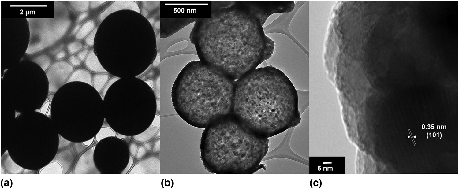
From the TEM characterization, it is observed that the morphology from the synthesized CS is a well-defined spherical shape with sizes of approximately 2.5 µm, from cyclodextrin precursor, as shown in Fig. 1(a).
TEM images of (a) CSs synthesized at 200 °C from cyclodextrin carbon precursor at 16 h synthesis time, (b) HTiO2NS calcinated at 550 °C for 4 h, from cyclodextrin carbon precursor template, and (c) high-resolution TEM (HRTEM) of TiO2 with an anatase crystal lattice.

HTiO2NS hollowness and size
The formation of the hollow HTiO2NS takes places between the burning of the CSs at approximately 300 °C and the crystallization of TiO2 anatase phase at 550 °C. The resulting HTiO2NS decrease in volume by approximately 70% from the CS templates [Fig. 1(b)]. Raman spectroscopy analysis (Supplementary Fig. S1) shows the characteristic D- and G-bands for CSs (inset of Supplementary Fig. S1). The D- and G-bands do not appear after the crystallization of TiO2, which is indicative of no residual CS material left behind after the crystallization.
The obtained hollow structures are composed of smaller TiO2 crystals (approximately 25 nm) that make the outer shell of the structure, leaving a void in the center, as shown in Fig. 1(b).
Crystallinity
The obtained narrow peaks of Bragg reflections at 2θ = 25.53°, 37.99°, 48.24°, 62.88°, and 76.64° are attributed to (101), (004), (200), (204), and (215), respectively. These peaks are indicative of anatase TiO2 crystal phase, as shown in Supplementary Fig. S2. To further confirm the crystallinity of the TiO2, HRTEM was performed. Figure 1(c) shows the surface crystal lattice spacing of 0.35 nm corresponding to TiO2 (101) facet. Due to the presence of more functional groups on its surface than other crystal phases of TiO2, the anatase phase has been shown to have a higher photocatalytic activity.[Reference Luttrell, Halpegamage, Tao, Kramer, Sutter and Batzill29] By synthesizing anatase HTiO2NS and exposing it to UV light, we expect to produce a higher quantity of ROS for PDT.
Functionalization of HTiO2NS with FA and ZnPc
FTIR study and analysis
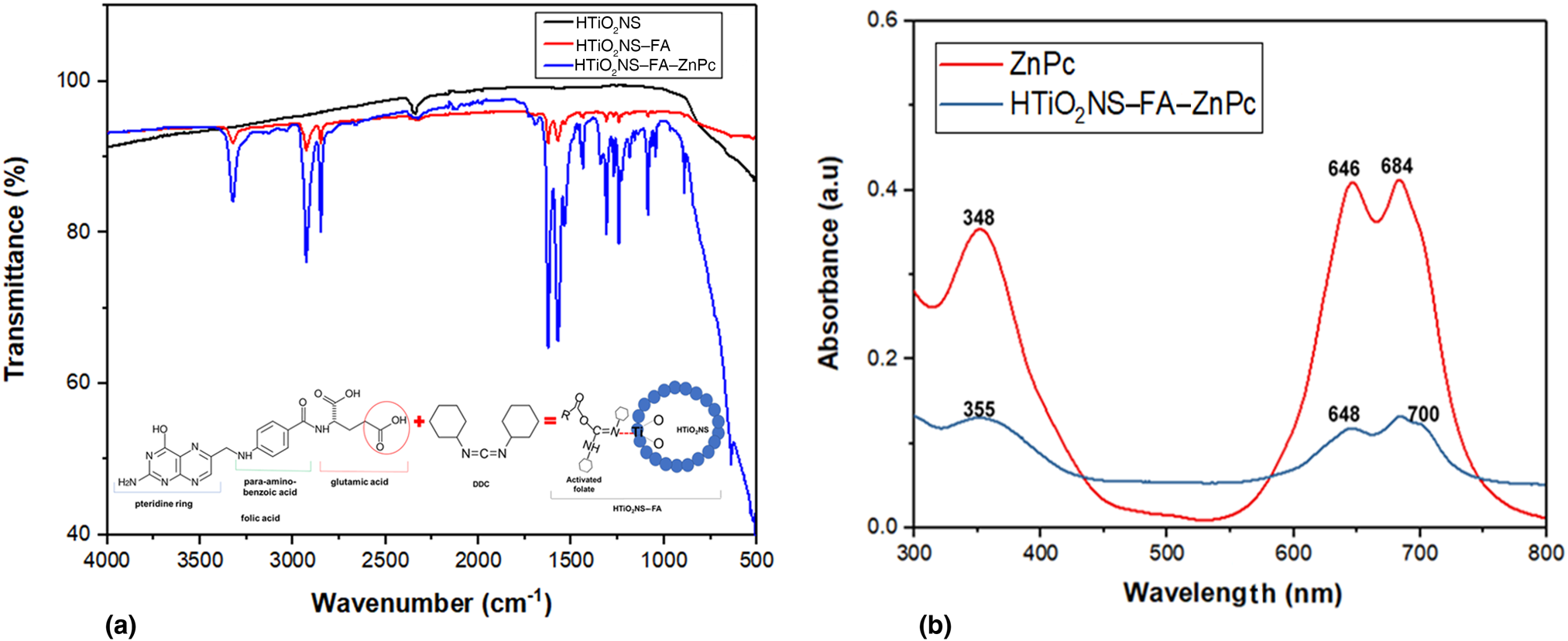
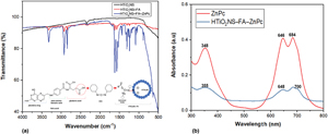
The IR spectra of HTiO2NS shows two peaks [Fig. 2(a)]. The peak at 2342 cm−1 is related to the O=C=O bonds and is commonly observed from CO2 from the environment. The broad peak below 1000 cm−1 is a peak due to the formation of Ti–O bonds from the network structure of TiO2. The HTiO2NS–FA spectrum shows the characteristic absorption peaks of FA. The peak at 3329 cm−1 corresponds to the N–H stretching vibration from the pteridine ring. The peak at 2932 cm−1 is attributed to vibrational C–H of CH2 groups from the pteridine ring and glutamic acid present in the FA. The peaks at 1625 and 1571 cm−1 belong to C=O and C=C vibrations in the FA, respectively. While the peak at 1533 cm−1 confirmed the formation of an N–Ti bond during the functionalization of HTiO2NS with the DDC-activated folate, as shown in the schematic of Fig. 2(a).[Reference Zhang, Rana, Srivastava and Misra30]
(a) FTIR spectra of HTiO2NS–FA–ZnPc and schematic of HTiO2NS–FA functionalization. (b) UV–Vis spectra of ZnPc and HTiO2NS–FA–ZnPc solutions in DMSO.

Although the HTiO2NS–FA and HTiO2NS–FA–ZnPc spectra are very similar, experimentally during functionalization a change in color from white to yellow (characteristic of FA) is observed after attaching FA to the HTiO2NS, and a change from yellow to blue is observed after loading ZnPc to HTiO2NS–FA. UV–Vis spectroscopy is used to further investigate the functionalization of HTiO2NS–FA by ZnPc.
UV–Vis study
The UV–Vis absorption spectra of ZnPc and HTiO2NS–FA–ZnPc are shown in Fig. 2(b). In the ZnPc spectrum, the typical energy bands for ZnPc: B-band (γ or Soret band) and Q-band (α-band in porphyrin) are observed,[Reference Shimizu, Kobayashi, Akasaka, Osuka, Fukuzumi, Kandori and Aso31,Reference Machado, França, Velani, Magnino, Velani, Freitas, Müller, Sattler and Schmücker32] with a maximum at 348 and 684 nm, respectively. Those bands result from the π–π electronic transitions of the phthalocyanine rings. In the HTiO2NS–FA–ZnPc spectrum, the B- and Q-bands appeared slightly displaced from the ZnPc spectra. Both peaks are broadened and red shifted. These changes are indicative of the interaction between the ZnPc and the HTiO2NS–FA.
ROS detection
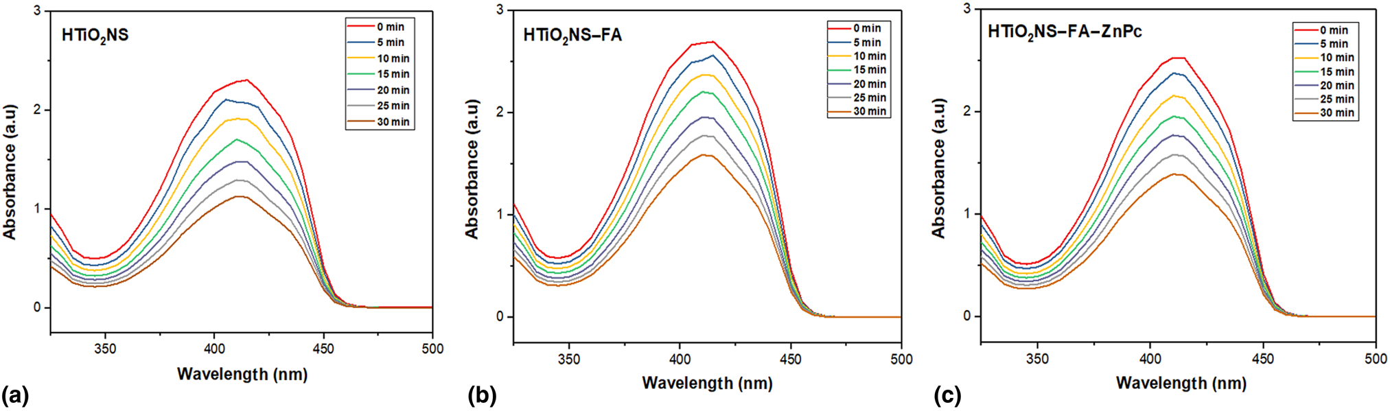
ROS detection was performed using DPBF as a chemical probe [Figs. 3(a)–3(c)]. To generate ROS (1O2), HTiO2NS, HTiO2NS–FA, and HTiO2NS–FA–ZnPc were exposed to irradiation from a 365 nm UV lamp for different times in the presence of the chemical probe. DPBF is a reductant material that can react with 1O2. It has a typical UV absorption at 415 nm when irradiated by the 365 nm UV light. An indication of ROS presence is the decrease of DPBF absorption, which reduces in concentration as it reacts with the 1O2. The observable reduction of DPBF absorption as the irradiation time increases is seen in Figs. 3(a)–3(c), demonstrating the successful generation of 1O2 by our proposed structures.
(a–c) Detection of ROS generation via a DPBF fluorescent probe.

Cytotoxicity
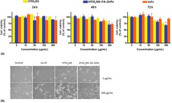
GBM cell lines are commonly used as an in vitro brain tumor model to evaluate the anti-cancer effect of novel therapeutics. In vitro cytotoxicity was assessed using MTT as a cell viability indicator after treatment with ZnPc, HTiO2NS, and HTiO2NS–FA–ZnPc at different concentrations [Fig. 4(a)]. Strikingly, nanoparticles at 1–100 µg/mL concentrations had no significant effect on cell viability when compared to the control. However, in the case of HTiO2NS–FA–ZnPc, significant viability decreases were found for 500 µg/mL concentration. The data indicate that nanoparticles are biocompatible with MK cell line. As ZnPc and HTiO2NS are photoresponsive to UV–Vis light, further experiments of nanoparticles activated by irradiation will be necessary to measure the photodynamic effects on cell.
(a) Dose and time cytotoxicity effect of ZnPc, HTiO2NS, and HTiO2NS–FA–ZnPc nanoparticles over cell viability in M059K cell line. Cells were treated at 5, 10, 50, 100, and 500 µg/mL and assayed at 24, 48, and 72 h. Results are the mean ± SEM, n = 3. *P < 0.01 versus control. (b) Representative images of the effects of nanomaterials on M059K cellular morphology after 24 h incubation time at 1 µg/mL (top row) and 100 µg/mL (bottom row).

Cell morphology
To asses the effect of the nanomaterials over MK cell line, we used untreated cells as control and Cis-Pt as a positive cytotoxicity control for comparison [Fig. 4(b)]. After incubation for 24 h with HTiO2NS and HTiO2NS–FA–ZnPc concentrations at 1 and 100 µg/mL, no grow inhibition or changes in morphology were observed. However, in the presence of Cis-Pt, at a concentration of 100 µg/mL, the cells had shrinken, had irregular shapes, and were detached from culture dishes. We also recognized the accumulation of nanoparticles aggregates mostly in the cell cytoplasm (green fluorescence) and differentiated from the cell nucleus (blue fluorescence) (Supplementary Fig. S3). However, further experiments will be necessary to determinate the exact organelle cell localization of the HTiO2NS–FA–ZnPc nanostructures.
Conclusion
In summary, HTiO2NS were synthesized using a facile and low-cost method. Functionalization of these hollow structures with FA and ZnPc for targeted PDT applications was achieved by modification of their surface. Viability of the HTiO2NS–FA–ZnPc nanostructures on MK cancer cells was demonstrated. Additionally, the in vitro activation of HTiO2NS–FA–ZnPc by UV irradiation was successful. In the future, further experiments will be neccesary to study the photodynamic effects of the proposed nanostructures on cells. These results show promising use of HTiO2NS as a nanostructure for targeted treatment of GBM cancer by PDT, as well as their potential as a delivery mechanism for drug delivery due to their hollow structure.
Supplementary material
The supplementary material for this article can be found at https://doi.org/10.1557/mrc.2019.142.
Acknowledgments
This material is based upon work supported by a grant from the University of California Institute for Mexico and the United States (UC MEXUS) and the Consejo Nacional de Ciencia y Tecnologia de Mexico (CONACYT; UCR-17091220).