Introduction
With advances in systemic therapy for breast cancer, brain metastases (BM) have emerged as a major source of morbidity and mortality. There has been increasing recognition that the natural history of breast cancer and propensity for BM is variable and highly influenced by breast cancer sub-type; with HER2-positive and triple-negative tumours most likely to develop BM. Reference Hicks, Short and Prescott1–Reference Smid, Wang and Zhang3
Furthermore, a number of patient’s tumour and treatment-related characteristics have emerged as prognostic factors for survival in breast cancer patients with BM. Reference Nam, Kim and Han4–Reference Niwińska, Murawska and Pogoda7 This has led to identification of subsets of breast cancer patients with BM who survive longer and have a better prognosis. As a result, the management of BM in patients with breast cancer has become increasingly nuanced, with emphasis on locally ablative treatment options like resection or stereotactic radio surgery (SRS)/stereotactic radiotherapy (SRT), with an aim of achieving better tumour control with lesser neurocognitive decline.
SRS involves the delivery of single, high dose of radiation, which is considered tumouricidal, ablative and/or vascular sclerosing/thrombosing, and allows for a high probability of local control. The use of high doses of radiation yield tumour control rates similar to that of surgery, without the invasiveness of a surgical resection. Reference Schulder, Patil, Chin and Regine8,Reference Leksell9 With SRT, the same principles of SRS apply, although the treatment is fractionated, as opposed to single fraction delivery. Reference Marcrom, McDonald and Thompson10 SRS/SRT are well-recognised treatment option for patients with limited intracranial disease (1–4 BMs) with optimal prognostic factors. Reference Mazzola, Corradini, Gregucci, Figlia, Fiorentino and Alongi11 SRS may be used as a first line treatment option without whole-brain radiation therapy (WBRT) in patients with a limited number of metastases; two randomised studies were unable to demonstrate a survival benefit with the addition of WBRT. Reference Aoyama, Shirato and Tago12,Reference Kocher, Soffietti and Abacioglu13 An SRS boost after WBRT improves overall survival (OS) in patients with single BM. Reference Andrews, Scott and Sperduto14 SRS may also be used in salvage setting at the time of intracranial recurrence. Reference Kelly, Lin, Claus, Quant, Weiss and Alexander15
In patients treated with gamma-knife based SRS, head immobilisation is generally achieved by invasive head fixation using a stereotactic ring. Reference Schulder, Patil, Chin and Regine8,Reference Leksell9 With the advent of LINACs with image-guided radiotherapy (IGRT) capability, conventional frame-based radiosurgery for BM has largely been replaced by frameless delivery using the thermoplastic cranial mask immobilisation system. The advantage of the mask is that it is non-invasive, convenient, no admission required and treatment planning can be done one or more days before treatment delivery. Reference Hartgerink, Swinnen and Roberge16,Reference Opp, Feygelman and Sarangkasiri17
The majority of the clinical results with SRS in treatment of BM, especially in breast cancer are based on treatment with conventional frame-based radiosurgery, while data with LINAC-based SRS are limited to a few studies to date. Reference Kelly, Lin, Claus, Quant, Weiss and Alexander15,Reference Combs, Schulz-Ertner, Thilmann, Edler and Debus18–Reference Armstrong, Ward and Dunne20 In this retrospective study we aim to report clinical outcomes such as local control, PFS and OS with LINAC-based SRS/SRT alone or as a boost after WBRT for BM in patients of breast cancer and to also explore prognostic factors affecting these outcomes.
Methods
Clinical and dosimetric records of breast cancer patients treated with LINAC-based SRS/SRT for BM at our institute between May, 2015 and December, 2019 were retrospectively reviewed. Patients who received WBRT or underwent surgery for BM prior to SRS/SRT delivery were also included for analysis. Departmental Ethics Committee approved the study.
Standard patient demographics, disease and treatment characteristics were recorded.
Patient selection
Patients of previously treated or newly diagnosed breast cancer with at least a radiological diagnosis of BM; 1–4 in number, ≤3·5 cm in maximum dimension, with a Karnofsky Performance Score (KPS) of ≥60 were taken up for treatment with SRS. SRT was generally considered if a tumour was >3·5 cm in diameter, near a critical or eloquent structure, or if the proximity of moderately sized tumours would lead to dose bridging in a single-fraction SRS plan.
Radiotherapy planning and delivery
Radiotherapy planning was based on contrast-enhanced simulation computed tomography (CT) with 1 mm slice thickness. Patients were immobilised in a thermoplastic mask. In each patient, the gross tumour volume (GTV) was delineated using post contrast thin-slice (1 mm) gadolinium-enhanced T1-weighted axial magnetic resonance imaging (MRI) sequences fused with contrast-enhanced simulation CT scan. The clinical target volume (CTV) was a zero-margin-expansion of the GTV. The Planning Target Volume (PTV) was defined as GTV plus margin which ranged from 3 to 5 mm while keeping in mind the set-up uncertainty and time elapsed between planning and treatment delivery. The organs at risk (OAR) such as optic nerves, chiasm and brainstem were also contoured.
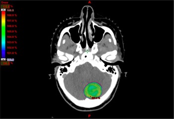
The prescribed SRS dose ranged from 7 to 24 Gy (prescribed at 90% isodose) depending upon the tumour size (24 Gy for <2 cm, 20 Gy for 2–2·5 cm, 18 Gy for 2·5–3 cm, 15 Gy for 3–3·5 cm and 12 Gy for 3·5–4 cm) Reference Shaw, Scott and Souhami21 , proximity to critical structures and prior WBRT dose (in cases where SRS was given after WBRT as boost or salvage). A dose of 27 Gy in 3 fractions was prescribed for SRT. Reference Minniti, Scaringi and Paolini22 The prescribed dose covered 100% of the target volume (Figure 1). Doses to the optic nerves, chiasm and brainstem were limited to 8–10 Gy. Reference Milano, Usuki, Walter, Clark and Schell23 Conformity index (defined as the coverage ratio multiplied by the selectivity ratio) and gradient index (defined as the ratio of the volume of half the prescription isodose to the volume of the prescription isodose) were calculated for plan evaluation. Reference Paddick24,Reference Paddick and Lippitz25 Varian Medical system Eclipse treatment planning system was used to make single/multiple isocenter Volumetric Modulated Arc Therapy (VMAT) with RapidArc® (Varian, Mumbai, India) plans and treatment was delivered using Trilogy cLINAC (Varian) with Brain LAB 6D couch (Varian). Cone beam CT was used for image guidance.
SRS plan with dose color wash image showing PTV coverage with 100% of the prescribed dose.

Follow up
The standard follow-up consisted of clinical examination and MR imaging at 6 weeks post-SRS/SRT and then every 3 months thereafter. Tumour response was classified into categories (complete response, partial response, progressive disease or stable disease) based upon the percent change in tumour dimension evaluated on contrast-enhanced MRI, corticosteroid use and clinical status in accordance with the RANO criteria for brain metastases (RANO-BM). Reference Eisele, Wen and Lee26 Recurrence was defined as either local or distant intracranial. Local recurrence at the site of SRS was defined as an increase in the diameter of the contrast-enhancing lesion of at least 25% on follow-up MRI. Distant intracranial recurrence was defined as the presence of a new enhancing lesion consistent with a BM or leptomeningeal enhancement outside the SRS target volume. Subsequent recurrence events after the first recurrence event were not recorded.
Statistical analysis
The initial analysis included descriptive statistics of clinical and demographic information. Estimates of intracranial progression-free survival and all OS were calculated using Kaplan–Meier method and subgroups compared using log rank test. Intra-cranial progression free survival was calculated from the date of SRS/SRT and was defined as time to local recurrence, distant intracranial progression or death. OS was calculated from the date of SRS/SRT to death due to any cause. The effect of clinical and demographic covariates on survival was estimated using a Cox proportional hazards model. Variables with a p value <0·1 on univariate analysis were used to construct a multivariate model. All statistical tests were 2-sided and used a significance level of <0·05. All analyses were performed using SPSS version 23.
The following potential prognostic factors were analysed: patient age at the date of SRS (dichotomised at 50 years); KPS at the time of SRS (<70 versus ≥70); recursive partitioning analysis (RPA) class (1 versus 2 versus 3); the number of metastases treated with SRS (1 versus ≥2); size of BM (<2 versus 2–3 versus ≥3 cm for first lesion and <2 versus ≥2 cm for second lesion), PTV margin (3 versus 5 mm), PTV volume (<35 versus ≥35 cc for first lesion and <10 versus ≥10 cc for second lesion), the interval between initial diagnosis of breast cancer and development of BM(<2 versus ≥2 years); the status of primary disease (controlled versus uncontrolled), presence and number of extracranial metastatic sites (1 versus ≥2); administration of palliative chemotherapy at the time of BM; ER/PR status; HER2 status; and triple-negative status (assigned on the basis of immunohistochemistry carried out on the primary tumour at the time of original diagnosis).
Results
Patient and disease characteristics
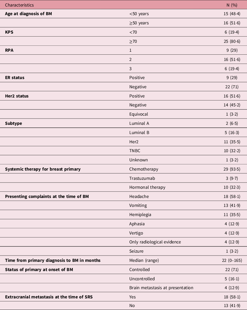
In total, 40 tumours were treated by SRS/SRT in 31 patients. All patients were females. The median age at presentation of BM was 50 years (range 29–75 years). The median interval from diagnosis of breast cancer to the development of BM was 22 months (range 0–165 months). 4 patients had BM at presentation. 18 (58·1%) patients had extracranial metastases as well; the number of metastatic sites varying from 1 to 4. Primary disease was controlled in most (71%) of the patients at the time of diagnosis of BM. Notably, 16 of the 31 patients (51·6%) had Her2 receptor positivity and 10 (32·3%) patients had triple negative status. The most common symptoms at presentation were headache (58·1%) and vomiting (41·9%). Perilesional oedema was noted in 24 (77·4%) patients. The metastatic lesions were close to critical structures (brain stem and optic tract) in 11 (35·5%) patients. Patient, disease and baseline treatment characteristics are listed in Table 1.
Patient, disease and baseline treatment characteristics

Abbreviations: TNBC, triple negative breast cancer; BM, brain metastases; KPS, Karnofsky Performance Score; RPA, recursive partitioning analysis; ER, estrogen receptor.
Treatment characteristics
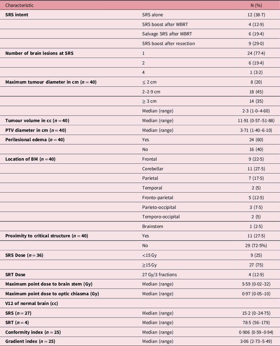
SRS was delivered in 27 patients and SRT in 4 patients. The number of BM ranged between 1 and 4, with a majority (77·4%) of patients having one lesion. Six patients (19·4%) had two lesions and only one patient (3·2%) had 4 lesions at the time of SRS/SRT. Descriptive analysis for characteristics of BM was done for each lesion separately. The median tumour diameter was 2·3 cm (range 1·0–4·6 cm) and the median tumour volume was 11·91 cc (range 0·57–51·88 cc). The median PTV diameter was 3·71 cm (range 1·4–6·10 cm).
Ten (32·6%) patients received prior WBRT. SRS/SRT was given as a boost to WBRT in 4 patients and as salvage for progression after WBRT in 6 patients. In this subgroup of patients, the median interval between WBRT and SRS/SRT was 6·5 months (3–24 months). Nine patients underwent prior surgery, of which, 8 patients had a gross total excision (GTE). Stereotactic Biopsy (STB) was performed in one patient. The WBRT dose was 30 Gy in 10 fractions over 2 week (n = 29) or 20 Gy in 5 fractions over 1 week (n = 2).
The median prescribed SRS dose was 15 Gy (range 7–24 Gy) and all four patients received SRT with a dose of 27 Gy in 3 fractions. The median maximum point dose to the brain stem was 5·59 Gy and that to the optic chiasm was 0·97 Gy. The calculated median V12 for the normal brain (minus PTV) was 15·92 cc in patients receiving SRS and 78·5 cc in patients who received SRT. The median conformity index (available for 25 lesions in 21 patients) was 0·906 (range 0·59–0·94) and the median gradient index was 3·06 (range 2·73–5·49) (Table 2).
SRS/SRT treatment parameters and dosimetric characteristics

Abbreviations: SRS, stereotactic radiosurgery; WBRT, whole brain radiotherapy; PTV, planning target volume; SRT, stereotactic radiotherapy; BM, brain metastases.
Systemic therapy
Total 18 (58·1%) patients also received palliative chemotherapy for systemic metastases, 8 (25·8%) patients received trastuzumab and hormonal therapy was given to 5 (16·1%) patients.
Side effects
SRS treatment was very well tolerated in nearly all of the patients. Three patients developed perifocal edema evidenced by clinical symptoms; and nausea and vomiting were reported in three patients. One patient developed radionecrosis within the irradiated volume demonstrated on follow up MRI. This patient had received trastuzumab concurrently with SRS. No focal neurological deficit or treatment-related deaths were noted.
Local and distant intracranial control
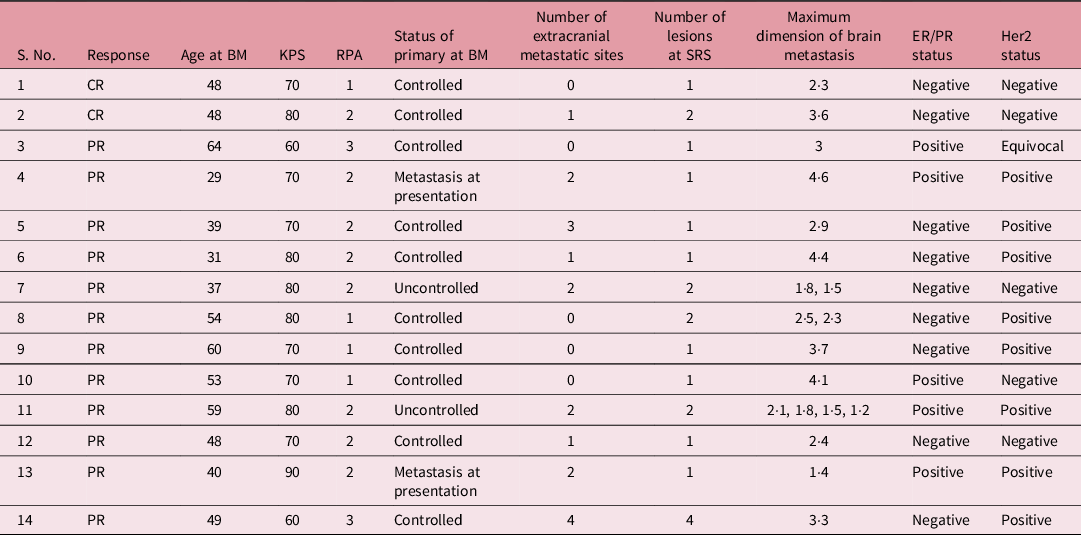
Two patients had complete response (CR), 12 patients had partial response (PR), 5 patients had stable disease (SD) and 12 patients developed progressive disease (PD) after SRS/SRT. Of these, four patients presented with clinical signs of progression even before the first follow-up date, which was then confirmed with a contrast-enhanced MRI.
Median follow-up was 7·9 months (0·2–34 months). Twenty (64·5%) patients developed local recurrence and 10 (32·3%) patients developed distant intracranial relapse. Of these, seven patients had both local and distant intracranial relapse.
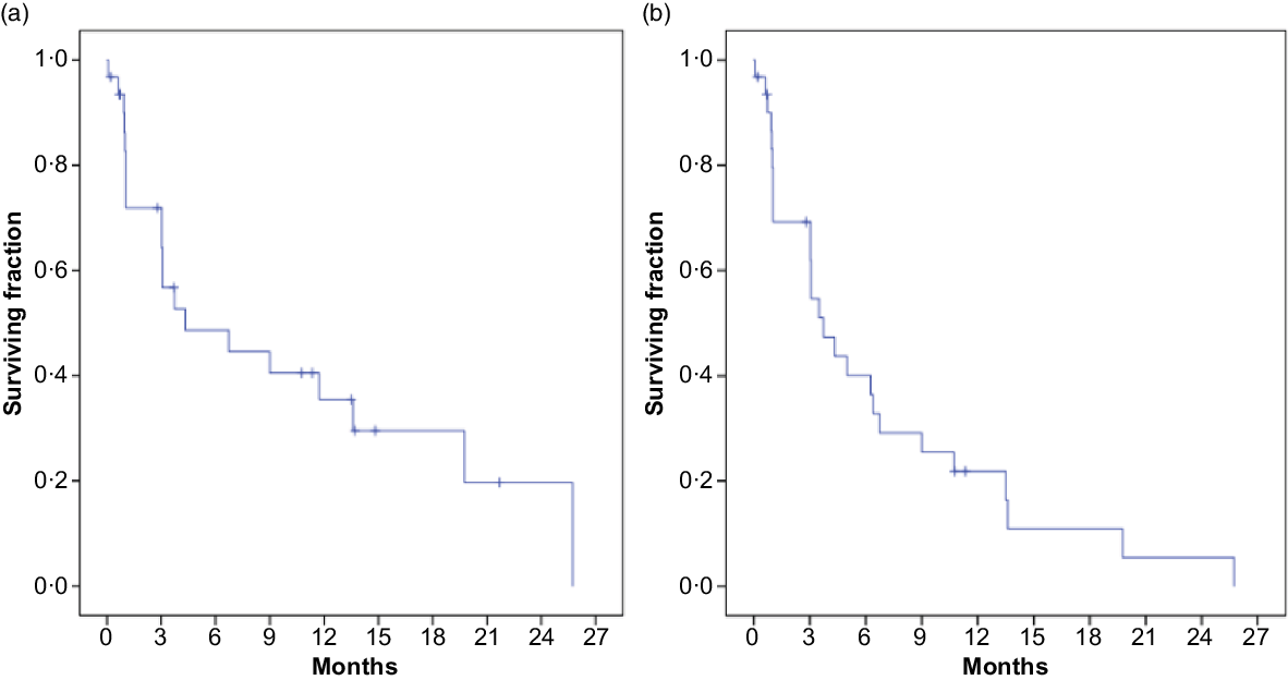
The estimated local control at 6 months and 1 year was 48 and 35%, respectively. The median time to local recurrence was 4·33 months (Figure 1a). The median time to development of distant intracranial relapse was 13·6 months in patients who received prior WBRT versus 6·73 months in patients who did not (p = 0·395). Six patients developed systemic relapse. Only four patients received salvage therapy in the form of WBRT and three received chemotherapy at the time of relapse.
Survival and prognostic factors
The median intracranial PFS was 3·73 months (range 0·2–25 months) (Figure 1b). On subgroup analysis, the median intracranial PFS was 3·02 months in patients who received SRS alone or as boost after WBRT, while it was 4·27 months in those who received SRS as salvage after WBRT (p = 0·793). No difference in intracranial PFS was observed in patients who underwent prior surgery versus patients who did not (p = 0·410).
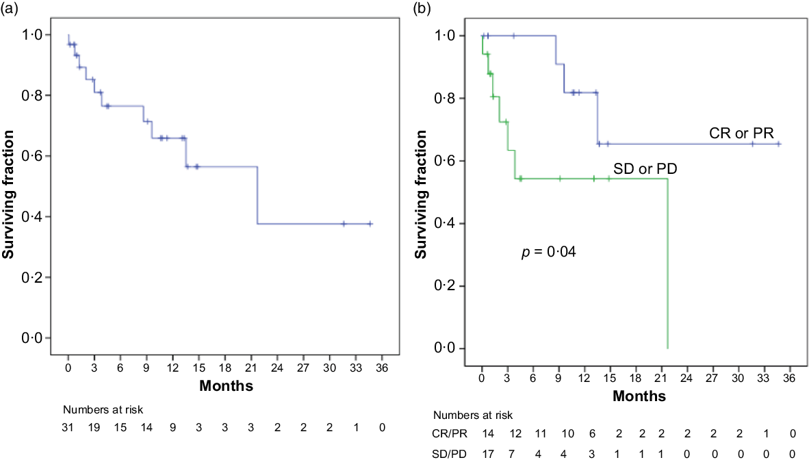
In total, 10 (32%) of the 31 patients died during follow-up. The median OS was 21·7 months (range 0·2–34 months) for the entire cohort (Figure 2a). Patients who received prior WBRT had a poor OS (13·31 months), as compared to those with SRS alone (21·4 months) (p = 0·699).
(a) Local control (b) Intracranial progression free survival

(a) Overall survival (b) Overall survival stratified by response to SRS, significant superior OS in patients with CR or PR to SRS/SRT

Univariate and multivariate analysis of the potential prognostic factors did not yield any significant factor affecting PFS or OS. However, it was observed that patients who had a CR or PR to SRS/SRT had a significant better OS as compared to those who had SD or PD (p = 0·04) (Figure 2b). Median OS in patients with SD/PD was 21·7 months while it was not reached in patients with CR/PR.
Discussion
This study describes the characteristics and clinical outcomes of breast cancer patients with BM who were treated with LINAC based SRS/SRT either alone or sequentially after WBRT and/or surgery. It was observed that none of the treatment strategies was superior to the other in terms of disease control. Patients with better response to SRS/SRT had a better survival.
SRS/SRT with or without WBRT is an evolving paradigm in the management of patients with limited BM with optimal prognostic factors. With advances in modern systemic therapy for breast cancer, an increasing number of patients are experiencing central nervous system (CNS) progression events for which CNS-directed treatment modalities are being considered. Quality of life and prevention of neurologic deficit are important goals of care in this population. SRS is a single-visit outpatient procedure that is associated with minimal acute toxicity and good local control rates. It is a feasible treatment option particularly in the era of frameless radiosurgery. However, breast cancer is not well represented in published phase 3 randomised studies of SRS, comprising only 6·8 to 11·7% of the study populations. Therefore, the direct applicability of data from these trials to patients with breast cancer, for whom prognostic considerations, systemic therapy options, and competing risks may be unique, remains a limitation to current practice.
The current study focused on BM only in patients with breast cancer. The distribution of biological and tumour subtype in our study cohort is consistent with published reports. Reference Hicks, Short and Prescott1,Reference Heitz, Harter and Lueck2 HER2 overexpressing and triple negative breast cancers comprised 51·6 and 32·3% of the total number of patients. Several studies have observed that HER2+ breast cancer is associated with a higher incidence of subsequent BM. Hicks et al. reported HER2 overexpression as a strong predictive factor for the development BM in breast cancer. Reference Hicks, Short and Prescott1 Similarly, Heitz et al. found that patients with triple-negative or HER2+ breast cancer have a higher risk for BM compared with patients bearing the ER+/HER2− phenotype and develop BM earlier in the course of disease. Reference Heitz, Harter and Lueck2
The median time to development of BM in our study population was 22 months, largely due to over representation of HER2+ and triple negative breast cancers in our cohort. Sperduto et al. in an analysis of 865 breast cancer patients with newly diagnosed BM found that basal and HER2+ tumour subtypes have shorter time to development of BM than patients with luminal A and B subtypes (27·5 and 35·8 months versus 54·4 and 47·4 months respectively); though it was not a significant prognostic factor for survival. Reference Sperduto, Kased and Roberge27 Similarly, we could not demonstrate any significant difference in OS among patients who were diagnosed with BM within and after 2 years of the primary diagnosis (p = 0·838).
Our outcomes are also comparable to those demonstrated in previous studies. The median time to local recurrence in our patients was 4·33 months and the median intracranial PFS was 3·73 months. In a retrospective analysis of 48 patients treated with LINAC-based SRS for BM, the median time to local failure was 4·8 months and median intracranial PFS was 3·73 months. Histology of breast cancer was associated with significantly worse PFS (p = 0·004, median PFS 2·27 versus 4·37 months). Reference Bilger, Frenzel and Oehlke19 In another study by Kelly et al. they observed a median CNS recurrence-free survival time of 5·7 months after LINAC-based salvage SRS for initially treated BM in breast cancer patients, with an associated median survival in excess of 9 months. Reference Kelly, Lin, Claus, Quant, Weiss and Alexander15
In a retrospective study of breast cancer patients treated with LINAC-based SRS for BM, Combs et al. reported a median overall loco-regional brain control of 6·5 months for the SRS only group, 4 months for WBRT+SRS boost group and 7 months for WBRT and salvage SRS group, the difference not being statistically significant. Reference Combs, Schulz-Ertner, Thilmann, Edler and Debus18 In our study also, there was no significant difference in intracranial PFS between these groups, though the WBRT and salvage SRS group had a slightly higher PFS (4·27 versus 3·02 months, p = 0·793). Patients who received prior WBRT had a longer time to distant intracranial relapse (13·6 months versus 6·73 months, p = 0·395). In a randomised controlled trial by Aoyama et al., it was demonstrated that the use of WBRT + SRS did not improve survival in patients with 1 to 4 BM as compared with SRS alone, but intracranial relapse occurred more frequently in those who did not receive WBRT and salvage treatment was frequently required. Reference Aoyama, Shirato and Tago12 The results of the EORTC phase III trial also suggested that after SRS or surgery of a limited number of BM, adjuvant WBRT reduces intracranial relapses and neurologic deaths but fails to improve the duration of functional independence and OS. Reference Kocher, Soffietti and Abacioglu13
In our study nine patients underwent prior surgery. There was no difference in local control or intracranial PFS among patients who underwent surgery and the rest of the cohort. In patients with resected BM, post-operative SRS/SRT appears to further reduce the risk of local disease recurrence within the surgical cavity. Mahajan et al. reported a randomised trial of post-operative cavity SRS versus observation after brain tumour resection, in which SRS improved the 1-year freedom from local failure (72 versus 43%, p = 0·015). Reference Mahajan, Ahmed and McAleer28
SRS/SRT was well tolerated by our patients with only a few side effects. One patient developed radionecrosis within the irradiated volume, which was demonstrated on follow-up MRI and was not clinically significant. This patient had received trastuzumab concurrently with SRS. Studies have reported strong correlation between the development of clinically significant radionecrosis (CSRN) after SRS and trastuzumab possibly due to upregulation of Aquaporin-4 resulting in astrocyte swelling and increased radiation-induced cytotoxicity. Reference Stumpf, Cittelly and Robin29
The median OS in our study was 21·7 months, with no significant difference between patients who received prior WBRT and patients who did not. Notably, patients who had CR or PR to SRS had a significantly better OS than patients who had SD or PD (p = 0·04, Figure 3), suggesting that response to SRS translates into a better OS in breast cancer patients with BM. This reflects the importance of careful identification of subgroups of breast cancer patients who are likely to benefit from locally ablative treatment options like SRS/SRT. The preservation of neurocognitive function and quality of life in this group of patients who survive longer is of paramount importance after the treatment of BM. Patient-wise characteristics of this subset have been illustrated in Table 3.
Characteristics of patients with complete and partial response to SRS/SRT

Abbreviations: CR, complete response; PR, partial response; BM, brain metastases; SRS, stereotactic radiosurgery; KPS, Karnofsky Performance Score; ER, estrogen receptor; PR, progesterone receptor; RPA, recursive partitioning analysis.
Several studies have reported patient and disease-specific prognostic factors such as age, performance status, RPA class, presence and burden of extracranial disease, Her2 overexpression, use of systemic therapy, size of BM etc., that might correlate with OS in patients of BM. Reference Niwińska, Murawska and Pogoda7,Reference Kelly, Lin, Claus, Quant, Weiss and Alexander15,Reference Melisko, Moore, Sneed, De Franco and Rugo6,Reference Tanguturi and Warren30 These factors can then be used to direct treatment or predict prognosis in the future. We could not demonstrate a significant impact of any of these prognostic factors on the survival end-points or treatment response in our study.
Our study has some limitations such as small patient number, retrospective nature and single institutional. Therefore, intention to treat as well as patient selection bias cannot be ruled out. Assessment of neurocognitive function before and after therapy was not performed and toxicities may be under-reported.
Nonetheless, this analysis provides a comprehensive overview of the characteristics and outcomes of breast cancer patients treated with LINAC-based SRS/SRT for brain BM, along with a detailed description of the dosimetric analysis. To the best of our knowledge, this is the first study to report the clinical outcomes of Indian breast cancer patients who were treated with LINAC-based SRS/SRT for BM. This study also supports the considerable available literature suggesting that BM from breast cancer are heterogeneous and differ in terms of tumour biology and prognosis. All treatment strategies were equally represented in the study cohort, though no definite conclusion could be made with respect to the ideal treatment strategy or subgroup of patients who are likely to benefit with this technique. It may be suggested that there may not be any difference in outcomes with WBRT ± SRS or SRS/SRT with or without surgery, but it should be interpreted with caution
Conclusion
SRS/SRT is an effective treatment option for upfront and salvage treatment of BM in breast cancer. Further prospective studies with larger patient numbers should be performed to define subpopulations of breast cancer patients with BM who are most likely to benefit or not benefit from this technique and who may be better served by trials of systemic therapy or by a transition to supportive care.
Acknowledgements
We thank the patients who participated in the study.