Supraventricular tachycardia (SVT) occurs in 6.7% of children with congenital heart disease (CHD).Reference Tripathi, Black, Park and Jerrell 1 It presents most commonly in children <12 months of age with a secondary spike around adolescence. Supraventricular tachycardia in this patient population may be associated with the underlying structural abnormality of the heart or with surgical/interventional procedures.Reference Kanter 2 In most instances, supraventricular tachycardia can be adequately controlled with medications. In infants, catheter ablation is reserved as a secondary treatment option because of the technical difficulty and the procedure’s higher risk of complications. Such factors are compounded in the infant with CHD. Patients with significant CHD, however, may be less tolerant of the haemodynamic consequences of supraventricular tachycardia, making catheter ablation a more favourable treatment option. When ablation is attempted in infants, steps are taken to minimise risk, including radiation exposure.
We report a case series of two neonates with significant CHD in whom catheter ablation was performed without fluoroscopy, using three-dimensional mapping. The first infant had pulmonary atresia with intact ventricular septum with SVT. The second infant presented with Ebstein’s anomaly of the tricuspid valve with SVT. These cases suggest that current approaches to minimising fluoroscopy may be useful even in challenging patients such as neonates with CHD.
Case number one
A full-term, 3.6-kg, female infant was cyanotic at birth and was noted to have a murmur. On physical examination, she was a well-developed, non-dysmorphic infant in no distress. Her room air arterial saturation was 78%. Cardiac examination revealed a normal S1, a single S2, and a 2/6 holosystolic murmur heard at the left lower sternal border. Peripheral pulses and perfusion were adequate. Her echocardiogram showed pulmonary atresia with intact ventricular septum, a large patent duct arteriosus, moderate tricuspid regurgitation, and coronary sinusoids. Wolff–Parkinson–White (WPW) patterns was noted on electrocardiogram (EKG).
To maintain ductal patency, prostaglandin treatment was initiated. A diagnostic catheterisation showed right ventricle (RV)-dependent coronary circulation, and therefore no attempt at pulmonary valvuloplasty was made. In the post-catheterisation period, she had recurrent narrow complex atrioventricular re-entrant tachycardia at 340 bpm, which was poorly tolerated with systolic blood pressures of 30 mmHg and poor perfusion. The episodes were terminated with intravenous (IV) adenosine. Even after treatment with intravenous amiodarone, she continued experiencing episodes of tachycardia with haemodynamic compromise. With the need for a surgical shunt, there was concern that continued episodes in the postoperative period would be associated with greater haemodynamic consequences, and potential for clotting of her shunt. Therefore, the decision was made for a cardiac catheter ablation before surgery.
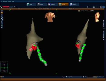
At 9 days of age, an electrophysiology (EP) study was performed under general anaesthesia. The EnSite Velocity System (St. Jude Medical, St. Paul, Minnesota, United States of America) was used for catheter guidance. The cutaneous patches required modification in order to fit the infant’s torso. A decapolar catheter was placed in the oesophagus for atrial pacing and sensing. A 6 Fr sheath was inserted into the left femoral vein. A 5 Fr Marinr SCXS RF ablation catheter (Marinr®; Medtronic Inc., Minneapolis, Minnesota, United States of America) was advanced into the right atrium. The radiofrequency (RF) catheter was used to create the geometry (Fig 1). We identified the location of the coronary sinus and His bundle to function as anatomical landmarks. The SVT could be easily induced and terminated with atrial pacing protocols. This permitted mapping of her pathway both anterograde and retrograde under controlled, stable circumstances, without the need for inotropic support or extracorporeal membrane oxygenation, although vasoactive medications were in line at the bedside. During her tachyarrhythmia, the earliest atrial activation was just anterior to the coronary sinus ostium. During sinus rhythm, the earliest ventricular activation mapped to the same area. Ablation was performed during sinus rhythm. Radiofrequency energy was delivered for 30 seconds with loss of accessory pathway (AP) conduction at 4 seconds. The temperature was set at 60ºC, and power was capped at 40 W, although the actual delivered power was closer to 20 W. The P-R interval was stable; three additional radiofrequency lesions were delivered for 30 seconds each. Fluoroscopy was available but not needed. The total procedure time was 112 minutes with no complications. Antiarrhythmic medications were discontinued at the end of the procedure. A central shunt was successfully placed the following day. At the 14-month follow-up, she has had no recurrence of her Wolff–Parkinson–White syndrome or supraventricular tachycardia.
Geometry of patient 1. Green catheter is the esophageal catheter. Bright red catheter is the ablation catheter. Dark red catheter marks the His bundle.

Case number 2
A 39-week gestation, 3.7-kg, female was born with meconium-stained amniotic fluid and severe distress. Her apgar scores were 0, 6, and 7 at one, five and ten minutes, respectively. Following resuscitation, her arterial saturations were 70–80% on 100% FiO2 by endotracheal tube. A chest X-ray revealed extreme cardiomegaly. Transferred for probable CHD, the physical examination indicated a non-dysmorphic infant. Cardiac examination showed a normal S1, whereas S2 splitting could not be appreciated. She had a 2–3/6 holosystolic murmur at the left lower sternal border and a 2/4 diastolic murmur in the same location. On echocardiogram, Ebstein’s anomaly was noted with an 18-mm displacement of the tricuspid valve towards the RV apex with severe tricuspid insufficiency. Prostaglandin was initiated to maintain ductal patency. She was also noted to have intermittent Wolff–Parkinson–White syndrome on telemetry.
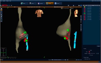
At 2 weeks of age, she began experiencing episodes of supraventricular tachycardia, atrioventricular re-entrant tachycardia, and was treated initially with atenolol. With recurrent episodes, amiodarone was added. She eventually progressed to incessant supraventricular tachycardia, which did not respond to various combinations of β blocker therapy, amiodarone drip, flecainide, and digoxin. At 28 days of life, she was urgently taken to the cardiac catheterisation laboratory. Under general anaesthesia, an oesophageal decapolar catheter was placed for atrial sensing and pacing, and a 5 Fr sheath was placed into the left femoral vein. The patient had adequate peripheral IV access for isuprel infusion, and therefore a 5 Fr sheath was used instead of a 6 Fr sheath. A 5 Fr Marinr SCXS RF ablation catheter was used to draw geometry (Fig 2). As in the earlier case, the cutaneous patches required modification to fit the infant’s torso. The His bundle was identified and marked, and the coronary sinus was located. Overdrive pacing was used to repeatedly terminate SVT each time it recurred, allowing maintenance of sinus rhythm throughout the case. Mapping identified a right posteroseptal accessory pathway just anterior to the coronary sinus ostium. Ablation was performed during sinus rhythm. The temperature was set at 60ºC, and power was limited to 40 W. Again, the actual delivered power was much less. Pathway conduction was eliminated on the third lesion. A total of seven lesions were delivered for 30 seconds each with no complications. Fluoroscopy was available, but not required to complete the procedure. The total procedure time was 110 minutes. All antiarrhythmic medications were discontinued at the completion of the procedure.
Geometry of patient #2. Blue catheter is esophageal catheter. Green sphere marks location of accessory pathway. Red ablation catheter is marking location of His bundle.

After prolonged hospitalisation, she was eventually weaned off prostaglandin and maintained adequate pulmonary blood flow without need for surgical intervention. At the 7-month follow–up, she has had no recurrence of her Wolff–Parkinson–White syndrome or supraventricular tachycardia.
Discussion
At present, three-dimensional mapping is emerging as an important tool for the management of arrhythmias in the catheterisation laboratory. It can decrease fluoroscopy time or, in many instances, eliminate the need for fluoroscopy. The Prospective Assessment after Pediatric Cardiac Ablation study reported that the average fluoroscopy time for ablation of supraventricular tachycardia in children was 38.3 minutes.Reference Van Hare, Javitz and Carmelli 3 This time can be significantly increased when performing the procedure on newborns and infants.Reference Blaufox, Felix and Saul 4 – Reference Erickson, Walsh, Triedman and Saul 6 In many institutions, catheter ablation of supraventricular tachycardia in the adolescent population with a normal heart is routinely performed without fluoroscopy.Reference Smith and Clark 7 , Reference Tuzcu 8 This is especially relevant when caring for children who, because of a long life expectancy, are at risk of demonstrating potential stochastic effects of radiation for many years to come. In addition, children with significant CHD often require multiple catheter procedures over time, resulting in an accumulation of significant radiation doses at young ages.
Several studies, however, have demonstrated that radiofrequency ablation in children <15 kg carries increased risk compared with children weighing>15 kg.Reference Kugler, Danford, Houston and Felix 9 This is because of the small size of the heart, limited vascular access, thin walls of vessels and cardiac chambers, as well as catheter designs unsuited to this population. Of the >100,000 ablations performed in the United States of America each year, <1% are performed in children, with <0.01% in infants, which demonstrates the infrequency of ablations in this population.Reference Blaufox, Felix and Saul 4
Electroanatomic mapping has proven successful in similar challenging procedures. These include adults with CHD and pregnant women with supraventricular tachycardia.Reference Leonelli, Tomassoni, Richey and Natale 10 – Reference Ferguson, Helms, Mangrum and DiMarco 12 To our knowledge, this report represents the first published cases of catheter ablation successfully performed without fluoroscopy in neonates with significant CHD. Both patients had haemodynamically compromising supraventricular tachycardia before the procedure. The cases bring to light several issues facing paediatric electrophysiologists: (1) the patients’ diminutive size and CHD both increase catheter ablation risks; (2) the tools available for catheter ablation are not designed for infants or young children. The smallest ablation catheter presently available (5F) is still far larger and stiffer than is ideal for use in infants. A cryoablation catheter is significantly larger, stiffer, and less manoeuverable than a 5 Fr radiofrequency catheter. It therefore cannot be used to draw geometry in a small patient. If cryoablation is necessary, a smaller, more manoeuverable catheter could be used to draw geometry and map the target, and then switched for a cryocatheter. Cryoablation would only be needed for substrates close to the atrioventricular (AV) node or the His bundle, which does not require much catheter flexibility to reach.
Given the patient’s small body surface area, use of Ensite system in this population is also difficult because of the large surface area of cutaneous patches. The average newborn has a body surface area of 0.25 m2. On the basis of burn center graphs, the torso accounts for approximately 32–36% of this. Therefore, a typical newborn will have a torso surface area of about 900 cm2. The total surface area of all the patches needed, including grounding pads, defibrillation patches, Ensite patches, and EKG leads, is 1380 cm2. Thus, there is 480 cm2 more patch than available skin. The Ensite patches have a surface area of 570 cm2 out of the package. As most of the patch is gel, only a small portion contains the actual wire mesh, which functions as the sensor. In these two cases, we trimmed the entire patch not containing wiring. This effectively removed 80–90% of the surface area of each patch. In this manner, all necessary electrodes were applied to the skin to complete the procedure.
It is our current practice to utilise three-dimensional mapping as much as possible for all ablation procedures and utilise fluoroscopy only when necessary. This may be more important in the newborn with CHD than in the teenager with a normal heart. Children with significant CHD will have to undergo many radiological procedures in their lifetime, and although we strongly agree with using fluoroscopy when needed, we also strive to eliminate unnecessary radiation exposure. Owing to the complexity of these cases, both procedures were performed in the cardiac catheterisation laboratory, so that fluoroscopy could be used if needed. Fluoroscopy was immediately available during each case, as it would facilitate therapy if a complication were to occur, such as tamponade. At the end of the procedures, however, we found that it was not needed. There are a few clinical situations where fluoroscopy is still necessary: patients with transvenous pacing leads or prosthetic valves, which could become entangled or entrapped by EP catheters. Moreover, we have had one patient requiring no sedation, who needed a transseptal puncture. In that case, fluoroscopy was used, as transesophageal echocardiogram (TEE) was not an option in a non-sedated child.
Details of three-dimensional images are limited in the smallest patients and in those with significant CHD. Therefore, in either situation, intracardiac electrograms are important to guide the procedure. The most important step is identifying and localising the His bundle. Once this is accomplished, location of the accessory pathway can be assessed relative to its distance from the His bundle. In each case, the His bundle was easily localised, and intracardiac electrograms predicted that the accessory connection was sufficiently distant from the His bundle to permit ablation.
Conclusion
Catheter ablation without fluoroscopy can be challenging in the smallest patients, especially those with CHD. Nevertheless, minimising radiation exposure may be quite important in this population. They often require multiple procedures throughout life, and their cumulative radiation exposure can become significant. With careful planning and modification of the three-dimensional mapping system patches, procedures without fluoroscopy may be accomplished safely and effectively. Availability of paediatric-sized surface patches would improve the ease and safety with which these procedures can be performed.
Acknowledgements
The authors thank the Rebecca D. Considine Research Institute for their administrative support. A.M.B. presented this abstract at the 2014 Cardiostim Congress in Nice, France. Authors’ Contribution: A.M.B., B.S.A., G.C.P., J.M.C. – study design, data collection, data analysis, manuscript preparation, and editing.
Financial Support
This research received no specific grant from any funding agency or from commercial or not-for profit sectors.
Conflicts of Interest
None.