INTRODUCTION
Intestinal helminths generally provoke changes in the morphology of the host tissues, which can in turn induce structural and functional alterations in the alimentary canal physiology of the vertebrates (Hoste, Reference Hoste2001). Several acanthocephalans species cause extensive damage to the gut and their pathogenicity is generally related to the depth of proboscis penetration (Taraschewski, Reference Taraschewski2000). Moreover, acanthocephalan species with trunk spines (e.g. Dentitruncus truttae) induce notable levels of damage by destroying the intestinal folds. Histopathology in salmonid fish as a result of helminth infection has appeared in a few records, and with reference to the cestodes, reports are available (see Reite, Reference Reite1997; Dezfuli et al. Reference Dezfuli, Pironi, Simoni, Shinn and Giari2007). Regarding the acanthocephalans, information has been provided by Hamers et al. (Reference Hamers, Jens Stürenberg and Taraschewski1992) and Bosi et al. (Reference Bosi, Arrighi, Di Giancamillo and Domeneghini2005a). Enteric parasites can induce inflammation of the host alimentary canal. The inflammatory response is the most primitive of the protective mechanisms (Reichlin, Reference Reichlin1999). The cellular involvement in the inflammatory response in teleostean fish could be biphasic, initiating with an influx of neutrophils followed by the subsequent arrival of monocytes/macrophages (Reite and Evensen, Reference Reite and Evensen2006). In fish, mast cells (MCs) and rodlet cells (RCs) are considered to be components of the teleost innate immune system and are closely linked to other piscine inflammatory cells (Reite, Reference Reite1997, Reference Reite2005; Silphaduang et al. Reference Silphaduang, Colorni and Noga2006).
Mast cells, also known as eosinophilic granular cells (EGCs), have been described in all classes of vertebrates and seem to be morphologically and functionally similar (Mulero et al. Reference Mulero, Sepulcre, Meseguer, Garcia-Ayala and Mulero2007). In fish exposed to pathological challenge, mast cells tend to accumulate in large numbers at the site of invasion (Reite, Reference Reite1997; Reite and Evensen, Reference Reite and Evensen2006). Fish mast cells, like their mammalian counterparts, exhibit partial or complete degranulation whenever they are exposed to pathogens (Vallejo and Ellis, Reference Vallejo and Ellis1989; Flaño et al. Reference Flaño, Lopez-Fierro, Razquin and Villana1996; Dezfuli and Giari, Reference Dezfuli and Giari2008) and/or to known degranulation agents (i.e. Compound 48/80) (Murray et al. Reference Murray, Leggiadro and Douglas2007). A plethora of studies in fish have shown that mast cells produce specific anti-microbial peptides and therefore are presumed to be directly involved in killing microbes (Silphaduang and Noga, Reference Silphaduang and Noga2001; Murray et al. Reference Murray, Leggiadro and Douglas2007; Silphaduang et al. Reference Silphaduang, Colorni and Noga2006). Recently it was reported that the mast cells of perciform fish are endowed with histamine, a substance which has been shown to regulate the fish inflammatory response (Mulero et al. Reference Mulero, Sepulcre, Meseguer, Garcia-Ayala and Mulero2007). Rodlet cells (RCs) are exclusive to fish and characterized by a thick fibrous capsule, a basal nucleus, and conspicuous inclusions called rodlets (Manera and Dezfuli, Reference Manera and Dezfuli2004). Despite the agreement on the development and stages of rodlet cells, their location, migration, origin and role are still controversial topics (Manera and Dezfuli, Reference Manera and Dezfuli2004; Jordanova et al. Reference Jordanova, Miteva and Rocha2007). The possible function of RCs as immune cells was derived from studies which reported an increase in the number of these cells in fish parasitized with protozoan (Leino, Reference Leino1996) and metazoan parasites (Reite, Reference Reite1997; Dezfuli and Giari, Reference Dezfuli and Giari2008).
Intestinal mucus is a glycoprotein secreted by specialized intestinal epithelial cells and covers the mucosa with a continuous adherent blanket or gel, which provides a physical barrier for microorganisms and their toxins (Lamont, Reference Lamont1992). In mammals, hyperplasia and hypertrophy of intestinal mucous cells in response to the presence of enteric helminths has been documented in several papers (Fairweather, Reference Fairweather and Beckage1997; Manjili et al. Reference Manjili, France, Sangster and Rothwell1998). Presently, little is known concerning parasitized fish and our results provide further evidence on the hyperplasia of mucous cells in the intestine of infected powan.
Several well-documented records have dealt with the influence of intestinal worms on the host-gut neuroendocrine system (Fairweather, Reference Fairweather and Beckage1997; Dezfuli et al. Reference Dezfuli, Arrighi, Domeneghini and Bosi2000a). Signal molecules, namely neuromodulators, are involved in the correct functioning of the neuroendocrine system (O'Dorisio and Panerai, Reference O'Dorisio and Panerai1990). Some records on fish presented the important role of immune and enteric neuromodulators in the inflammation process induced by enteric helminths (Dezfuli et al. Reference Dezfuli, Arrighi, Domeneghini and Bosi2000a; Bosi et al. Reference Bosi, Domeneghini, Arrighi, Giari, Simoni and Dezfuli2005b). High levels of serotonin (5-HT) and met-enkephalin in fish (Dezfuli et al. Reference Dezfuli, Arrighi, Domeneghini and Bosi2000a, Reference Dezfuli, Pironi, Giari, Domeneghini and Bosi2002 respectively) and high content of serotonin (5-HT) in mammals (Fairweather, Reference Fairweather and Beckage1997) were associated with the occurrence of intestinal worms. With regard to mammals, one of the major substances produced by mast cells is serotonin; nevertheless, little is known about the role of this neuromodulator in the fish immune system (Dezfuli et al. Reference Dezfuli, Arrighi, Domeneghini and Bosi2000a). Furthermore, it is documented that met-enkephalin in mammals has a role in the regulation of gut contractile activity (Fairweather, Reference Fairweather and Beckage1997) but there is lack of the same information in fish. Therefore, this study was undertaken to provide data describing the involvement of serotonin (5-HT) and met-enkephalin in the powan gut inflammation induced by Dentitruncus truttae.
The results from this investigation will be discussed alongside the observations made on the histopathology and ultrastructure of the infected powan immune cells and their role in the intestinal inflammatory response. Moreover, an insight into the ultrastructure of intestinal mucous cells, their high presence at the sites of infection and the roles of mucus will be presented.
MATERIALS AND METHODS
The digestive tracts of 30 Coregonus lavaretus (39·13±5·11 cm total length±s.d. standard deviation; 580·67±302·30 g total body weight±s.d.) were analysed from fish collected in 3 gill net samples (September 2006, April and October 2007) taken in Lake Piediluco (Province of Terni, Central Italy). The powan were removed from the nets, given a lethal dose of the anaesthetic MS222 (Sandoz), weighed and measured. The spinal cords of the anaesthetized fish were severed, and then the powan were dissected ventrally and sexed. The alimentary canals were removed, opened longitudinally and screened for helminths, which were recorded by number and position. Comparable intestinal regions were examined from healthy and parasitized C. lavaretus.
Pieces of powan intestine measuring up to 15×15 mm, with attached acanthocephalans, were excised from 10 infected fish and fixed in chilled (4°C) Bouin's for 8 h. The samples were then processed routinely for paraffin embedding, cut in 5 μm thick sections and then stained either with haematoxylin-eosin, Mayer's haematoxylin-alcian blue, toluidine blue, Giemsa or alcian blue/PAS, or used for immunohistochemistry. The latter was done according to the peroxidase-antiperoxidase method detailed in Dezfuli et al. (Reference Dezfuli, Pironi, Giari, Domeneghini and Bosi2002). Briefly, powan-acanthocephalan and uninfected tissue sections were processed using the indirect immunohistochemical method (peroxidase-anti-peroxidase immunocomplex). The sections were incubated with the primary antisera (met-enkephalin 1:300 and serotonin 1:500 raised in rabbit (codes AB 1975 and AB 938, Chemicon Int., Temecula, CA, USA) for 24 h at room temperature. Controls for the specificity of the immunohistochemical reactions (to exclude the occurrence of both cross- and aspecific-reactivity on tissue sections) were performed by pre-absorption of each antiserum with the corresponding antigen (met-enkephalin, code H 2785, Bachem AG, Bubendorf, Switzerland; serotonin, code H 9523, Sigma Chemicals, St Louis, MO, USA). Mammalian (swine and rat) tissue sections were used as positive controls.
To quantify differences in mast cell, rodlet cell and mucous cell numbers in the intestines of both uninfected and parasitized fish, paraffin sections were analysed at 400× magnification. Two intestine sections were examined from each fish with a total of 10 uninfected and 10 infected powan being assessed. Cell counts were based on the assessment of the same area of tissue (35 000 μm2) within the same region of each intestine within each fish being looked at. Results were analysed using a Student's two-tailed t-test, where the significance was set at P<0·05. Evaluation of the distribution and frequency of the immunoreactive elements were based on subjective estimates after the examination of 5 sections of the intestine of 20 powan (10 parasitized and 10 uninfected conspecifics) with a×400 objective.
For light and electron microscopy, infected intestine tissues measuring up to 7×7 mm in diameter were fixed for 2·5 h in a chilled (4°C) 2% glutaraldehyde solution buffered at pH 7·2 with 0·1 m sodium cacodylate. Thereafter, the pieces were rinsed for 12 h with 0·1 m sodium cacodylate buffer containing 6% sucrose. The tissues were then post-fixed in 1% osmium tetroxide in the same buffer for 2 h, dehydrated through a graded ethanol series, transferred to propylene oxide and then embedded in an Epoxy-Araldite® mixture (Fluka, Switzerland). Semi-thin sections (1·5 μm) were cut on a Reichert Om U2 ultramicrotome and stained with toluidine blue. Ultra-thin sections (90 nm) were stained with a solution of 4% uranyl acetate in 50% alcohol and Reynold's lead citrate and examined using a Hitachi H-800 electron microscope. For comparative purposes, pieces of the uninfected intestine of 10 C. lavaretus were also processed. Light photomicrographs were taken using a Nikon microscope ECLIPSE 80i. Images of the intestine sections showing the overall distribution of mast cells, rodlet cells and mucous cells were obtained using computerized image analysis software (Lucia G 4.8, Laboratory Imaging, Praha, Czech Republic).
RESULTS
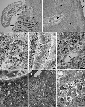
From the 30 Coregonus lavaretus examined, the intestines of 10 (33·3%) were parasitized by D. truttae. A total of 120 acanthocephalans were counted (12·10±16·26, mean intensity±s.d.) with the majority being found in the median intestine (Fig. 1a). All infected powan had more than 1 worm per host, in other words, the results referred to fish with multiple infections. The intensity of damage appeared to be the same in fish with a number of attached worms between 2 and 8, whilst it was higher in the 3 powan with a number of parasites ⩾14.
Dentitruncus truttae-infected intestine of Coregonus lavaretus. (a) Sagittal section through a powan intestine with a D. truttae attached in situ. The acanthocephalan with a fully everted proboscis (P) has penetrated the intestinal wall; note the destruction of the folds around the proboscis. Note the high number of mucous cells (thin arrows) and the adherent mucous gel (thick arrows); scale bar=200 μm. (b) Micrograph showing the deep penetration of the D. truttae proboscis (P) within the gut muscle layer (asterisks). The arrows denote the serosa; scale bar=200 μm. (c) Numerous detached epithelial cells can be seen around a trunk spine (empty arrow), as can several free rodlet cells (arrows); scale bar=20 μm. (d) Intact epithelia located at a distance from the site of acanthocephalan attachment. Some rodlet cells (arrows) are also evident; scale bar=20 μm. (e) Mast cells (thick arrows) in close proximity to the proboscis of the parasite with a mast cell inside the vessel lumen (thin arrow); scale bar=10 μm. (f) Several mast cells (arrows) near the proboscis (P) of the parasite; scale bar=10 μm. (g) The trunk (T) of the parasite is surrounded by mast cells (arrows); scale bar=20 μm. (h) A higher magnification of the rectangle featured in Fig. 1g, which shows mast cells (black arrows) inside the vessel lumen and in the tissues outside it (empty white arrows). Note also the free granules in the vessel lumen; scale bar=10 μm.

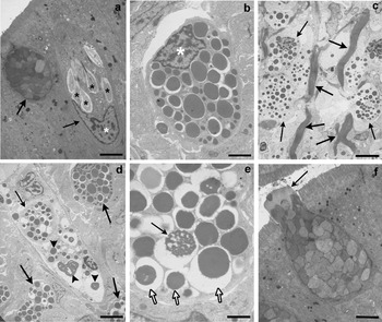
Most of the worms did not cross the stratum granulosum (Fig. 1a), but on some occasions the proboscis penetrated the muscularis layer (Fig. 1b). Dentitruncus truttae with its numerous trunk spines were observed to make contact with the intestinal epithelium where they damaged the villi. The spines detached the epithelial cells (Fig. 1c) and reduced the numbers of mucosal folds (Fig. 1a). Folds more distant from the worm's body remained intact, which were found to possess rodlet cells (RCs) in their epithelial layer (Figs 1d and 2a). The RCs (Fig. 2a) were characterized by a distinctive cell cortex, a basal nucleus and several inclusions (rodlets). The number of RCs in 40 microscopic fields found that there was no significant difference in the number of RCs found in parasitized intestines (1·95±1·67, mean±s.d., n=20) versus the number found in uninfected intestines (2·35±2·18, mean±s.d., n=20) (t-test, P>0·05).
Electron micrographs of powan cells. (a) Mucous cell (thick arrow) close to a rodlet cell (thin arrow), note the basal nucleus (white asterisk) and some rodlets (black asterisks) inside the cytoplasm of the rodlet cell; scale bar=3·4 μm. (b) Intact mast cell with an eccentric nucleus (white asterisk); the cytoplasm is filled with numerous granules; scale bar=1·9 μm. (c) Mast cells (thin arrows) scattered among the collagen fibres (thick arrows); scale bar=5·4 μm. (d) Mast cells (thick arrows) in close proximity to a vessel. A mast cell (thin arrow) and several free granules (arrow heads) inside the lumen are evident; scale bar=4·6 μm. (e) Mast cell in degranulation. The reticulated appearance of a granule (arrow) and electron-lucent halos (empty arrows) around the granules can be seen; scale bar=1·1 μm. (f) Mucous cell close to the body of an acanthocephalan releases its contents (arrow) in the gut lumen; scale bar=2·8 μm.

The mucosa, lamina propria, stratum granulosum and the muscularis layer were disrupted at the point of attachment of the parasite's proboscis. Numerous mast cells were noted in the tissues near the proboscis (Fig. 1e) and the trunk/body (Fig. 1f). A comparison of the number of intact mast cells in uninfected (35·10±8·70, mean±s.d., n=20) and infected powan (106·00±54·43, mean±s.d., n=20) in 40 microscopic fields found that the intestines of parasitized fish had a significantly higher number of mast cells (t-test, P<0·01). The mast cells were irregular in shape with an eccentric, polar nucleus (Fig. 2b), and a cytoplasm characterized by numerous large, electron-dense, membrane-bounded granules (Figs 1h and 2b). The cytoplasm typically contained 2–3 mitochondria and an inconspicuous Golgi apparatus. Mast cells were frequently surrounded by collagen fibres of the stratum granulosum (Fig. 2c) or by fibroblast-like ensheathing cells. In both infected and uninfected C. lavaretus the stratum granulosum was rich in mast cells but numbers were higher in parasitized powan. In the stratum granulosum (Figs 1h and 2d) and in the muscularis layer, numerous mast cells were in close contact with the capillaries, and the cells were observed in the outer layer of the endothelia (Figs 1h and 2d) as well as inside the blood vessels (Figs 1e, h and 2d).
Mast cells were observed scattered throughout the muscle fibres. Degranulation of the mast cells was common in the lamina propria and the stratum granulosum (Fig. 2c, e). The cells exhibited higher rates of degranulation in areas near the acanthocephalan body. Degranulation was visible by light microscopy and characterized by the conspicuous swelling of granules and often by the presence of free granules adjacent to the mast cells. Transmission electron microscopy revealed several stages of mast cell degranulation. The matrix of the granules had an extensively reticulated aspect, and prominent electron-lucent halos which encircled the granules (Fig. 2e). In some instances, the granules were fused (not shown).
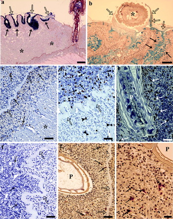
During dissection, excessive yellowish mucus was noticed around many worms. In histological sections, numerous mucous cells were noted in the intestinal epithelium (Figs 1a, 2f, 3a, b). A comparison of the number of mucous cells in uninfected (32·15±11·03, mean±s.d., n=20) and infected powan (60·45±28·79, mean±s.d., n=20) in 40 microscopic fields found that the intestines of parasitized fish had a significantly higher number of mucous cells (t-test, P<0·01). In the histological sections of the parasitized intestine stained with alcian blue/PAS or Mayer's haematoxylin-alcian blue taken near the site of infection (Fig. 3a, b), the mucous cells were filled with mucous which appeared as a blue colour (Fig. 3a, b). Moreover, the surface of the intestinal folds was found to be covered with a mucous gel (Figs 1a and 3a, b). Where the acanthocephalan trunk was in close proximity to the intestinal folds, the adherent mucous gel was seen to encircle the worm's body (Fig. 3b). Figure 3b clearly shows that the adherent mucous gel is formed by epithelial mucous cells.
Histochemical and immunohistochemical tests applied to tissue sections taken from the intestines of infected and uninfected powan. (a) The lamina propria close to the proboscis of the acanthocephalan (short arrows) is thickened (asterisks). Note the high presence of mucous cells (thin arrows) and an adherent mucous gel (empty arrows) seen covering the apices of the villi, stained with alcian blue/PAS; scale bar=200 μm. (b) Adherent mucous gel (empty arrows) produced by mucous cells (thin arrows) encircling the body of a Dentitruncus truttae (asterisk), stained with Mayer's haematoxylin-alcian blue; scale bar=100 μm. (c) Uninfected intestine of a powan seen to possess a comparatively lower number of cells positive to the met-enkephalin antiserum (arrows). Empty arrows denote the stratum compactum, asterisk shows muscle layer; scale bar=50 μm. (d) A high number of mast cells (arrows) close to the parasite proboscis that were positive to the met-enkephalin antiserum. The arrow heads highlight a small number of positive cells located at a distance from the parasite and underneath the stratum compactum (empty arrows); scale bar=50 μm. (e) D. truttae proboscis (P) surrounded by numerous mast cells (arrows) reacting positively to the met-enkephalin antiserum; scale bar=50 μm. (f) Tissue section taken from the intestine of an uninfected powan. A low number of mast cells (arrows) staining positively with the anti-serotonin serum. The empty arrows denote the stratum compactum; scale bar=50 μm. (g) Proboscis (P) embedded within the stratum granulosum. Several mast cells (arrows) appear to stain positively with the anti-serotonin serum. The empty arrows denote the stratum compactum; scale bar=50 μm. (h) A higher magnification of the mast cells (arrows) close to the proboscis (P) of the parasite observed to be immunoreactive to the anti-serotonin serum; scale bar=20 μm.

Immunohistochemical tests with the antiserum against met-enkephalin revealed a low number of mast cells in uninfected powan (Fig. 3c). In contrast, a larger number of mast cells was observed near the body of D. truttae in the lamina propria-stratum granulosum (Fig. 3d), especially close to the proboscis (Fig. 3e). Additionally, all the mast cells in the intestinal layers of the host appeared to be positive to the met-enkephalin antiserum. In the uninfected fish, very few serotonin (5-HT) immunopositive cells (mast cells) were observed (Fig. 3f), whereas a larger number of these cells that were positive to the anti-serotonin serum were found in the lamina propria, principally around the proboscis of D. truttae (Fig. 3g, h). A comparison between the number of cells positive for met-enkephalin (Fig. 3d) and serotonin (Fig. 3g) antisera showed that the cells that were positive for met-enkephalin were more numerous. Moreover, it was noticed that the number of positive cells was greater over the compact membrane, namely near the parasite proboscis. Furthermore, no immunoreactivity was observed on the sections treated with pre-absorbed antisera, and the mammalian (pig, rat) tissue sections, which were used as positive controls, gave their expected immunoreactivities.
DISCUSSION
Often intestinal helminths, via the use of their attachment organs, can induce inflammation of the host alimentary canal. The inflammation consists of a complex series of homeostatic mechanisms involving the circulatory, nervous and immune systems in response to the invading organism(s) (Sharkey, Reference Sharkey1992). Two inflammatory cell types, rodlet cells and mast cells, have been demonstrated to play a critical role in fish as part of the defence function against pathogens and evidence for their involvement in the immune system of fish is growing (Reite and Evensen, Reference Reite and Evensen2006; Silphaduang et al. Reference Silphaduang, Colorni and Noga2006; Dezfuli and Giari, Reference Dezfuli and Giari2008).
With reference to rodlet cells (RCs), although their structure is well established, their origin remains a subject of debate. The results of several recent investigations on wild and farmed fish suggest that RCs represent an immune cell type closely linked to other piscine inflammatory cells (Reite and Evensen, Reference Reite and Evensen2006; Silphaduang et al. Reference Silphaduang, Colorni and Noga2006; Jordanova et al. Reference Jordanova, Miteva and Rocha2007; Vigliano et al. Reference Vigliano, Bermúdez, Nieto and Quiroga2008). Accordingly, an increase in the number of RCs at the site of metazoan infection was reported in (Reite, Reference Reite1998; Dezfuli et al. Reference Dezfuli, Simoni, Rossi and Manera2000b). Moreover, RCs can proliferate in response to xenobiotics agents (Manera et al. Reference Manera, Simoni and Dezfuli2001). In the present survey, however, the number of RCs in infected and uninfected powan did not appear to differ significantly.
Most teleosts also appear to have within their alimentary canal, gills and other major organs, a type of cell that possesses structural and functional properties similar to those of mammalian mast cells (Vallejo and Ellis, Reference Vallejo and Ellis1989; Reite and Evensen, Reference Reite and Evensen2006). In fish infected with helminths, it was noticed that the mast cells tend to migrate and accumulate in large numbers at the site of infection (Reite and Evensen, Reference Reite and Evensen2006; Dezfuli and Giari, Reference Dezfuli and Giari2008). In powan parasitized with D. truttae, the mast cells were numerous at the site of acanthocephalan infection, although they were also present in lower numbers in uninfected fish.
Descriptive data exist detailing how mast cells degranulate in response to their exposure to parasites (Dezfuli and Giari, Reference Dezfuli and Giari2008). In parasitized C. lavaretus, intense mast cell degranulation was noticed at the site of acanthocephalan infection especially in close proximity to the parasite's proboscis.
The mast cell secretions may have a role in attracting other types of cells (i.e. neutrophils) involved in the inflammatory process, especially during the period of initial pathogen challenge (Reite and Evensen, Reference Reite and Evensen2006). The recent report of histamine stored in the granules of mast cells of the largest and most evolutionarily advanced order of teleosts, Perciformes, is significant (Mulero et al. Reference Mulero, Sepulcre, Meseguer, Garcia-Ayala and Mulero2007). The account of Murray et al. (Reference Murray, Leggiadro and Douglas2007) suggested that mast cells could be directly involved in the destruction of pathogens based on evidence of the multifunctional role of these cells in teleosts.
Immunohistochemical staining on C. lavaretus intestine demonstrated the occurrence and accumulation of elements positive for serotonin (5-HT) and met-enkephalin antisera in close proximity to the acanthocephalan body. The same was found in other fish species naturally infected with enteric worms (Dezfuli et al. Reference Dezfuli, Arrighi, Domeneghini and Bosi2000a). With reference to serotonin (5-HT) in fish, it has been established that this biogenic amine is stored in considerable amounts by mast cells and platelets (Khan and Deschaux, Reference Khan and Deschaux1997). Further information on the relationship between serotonin and the fish immune system, however, is needed (Khan and Deschaux, Reference Khan and Deschaux1997).
This study noted that the mast cells in the gut of C. lavaretus appeared to react positively to met-enkephalin antiserum, although a low number of mast cells were found in the intestines of uninfected powan. Enkephalins belong to the endogenous opiate system and in mammals they play an important role in the modulation of the inflammatory process (Radulovic et al. Reference Radulovic, Mancev, Stanojevic, Vsiljevic, Kovacevic-Jovanovic and Pesic1996) but, in fish, the same function has yet to be established. The presence of a large number of mast cells positive for met-enkephalin antiserum in infected powan makes it reasonable to suggest that by increasing the amount of the met-enkephalin-like substance secreted, inflammation caused by the acanthocephalan may be modulated.
It is well known that in all vertebrates, mast cells are strategically positioned at perivascular sites to regulate inflammatory responses (Mekori, Reference Mekori2004). In the present survey, mast cells were documented in close contact with capillaries of uninfected and infected intestine but, in parasitized fish and in the acanthocephalan site of attachment, their number was higher. Some data in the literature (Temkin and McMillan, Reference Temkin and McMillan1986), the results of our previous study on infected intestines of Salmo trutta (Dezfuli and Giari, Reference Dezfuli and Giari2008), and the present investigation support the following hypothesis. The abundance of mast cells at the site of parasite infection might be due to the migration of these cells via the circulatory system from the muscularis layer to the stratum granulosum and lamina propria.
Mast cells in the muscularis layer and the stratum granulosum of the intestine were in contact with fibroblasts and the same relationship between these two cell types has also been reported in other salmonids (Flaño et al. Reference Flaño, Lopez-Fierro, Razquin and Villana1996; Dezfuli and Giari, Reference Dezfuli and Giari2008). Interestingly, mast cell-fibroblast association was found in lesions in which an unusual mast cell proliferation was observed in coho salmon (Kent et al. Reference Kent, Powell, Kieser, Hoskins, Speare and Burka1993) and in brown trout (Dezfuli and Giari, Reference Dezfuli and Giari2008). In fish (Temkin and McMillan, Reference Temkin and McMillan1986) it is reported that fibroblasts can influence mast cell proliferation and motility. Further accounts suggest that within fish and mammals, mast cells are involved in fibrotic processes and in tissue remodelling (Rocha and Chiarini-Garcia, Reference Rocha and Chiarini-Garcia2007).
The mucous layer of the gut is a complex biofilm containing lipids, proteins and bacteria suspended in adherent mucous gel, and the ability of mucous to protect the underlying epithelial layer has been comprehensively discussed in the account of Lamont (Reference Lamont1992). In mammals, hyperplasia and hypertrophy of mucous cells has been found in response to intestinal parasites (Miller and Huntley, Reference Miller and Huntley1982; Fairweather, Reference Fairweather and Beckage1997; Manjili et al. Reference Manjili, France, Sangster and Rothwell1998). In the intestines of Salmo trutta infected with helminths, the number of mucous cells was found to have increased significantly and the composition of the mucous was different from that produced in the guts of uninfected conspecifics (Bosi et al. Reference Bosi, Arrighi, Di Giancamillo and Domeneghini2005a). The results of the present study regarding the higher number of mucous cells observed in parasitized powan agrees closely with the observations of Bosi et al. (Reference Bosi, Arrighi, Di Giancamillo and Domeneghini2005a). Histological sections of C. lavaretus-infected intestines suggest that all D. truttae specimens were surrounded by an adherent mucous gel which is produced to protect the underlying intestinal mucosa (Miller and Huntley, Reference Miller and Huntley1982; Lamont, Reference Lamont1992).
Enteric worms cause intense involvement of vertebrate immune and neuroendocrine systems (Castro, Reference Castro1992; Fairweather, Reference Fairweather and Beckage1997). The evidence presented here and within a number of earlier accounts (i.e. Dezfuli et al. Reference Dezfuli, Arrighi, Domeneghini and Bosi2000a, Reference Dezfuli, Pironi, Simoni, Shinn and Giari2007; Bosi et al. Reference Bosi, Domeneghini, Arrighi, Giari, Simoni and Dezfuli2005b) is clearly on the side of this concept.
We are grateful to E. Simoni from the University of Ferrara for her technical assistance. Thanks are also due to A. P. Shinn from the University of Stirling, UK for help with our English.