Introduction
Platelet-rich plasma is a novel material that is being used more and more frequently in many surgical specialties. Its numerous advantages include safety (because it is an autologous material), the increased deposition of platelets and growth factors in the healing area, and a short preparation time.
Platelet-rich plasma was first used in 1987 in open-heart surgery and is now widely accepted as an effective means of promoting wound healing and tissue regeneration. Owing to its value in biostimulation and healing acceleration, this material has various applications in cardiothoracic surgery, orthopaedic surgery, neurosurgery, plastic surgery, dentistry, and oral and maxillofacial surgery.Reference Gopalakrishnan, Venkatasamy and Vivekanandamurthy 1 Examples include its use in treating epicondylitis, muscle tears, osteoarthritis, burns, cardiovascular surgical wounds and diabetic ulcers. New applications of platelet-rich plasma are also being investigated. This material is particularly interesting to the ENT surgeon because of its healing properties and as a potential packing or grafting material.
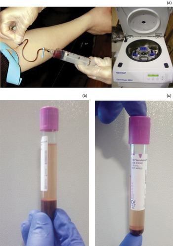
Platelet-rich plasma preparations have traditionally been produced by dual-speed centrifugation. Figure 1a shows the equipment needed for blood centrifugation. The first ‘soft spin’ separates blood into red blood cells, platelet-poor plasma and a buffy coat. The second ‘hard spin’ separates platelet-rich plasma from platelet-poor plasma. Figure 1b depicts the different layers produced by blood centrifugation. The isolated platelet-rich plasma is then mixed with thrombin and calcium chloride to form a gel-like product with five to eight times the platelet concentration of whole blood.Reference Gopalakrishnan, Venkatasamy and Vivekanandamurthy 1 , Reference Sclafani and Azzi 2
Photographs showing (a) blood sampling and centrifugation apparatus, (b) blood separation after ‘soft spin’ centrifugation and (c) platelet-rich plasma production after a second ‘hard spin’ to separate platelet-rich plasma from platelet-poor plasma.

Besides its high platelet concentration, platelet-rich plasma contains a number of growth factors that are useful for wound healing such as platelet-derived growth factor (PDGF), transforming growth factor alpha (TGF-α), transforming growth factor beta (TGF-β), epidermal growth factor (EGF), fibroblast growth factor (FGF), insulin-like growth factor and platelet-derived angiogenesis factors, along with white blood cells, phagocytic cells, a high native fibrinogen concentration, and vasoactive and chemotactic agents. In particular, PDGF and TNF-β facilitate rapid wound healing, haemostasis and decreased scarring.Reference Pomerantz and Dutton 3
Platelet-rich plasma is an established treatment that provides satisfactory outcomes for various clinical conditions. However, its use in ENT is not yet common and its application needs further investigation. This review describes the available evidence on the use of platelet-rich plasma in ENT surgery.
Materials and methods
A literature search of the PubMed database was conducted using the terms ‘platelet-rich plasma’ and/or ‘platelet-rich plasma in ENT’, ‘platelet-rich plasma in otolaryngology’, ‘platelet-rich plasma in otorhinolaryngology’, ‘platelet-rich plasma in rhinology’ and ‘platelet-rich plasma in otology’. Articles related to oral surgery, oral and maxillofacial surgery, and plastic surgery were excluded. As the study methodologies and outcomes varied widely, data extraction comprised a narrative summary of each article.
Results
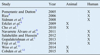
All results were evaluated for their relevance, and both abstracts and full articles were obtained. A total of 11 included studies were classified according to subspecialty (Table I).
Reports included in the review

Platelet-rich plasma in otology surgery
In 2009, Erkilet et al. reported the contribution of platelet-rich plasma to tympanic membrane healing in 44 rats following bilateral tympanic membrane perforation.Reference Erkilet, Koyuncu, Atmaca and Yarim 6 Platelet-rich plasma was applied to the right side, while the left side was used as a control. In a group of 20 rats, daily otomicroscopic assessment demonstrated a significant difference in healing time for the right side compared with the left (10.2 ± 2.1 days vs 13.0 ± 2.9 days; mean difference 2.8 days). The other 24 rats were divided into 4 groups of 6 rats and sacrificed on days 3, 7, 14 and 28, and then histopathological examination of the tympanic membranes was performed. No significant differences in healing were found for any time point.
In 2001, Navarrete Álvaro et al. reported the preliminary results of platelet-rich plasma assisted type I tympanoplasty for central perforations.Reference Navarrete Álvaro, Ortiz, Rodriguez, Boemo, Fuentes and Mateo 8 Three patients with dry perforations were treated by applying a platelet-rich plasma plug around the perforation, followed by overlay type I tympanoplasty. A satisfactory outcome was achieved by the complete closure of all three perforations.
Another study of platelet-rich plasma assisted tympanoplasty was conducted by Gopalakrishnan et al. in 2013.Reference Gopalakrishnan, Venkatasamy and Vivekanandamurthy 1 These authors examined 25 patients who underwent underlay tympanoplasty using platelet-rich plasma and 25 control patients who underwent tympanoplasty without platelet-rich plasma: closure rates of 72 per cent closure in the platelet-rich plasma group and 40 per cent closure in the control group were achieved at the end of the first month; 92 per cent in the platelet-rich plasma group and 72 per cent in the control group at the end of the second month; and 96 per cent in the platelet-rich plasma group and 80 per cent in the control group at the end of the third month. The authors concluded that applying platelet-rich plasma during tympanoplasty prevents graft displacement.
Cho et al. (2010) investigated the effect of platelet-rich plasma on facial nerve regeneration in 24 guinea pigs randomly divided into 4 treatment groups: group 1, 2 perineural 10-0 gauge microsutures only; group 2, perineural microsutures plus platelet-rich plasma; group 3, perineural microsutures plus mesenchymal stem cells; and group 4, perineural microsutures plus platelet-rich plasma and mesenchymal stem cells.Reference Cho, Jang, Lee, Jeong, Park and Han 7 Groups 2–4 had significant functional improvement compared with the control group, at 4 weeks after surgery. Electrophysiologic evaluation four and six weeks after the intervention showed significant improvement in groups 2–4, with group 4 having the best performance. Histological analysis showed that group 4 animals had more myelinated axon fibres than all other groups. The authors concluded that the combined use of platelet-rich plasma and mesenchymal stem cells had a beneficial effect compared with either material alone.
Platelet-rich plasma in rhinology surgery
Four reports describing the use of platelet-rich plasma in rhinology were identified. In 2014, Friji et al. reported a method for the autologous transplantation of fat and platelet-rich plasma to treat atrophic rhinitis.Reference Friji, Gopalakrishnan, Verma, Parida and Mohapatra 10 Five patients had an autologous lipoaspirate applied to both nasal cavities, specifically the inferior turbinate, middle turbinate, floor and septum. Platelet-rich plasma was injected into the same areas. All five patients described symptom improvement, with the disappearance of nasal crusting. Clinical examination confirmed the presence of glistening nasal mucosa and no signs of atrophy. Six months after the intervention, Sino-Nasal Outcome Test 20 scores had improved from an average of 36 to an average of 8. In addition, the nasal mucociliary clearance time was significantly shorter after (960 seconds) than before (1995 seconds) surgery. The authors concluded that this method achieved encouraging subjective and objective outcomes.
Pomerantz and Dutton (2005) evaluated the quality of life of 16 patients who underwent packing with platelet-rich plasma after endoscopic sinus surgery.Reference Pomerantz and Dutton 3 Their results were compared with retrospectively collected data from another group of 16 control patients who had undergone traditional packing. Each group completed two Sino-Nasal Outcome Test 16 questionnaires related to quality of life before and after surgery; the mean scores before and after surgery were converted into an absolute change score. The control group scored 0.938 and the platelet-rich plasma group scored 0.957, indicating more improvement in the platelet-rich plasma group. However, the difference between the groups was not significant because of the small population size.
Rice (2006) published the early results of a prospective study into the use of platelet-rich plasma as packing material after endoscopic sinus surgery.Reference Rice 4 The first 13 patients experienced no significant benefit of platelet-rich plasma, with some experiencing a worse post-operative course. As a consequence of these discouraging results, the study was not completed.
Salaheldin and Hussein (2012) treated a group of 30 patients with a platelet-rich plasma injection into the inferior turbinates following submucosal diathermy.Reference Salaheldin and Hussein 9 Patients treated with platelet-rich plasma showed significant improvements in crusting, bleeding and mucociliary clearance compared with a saline-treated control group (n = 30).
Platelet-rich plasma in laryngology surgery
In 2014, Woo et al. evaluated the healing properties of platelet-rich plasma in rabbits after vocal-fold stripping.Reference Woo, Jeong, Kim, Koh, Lee and Jin 11 Morphological analysis demonstrated less granulation tissue and histopathology findings showed well-organised collagen deposition on the side injected with platelet-rich plasma. In addition, levels of growth factors such as EGF, PDGF, TGF-β and vascular endothelial growth factor were increased at 14 days after surgery. However, at 12 weeks after surgery, these growth factors were not markedly expressed. Thus, the effect of platelet-rich plasma on growth factor expression did not appear to last for a long period. Overall, platelet-rich plasma appeared to maintain the structure of the injured vocal fold, while decreasing granulation tissue formation.
In 2015, Cobden et al. reported histopathological findings in 20 Wistar rats (10 each in experimental and control groups) after applying platelet-rich plasma to injured vocal folds.Reference Cobden, Oztürk, Duman, Esen, Aktan and Avunduk 12 The authors concluded that platelet-rich plasma increased EGF receptor, FGF and fibroblast growth factor receptor 1 (FGFR1) levels in the healing vocal fold and is thus beneficial for vocal fold scar healing.
Platelet-rich plasma in paediatric ENT surgery
In 2008, Sidman et al. reported the effects of applying platelet-rich plasma to the tonsillar fossae on tonsillectomy outcome in a cohort of 70 children (35 in treatment group, 35 in control group; drop-out rate 17.1 per cent).Reference Sidman, Lander and Finkelstein 5 No significant difference in pain levels, recovery time or number of post-operative office visits in patients who had platelet-rich plasma treatment. The authors suggested that platelet-rich plasma may have not been retained for sufficient time to benefit tissue healing. Moreover, pain after tonsillectomy originates from the inflamed underlying muscles and the surrounding mucosa of the healing wound, and this is unlikely to be affected by platelet-rich plasma application.
Discussion
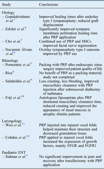
This literature review included various types of studies into platelet-rich plasma in ENT surgery. The number of studies included is rather small compared with the number of studies into the use of this material in other surgical fields. Each article used different experimental and control groups, and there were few commonalities among the techniques and evaluation methods. Therefore, safe conclusions cannot be made about the value of platelet-rich plasma in ENT surgery. Other limitations included the small number of participants, a failure to maintain close follow up and study design drawbacks. The conclusions of each study are summarised in Table II.
Significant findings in studies review, broken down by subspecialty

PRP = platelet-rich plasma; MSC = mesenchymal stem cell; EGFR = epidermal growth factor receptor; FGFR1 = fibroblast growth factor receptor 1
Overall, platelet-rich plasma is a promising material with a number of potential clinical applications. The studies included in this review confirmed that platelet-rich plasma benefits the healing process, mainly by promoting growth factor aggregation. The findings for platelet-rich plasma use in functional endoscopic sinus surgery are discouraging but more recent findings in vocal-fold, atrophic rhinitis and turbinate surgery show that it has potential applications in vocal-fold scar treatment and nasal mucosa healing. It was also a beneficial adjunct in tympanoplasty via speeding up graft adhesion while reducing the graft displacement risk. Injecting platelet-rich plasma may be beneficial for treating vocal-fold palsy (medialisation) and atrophic rhinitis. However, more studies are required to establish this material as a useful tool for the ENT surgeon.