Implications
Dietary energy utilization from starch is potentially limited by the inadequate synthesis of α-amylase, which is secreted by the pancreas in the dairy cow. The present study provides evidence that leucine acts as a signalling molecule to stimulate the synthesis of α-amylase and trypsin through the mTOR–messenger RNA (mRNA) pathway in pancreatic tissue, so that the digestibility of starch-based energy feeds could be improved in the intestine of the dairy cow. Therefore, leucine could act as a functional amino acid additive for dairy cow nutrient management.
Introduction
Dietary energy utilization of starch is potentially limited by starch digestion from the alimentary canal in cattle (Huntington et al., Reference Huntington, Harmon and Richards2006). Research suggests that when high-grain diets are fed, starch digestion is limited (Owens et al., Reference Owens, Zinn and Kim1986) and this limitation could be the result of the inadequate synthesis and secretion of α-amylase into the small intestine (Harmon, Reference Harmon1992). Therefore, an important strategy to improve small intestinal digestion of starch is to enhance the synthesis and secretion of α-amylase in the dairy cow.
Interactions between supplemental proteins and pancreatic α-amylase might be affected by post-rumen flows of α-glucosides (Swanson et al., Reference Swanson, Matthews, Woods and Harmon2002). The requirement for nutritionally functional amino acids (Wu, Reference Wu2013) to optimize pancreatic secretions may be proportional to the starch appearing in the lumen of the small intestine in cattle. However, increasing starch flow through the small intestines decrease α-amylase production and secretion, which subsequently results in the inefficiency of starch digestion in the small intestines. This indicates that only the intestinal amino acid flow affects pancreatic exocrine secretion. Unfortunately, information is lacking about the effects of functional amino acids on the protein synthesis or secretion of the pancreas in dairy cows.
Functional amino acids not only serve as substrates for protein synthesis but also function as signalling molecules that regulate the mammalian target of rapamycin (mTOR) pathways in several tissues and organs of mammals. As an important functional amino acid, leucine is the most effective signal for the mTOR protein, which is also known for stimulating protein synthesis via the mTOR signal pathway in skeletal muscle, liver, adipose tissue and mammary glands (Appuhamy et al., Reference Appuhamy, Knoebel, Nayananjalie, Escobar and Hanigan2012; Arriola Apelo et al., Reference Arriola Apelo, Singer, Lin, McGilliard, St-Pierre and Hanigan2014). The mTOR is a multidomain protein that is important in regulating several components of the translational machinery. It senses and responds to nutrient availability (amino acids), energy sufficiency, stress, hormones and mitogens to regulate protein synthesis. When activated by amino acids, mTOR, in turn, catalyses the phosphorylation of ribosomal protein S6 kinase (S6K1) and eukaryotic initiation factor 4E-binding protein 1 (4EBP1) (Wang and Proud, Reference Wang and Proud2006). The binding of mRNA to the 43S preinitiation complex is critical for translation initiation and is inhibited by hypophosphorylated 4EBP1 (Kimball and Jefferson, Reference Kimball and Jefferson2006). Hence, the phosphorylation of 4EBP1 by mTOR nullifies its inhibitory action, allowing increased rates of initiation (Soulard and Hall, Reference Soulard and Hall2007).
Although much is known about the signalling pathways involved in the regulation of mTOR by leucine, the molecular mechanism has not yet been well characterized in the dairy cow pancreas, particularly the regulatory effect of leucine on amylase synthesis. In addition, the effects of leucine on the digestive enzyme secretion in dairy goats and dairy heifers have not been explored (Yu et al., Reference Yu, Xu, Liu, Yao, Yu and Wang2014; Liu et al., Reference Liu, Liu, Liu, Xu, Yu, Wang, Cao and Yao2015). Therefore, the main goals of this study were to investigate the effects of leucine on the mTOR signalling pathway and to define the associations between these signalling activities and the synthesis of pancreatic enzymes using an in vitro model of cultured Holstein calf pancreatic tissue. The hypothesis was that leucine could stimulate the enzymes synthesis through the mTOR signal pathway in pancreatic tissue segments. We focussed on the main signal factors including the upstream of mTOR signal factor mitogen-activated protein kinase kinase kinase kinase 3 (MAP4K3), the mTOR kinase, the downstream of mTOR signal factors S6K1 and 4EBP1, as well as the phosphorylation state of these factors.
Material and methods
Animal management
All three experimental male Holstein calves were raised at Modern Farming (BaoJi, China), and their diet consisted of raw milk and calf starter (Purina, China). The raw materials of calf starter feed included maize, soya bean meal, wheat bran, cane molasses, dicalcium phosphate, mountain flour, salt, vitamin A, vitamin D3, vitamin E and so on. The chemical composition of the starter feed and the concentration of amino acids in the milk are shown in Supplementary Tables S1 and S2. The calves management was refer to Cao et al. (Reference Cao, Yang, Guo, Zheng, Wang, Cai, Liu and Yao2018). Briefly, calves were fed with about 3 to 4 l/day of milk. The calf starter was added from 3rd to 8th weeks and then calves (79.2±1.68 kg; mean ± SD) were slaughtered to collect the pancreatic tissue. The amount of feed offered to each calf was 210 g/day in the 3rd week and gradually increased to 1800 g/day in the 8th week to ensure that all calves consumed the feed without any refusal. The calves had free access to fresh drinking water. The method of slaughter was by injection of anaesthetic in the jugular vein and bleeding the calves to death. The animal care and procedures were approved and conducted under established standards of the College of Animal Science and Technology, Northwest A&F University, Yangling, China.
Tissue collection
Pancreatic tissue was obtained from the Holstein calves. All calves were starved for 12 h before slaughter to induce a responsive and homogenous phosphorylation state of signalling proteins. Slaughtering of animals was performed at the Animal Nutrition Laboratory of Northwest A&F University. Mesentery, fat and lymph were removed from the pancreas. Approximately 10 g of pancreas was rinsed with and transported to the lab in ice-cold saline (0.9% NaCl).
Tissue incubation and treatment design
Dulbecco’s modified Eagle’s medium (DMEM)/Ham’ F12 (HyClone, Logan, UT, USA) was used as the basal culture medium, which was supplemented with 10% foetal bovine serum (Invitrogen Trading Co. Ltd, Shanghai, China), 100 000 units of penicillin (Solarbio, Beijing, China), 100 mg of streptomycin (Solarbio), 10 ng/ml epidermal growth factor (Sigma, Louis, MO, USA) and 5 μg/ml bovine insulin (Sigma). The working leucine concentration in the leucine medium was 0.45 mM, which is consistent with the normal concentration in DMEM/F12 culture medium (two times the normal plasma leucine concentration of the dairy cow) (Mackle et al., Reference Mackle, Dwyer, Ingvartsen, Chouinard, Ross and Bauman2000). The control medium was the same as the treatment medium but contained no leucine (Special custom medium; HyClone).
The incubation techniques described were based on similar procedures used for other species or purposes but with some differences (Katoh and Tsuda, Reference Katoh and Tsuda1984; Katoh and Yajima Reference Katoh and Yajima1989; Swanson et al., Reference Swanson, Matthews, Woods and Harmon2002). The pancreatic pieces were transferred to ice-cold Krebs–Ringer bicarbonate buffer (KRB) and cut into small segments (~2×2 mm) with a pair of fine scissors. Tissue segments (~100 mg) were blotted dry on a filter paper, weighed using an electronic balance (Mettler Toledo, Shanghai, China) and transferred by rinsing the tissue with 1 ml DMEM/F12 culture medium into 25-ml flasks containing 2 ml of DMEM/F12 culture medium and various substrates (total volume of DMEM/F12 culture medium = 3 ml). Here, we used DMEM/F12 medium instead of KRB medium because the former was more similar to the physiological environment. The leucine treatment and the control each had three replicates. The flasks were gassed with a mixture of 95% O2 and 5% CO2, capped, and placed in a 39°C shaking water bath at 90 oscillations/min for incubation (Swanson et al., Reference Swanson, Matthews, Woods and Harmon2002).
Experiment on rapamycin-inhibited the mammalian target of rapamycin signalling pathway
Rapamycin was purchased from Selleck (Shanghai, China) and dissolved in dimethyl sulfoxide. A 10 mM stock solution of rapamycin was prepared. The working concentration of rapamycin in the leucine treatment was 40 nM (Phung et al., Reference Phung, Ziv, Dabydeen, Eyiah-Mensah, Riveros, Perruzzi, Sun, Monahan-Earley, Shiojima, Nagy, Lin, Walsh, Dvorak, Briscoe, Neeman, Sessa, Dvorak and Benjamin2006). There were three treatments including control (0 mM leucine), Rapa (40 nM rapamycin + 0.45 mM leucine) and Leu (0.45 mM leucine). The incubation time was 1 h, after which the samples were collected for subsequent analysis.
In vitro enzyme release
At 1, 2 and 3 h post-incubation, the incubated tissue segments were removed from the culture medium, rinsed with saline and then stored at −80°C until analysis. The activities of α-amylase, trypsin, lipase and chymotrypsin in tissue segments homogenates were determined using the respective commercially available kits (Nanjing Jiancheng Bioengineering Institute, Nanjing, China) according to the manufacturer’s instructions. Enzyme activity was expressed in units (one unit being defined as 1 µM product released per min at 39°C) per milligram protein. Sample quality control and testing parameter settings were strictly in accordance with the product specifications.
Ribonucleic acid extraction and quantitative real-time PCR
The expression levels of α-amylase, trypsin, lipase and chymotrypsin in tissue segments were measured using real-time quantitative PCR (RT-qPCR). Total RNA was abstracted from the tissue using RNAiso Plus (Takara, Dalian, China) according to the manufacturer’s protocols. The quality of RNA was measured using NanoDrop 2000 UV–Vis spectrophotometer (Thermo ScientificTM, Waltham, MA, USA) and all RNA samples were tested to have A260/A280 ratios of 2.0. Total RNA was converted to complementary DNA using the PrimeScript RT kit (Takara) according the manufacturer’s methods. The RT-qPCR was performed using SYBR Green (SYBR Premix Ex TAQII, Takara) on a Bio-Rad IQ5 Real-Time PCR Detection system (Bio-Rad, Hercules, CA, USA). The PCR was done at 95°C for 3 min, followed by 40 cycles at 95°C for 3 s and 60°C for 30 s. Melting curves and agarose gel electrophoresis were performed to insure a single specific PCR product for each target gene. All PCRs were performed in triplicate. Specific PCR primers were designed using the Primer 5.0 design software (PREMIER Biosoft, Palo Alto, CA, USA) and NCBI software. The accession numbers of the reference sequences used and the primers for each gene are listed in Supplementary Table S3. The 18S ribosomal RNA bovine gene of bovine was used as the housekeeping gene, and the mRNA expression of the target genes was compared with that of the housekeeping gene. The average cycle threshold (Ct) values were used for quantification by the 2−ΔΔCt method.
Protein extraction and Western blot
Proteins were extracted from pancreatic segments homogenates in radioimmunoprecipitation assay fluid (Solarbio) supplemented with inhibitors of protease and phosphatase (Roche, Mannheim, Baden-Wurttembery, Germany). The protein concentration was determined using a Pierce bicinchoninic acid assay kit (Thermo Scientific) following the manufacturer’ protocols. For Western blot analysis, the samples were boiled at 95°C for 7 min in 5×SDS-PAGE loading buffer (CWBIO, Beijing, China). The Western blot procedure was described our recently study (Guo et al., Reference Guo, Liang, Zheng, Liu, Yin, Cao and Yao2018). The specific methods were showed in Supplementary Material S1. Polyclonal rabbit anti-phosphorylated form of mTOR (Ser2488), 4EBP1 (Thr37/46), S6K1 (Thr389), and polyclonal rabbit anti-mTOR, 4EBP1, S6K1 and MAP4K3 antibodies (Catalogue No. 2971, 9459, 9205, 2972, 9452, 9202 and 9613; Cell Signaling Technology, Danvers, MA, USA, 1 : 1000) were used as the primary antibodies. Monoclonal mouse anti-β actin (Catalogue No. CW0096M; CWBIO, 1 : 3000) was used as the housekeeping protein. Polyclonal goat anti-rabbit HRP-IgG (Catalogue No.CW0103; CWBIO, 1 : 1000) and polyclonal goat anti-mouse HRP-IgG (Catalogue No.CW0102; CWBIO, 1 : 1000) were used as secondary antibody. Western blots were developed and quantified using Image J software (National Institutes of Health, Bethesda, MD, USA). The protein level was quantified by normalizing total or phosphorylated proteins with β-actin. For the target protein expression analysis, the normalized target protein expression level for each sample was compared with the positive control sample.
Statistical analysis
The data of digestive enzyme activities and gene expression were analysed using the GLM procedure of SPSS 19.0 software (SPSS Inc., Chicago, IL, USA) for the main effects on two different leucine concentrations (0 v. 0.45 mM) and different incubation times (1, 2 and 3 h), which resulted in a 2×3 factorial arrangement. Protein expression levels were calculated as the ratio of the band intensity of β-actin. Significant differences were declared at P <0.05, and data were presented as the means ± standard error of the mean. Every experiment was repeated with three biological samples, and each of the samples was run in triplicate.
Results
The animal performance of donor Holstein calves
Three donor Holstein calves were 8 weeks old and weighed ~79.2 ± 0.78 kg. No significant differences (P >0.05) were observed in the BW and feed intakes in all three Holstein calves during the 8 weeks. All calves were healthy and disease free.
Effects of leucine on digestive enzyme activity in pancreatic tissue
As shown in Table 1, leucine treatment increased the activity of α-amylase at 1 and 2 h (P <0.05), but not at 3 h (P >0.05) post-incubation. Leucine increased trypsin activity at all three time points (P <0.05). Lipase activity was not affected by leucine at 1 and 2 h (P >0.05) but was decreased at 3 h (P <0.05). The activity of chymotrypsin was decreased at 1 h (P <0.05) and 2 h (P <0.05), but not at 3 h (P >0.05).
Effects of leucine on the enzyme activity of α-amylase, trypsin, lipase and chymotrypsin in the pancreas tissue of Holstein dairy calves1

a,b,c,d,e,fMeans with different superscripts within a column differ significantly (P <0.05, n = 3).
1 One unit of enzyme is defined as 1 µM of the product formed per min at 39°C.
There were leucine effects on the activity of α-amylase (P <0.001), trypsin (P <0.001) and lipase (P <0.001) over time. The main effects showed that compared to lack of leucine, the leucine treatment increased the activity of α-amylase and trypsin (P <0.001) but decreased that of lipase (P <0.001) and chymotrypsin (P = 0.001). The activity of α-amylase decreased at 3 h (P = 0.03), while that of trypsin decreased with time (P <0.001), suggesting that the activities of the enzymes decreased over time.
Effects of leucine on digestive enzyme messenger RNA expression in pancreatic tissue
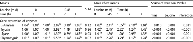
Gene expression of digestive enzymes in pancreatic tissue was affected by the leucine administration at different time points (Table 2). The leucine treatment elevated α-amylase mRNA expression at 1 and 2 h (P <0.05) but not at 3 h (P >0.05). The gene expression of the trypsin gene was upregulated at all three time points (P <0.05). For the lipase gene, its expression was higher at 1 and 3 h (P <0.05) but not at 2 h (P >0.05). The leucine treatment only increased chymotrypsin mRNA relative expression only at 1 h (P <0.05).
Effects of leucine on the mRNA expression of α-amylase, trypsin, lipase and chymotrypsin in the pancreas tissue of Holstein dairy calves1

a,b,c,dMeans with different superscripts within a column differ significantly (P <0.05, n = 3).
1 18s ribosomal RNA was used as the housekeeping RNA.
There were leucine interactions on gene expression of α-amylase, trypsin and lipase (P <0.001) over time. Gene expression of all the digestive enzymes decreased with time (P <0.001), which suggests that the leucine stimulation of digestive enzymes gene expression is transient.
Effects of leucine on the activation of the mammalian target of rapamycin signalling pathway factors in pancreatic tissue
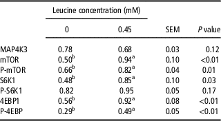
The results of the activity and mRNA expression of the digestive enzymes showed that the expression of α-amylase peaked at 1 h post-leucine treatment. Thus, we analysed the samples collected at 1 h for synthesis and phosphorylation state of the mTOR pathway proteins (Table 3). Total MAP4K3 and total/phosphorylated mTOR, S6K1 and 4EBP1 were readily detected in the pancreatic tissue of dairy calves. The abundance of both total and phosphorylated forms of mTOR, p70S6K and 4EBP1 was increased (P <0.05) by 0.45 mM leucine, while that of total MAP4K3 and phosphorylated p70S6K were not affected (P >0.05).
The effect of leucine on activation of mammalian target of rapamycin (mTOR) pathway factors in the pancreatic tissue of Holstein dairy calves cultured for 1 h1

MAP4K3 = mitogen-activated protein kinase kinase kinase kinase 3; P-mTOR = phosphorylated-mTOR; S6K1 = p70 ribosomal protein S6 protein kinase 1; P-S6K1 = phosphorylated-S6K1; 4EBP1 = eukaryotic initiation factor 4E binding protein 1.
a,bMeans with different superscripts within a row differ significantly (P <0.05, n = 3).
1 β-actin was used as the reference.
Effects of rapamycin on the enzymes synthesis and the mammalian target of rapamycin signalling pathway activation in pancreatic tissue
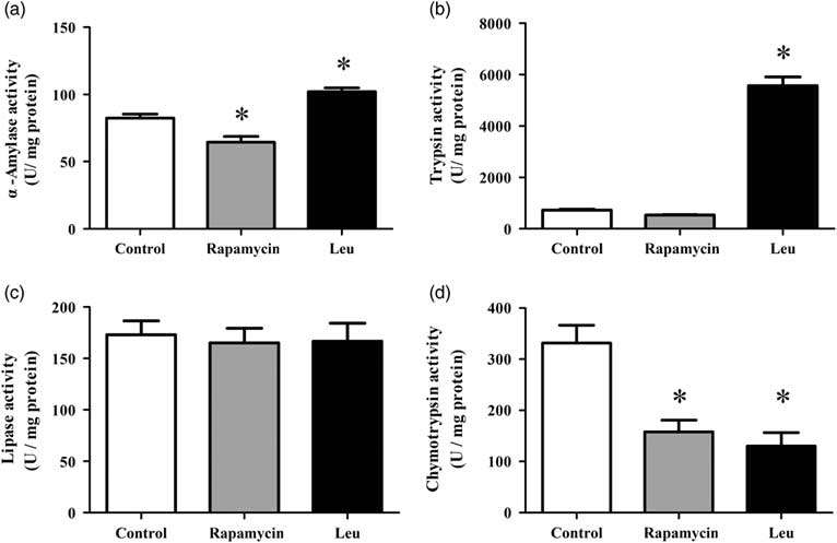
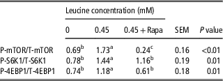
The results showed that leucine treatment with 40 nM rapamycin inhibited the activities of α-amylase (Figure 1a) and trypsin (Figure 1b). After checking expression of the mTOR pathway factors (Table 4), we found that rapamycin treatment reduced the phosphorylation of mTOR (P <0.05). Compared to leucine treatment, the rapamycin treatment significantly reduced the phosphorylation of p70S6K (P <0.05), and 4EBP1 (P <0.05). These results suggested that rapamycin inhibited the phosphorylation of mTOR, p70S6K and 4EBP1 after the addition of leucine.
The effects of rapamycin on the activity of α-amylase (a), trypsin (b), lipase (c) and chymotrypsin (d) in the pancreatic tissue of Holstein dairy calves cultured for 1 h. Error bar represent SEM. *Indicates significantly difference (P <0.05, n = 3). Leu = leucine.

The ratio of phosphorylated to total mammalian target of rapamycin (mTOR) signal pathway factors in the pancreatic tissue of Holstein dairy calves cultured for 1 h in the presence of 0 or 0.45 mM l-leucine or 0.45 mM l-leucine plus rapamycin (Rapa)

S6K1 = p70 ribosomal protein S6 protein kinase 1; 4EBP1 = eukaryotic initiation factor 4E binding protein 1; P- = phosphorylated-; T- = total-.
a,b,cMeans with different superscripts within a row differ significantly (P <0.05, n = 3).
Discussion
In ruminants, pancreatic α-amylase is the primary enzyme responsible for the initial hydrolysis of starch in the small intestinal lumen. It has been shown that increasing the small intestinal protein flow enhanced the pancreatic α-amylase secretion and small intestinal starch digestion (Richards et al., Reference Richards, Branco, Bohnert, Huntington, Macari and Harmon2002). In the present study, we found that leucine treatment increased the synthesis of α-amylase and trypsin during the in vitro incubation period and the highest activity level of these two enzymes was observed at 1 h, consistent with the in vivo physiological state of dairy calves. This suggested that pancreatic α-amylase and trypsin synthesis in vitro depends on a high concentration of leucine in a transient manner. For lipase and chymotrypsin, the leucine dosage showed inhibitory effects, but the time effect was not statistically significant. A possible explanation could be that leucine selectively activates the synthesis of proteins important for intracellular metabolism.
Our research team have founded that leucine could significantly affect the secretion of amylase in the pancreatic fluid of dairy goats (Yu et al., Reference Yu, Xu, Liu, Yao, Yu and Wang2014) and dairy heifers (Liu et al., Reference Liu, Liu, Liu, Xu, Yu, Wang, Cao and Yao2015). Besides, supplementation of leucine could also affect pancreatic growth and development in Holstein calves (Cao et al., Reference Cao, Yang, Guo, Zheng, Wang, Cai, Liu and Yao2018). The present results further confirmed that leucine could enhance the synthesis of α-amylase of the pancreas both at 1 and 2 h. In addition, we also found that leucine increased trypsin synthesis. However, there were also discrepancies in the response to leucine treatment with respect to dose and time, which is probably attributable to the difference between in vivo and in vitro experiments. As the interactions occurring in vivo complicate the interpretation of experimental data, in vitro systems (Katoh and Tsuda, Reference Katoh and Tsuda1984; Katoh and Yajima, Reference Katoh and Yajima1989; Swanson et al., Reference Swanson, Matthews, Woods and Harmon2002) can be better suited to understanding the direct interactions and molecular mechanisms that regulate the exocrine pancreas.
The regulation of pancreatic enzyme expression is very complex with multiple mechanisms. One such possible mechanism is the variation of the mRNA concentrations of pancreatic enzymes (Swanson et al., Reference Swanson, Matthews, Woods and Harmon2002). In monogastric animals, the mRNA expression of pancreatic enzymes is generally related to the amount of their specific substrates passing through the small intestine (Brannon, Reference Brannon1990). However, in lambs, the dietary adaptation of amylase mRNA expression appears to be regulated in part by post-transcriptional events (Swanson et al., Reference Swanson, Matthews, Matthews, Howell, Richards and Harmon2000). In our study, the expression level of α-amylase and trypsin genes and the synthesis of the respective enzymes showed similar trends. A possible explanation is that leucine probably mediates digestive enzyme synthesis by modulating the transcription of the coding genes and post-transcriptional regulation.
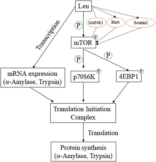
Amino acids regulate gene expression, which can occur at transcription and translation (Wu, Reference Wu2014). To further investigate possible transcriptional and post-transcriptional regulation that affected pancreatic α-amylase expression, we examined the main regulation of protein synthesis of the mTOR signalling pathway. Our results confirmed that leucine supplementation also enhanced the protein synthesis of p70s6k and 4EBP1. Based on the results of this study, we proposed a model to illustrate how leucine may regulate amylase and trypsin synthesis in the pancreatic tissue of dairy calves (Figure 2).
A proposed model of action by which leucine regulates synthesis of α-amylase and trypsin in the pancreatic tissue of Holstein dairy calves.  , stimulatory;
 , inaction;
 findings reported previously (Holz et al., Reference Holz, Ballif, Gygi and Blenis2005; Soulard and Hall, Reference Soulard and Hall2007; Wolfson et al., Reference Wolfson, Chantranupong, Saxton, Shen, Scaria, Cantor and Sabatini2016). Activation of leucine (Leu) did not influence the mitogen-activated protein kinase kinase kinase kinase 3 (MAP4K3) content; Leu enhanced the protein synthesis of α-amylase and trypsin, by stimulating the mammalian target of rapamycin (mTOR) pathway and increasing their messenger RNA (mRNA) expression. Rheb = Ras homolog enriched in brain; P = phosphorylation; p70s6k = ribosomal protein S6 protein kinase; 4EBP1 = eukaryotic initiation factor 4E-binding protein 1.

The mTOR kinase exists in two distinct multiprotein complexes, mTORC1 and mTORC2, which differ in their protein composition, function and regulation. The mTORC1 activity is regulated by several upstream pathways, allowing the coordination of cell growth with energy availability, growth factors and nutrients, particularly amino acids (Averous et al., Reference Averous, Lambert-Langlais, Carraro, Gourbeyre, Parry, B’Chir, Muranishi, Jousse, Bruhat, Maurin, Proud and Fafournoux2014). Some studies have shown that branched-chain amino acids (BCAAs), including leucine, can stimulate the mTOR pathway (Lang et al., Reference Lang, Frost, Deshpande, Kumar, Vary, Jefferson and Kimball2003). Furthermore, leucine is a well-known activator of the mTOR protein. In our study, the leucine-induced increase of mTOR kinase expression and phosphorylation in dairy calf pancreatic tissue suggests that leucine is probably involved in the activation of elements of the mTOR protein synthesis pathway. This premise agrees with the results in adipose (Layman and Walker, Reference Layman and Walker2006), breast (Jiang et al., Reference Jiang, Zhu and Thompson2008) and muscle tissue (Sugawara et al., Reference Sugawara, Ito, Nishizawa and Nagasawa2009).
The amino acid sensors upstream of mTORC1 have eluded researchers for many years. Soulard and Hall (Reference Soulard and Hall2007) suggested that leucine could affect MAP4K3 status first, after which the mTOR target protein could sensitize the signal. However, in our study, we did not detect any effect of leucine on the expression of MAP4K3. A previous report indicated that the Sestrins inhibited mTORC1 signalling through AMPK and TSC (Budanov and Karin, Reference Budanov and Karin2008). New evidence shows that Sestrin2 is a leucine sensor for the mTORC1 pathway (Wolfson et al., Reference Wolfson, Chantranupong, Saxton, Shen, Scaria, Cantor and Sabatini2016). Future studies should be directed towards investigating the underlying molecular mechanisms by which leucine regulates the mTOR pathway in pancreatic tissue of dairy calves.
The ribosomal S6 kinases (S6K1 and S6K2) are important translational regulators modulated by mTOR (Holz et al., Reference Holz, Ballif, Gygi and Blenis2005). S6 kinase 1 is activated by growth factors such as insulin and by mTOR, which is itself regulated by amino acids (Byfield et al., Reference Byfield, Murray and Backer2005). S6 kinase 1 phosphorylation is often used as an indicator of mTORC1 activity (Appuhamy et al., Reference Appuhamy, Knoebel, Nayananjalie, Escobar and Hanigan2012). In this study, we also revealed a similar trend in phosphorylation of mTOR proteins and S6K1 protein. Interestingly, the expression of total S6K1 was also markedly increased by the leucine treatment, which is consistent with that observed in skeletal muscle in rats (Ju and Chang, Reference Ju and Chang2015).
Previous studies have shown that the leucine-induced upregulation of protein synthesis occurs via the insulin-independent phosphorylation of the mTOR complex and downstream effectors, namely, S6K1 and 4EBP1 (Kimball and Jefferson, Reference Kimball and Jefferson2004). Once phosphorylated, 4EBP1 releases eIF4E from the inactive eIF4E–4EBP1 complex to form the active eIF4G–eIF4E complex that binds to mRNA and initiates translation (Kimball and Jefferson, Reference Kimball and Jefferson2004). In our research, leucine supplementation increased the phosphorylation of 4EBP1 but it did not alter its total amount. The evidence presented in this study suggests that the efficiency of translation initiation increased after leucine supplementation in pancreatic tissue of dairy calves.
Many researchers have found that rapamycin inhibit the phosphorylation of the mTOR signal pathway (Guba et al., Reference Guba, von Breitenbuch, Steinbauer, Koehl, Flegel, Hornung, Bruns, Zuelke, Farkas, Anthuber, Jauch and Geissler2002; Raman et al., Reference Raman, Nayak and Muller2014). In the present experiment, compared with leucine treatment, the treatment consisting of rapamycin mixed with leucine decreased both the activity of α-amylase and trypsin and the phosphorylation of mTOR signal pathway factors. These results demonstrated that leucine significantly increased mTOR-mediated signalling to enhance the translation initiation of amylase and trypsin in cultured pancreatic tissue segments. Our pervious study showed that leucine could stimulate pancreatic acinar cells and produce more amylase through PI3K/Akt–mTOR pathways (Guo et al., Reference Guo, Liang, Zheng, Liu, Yin, Cao and Yao2018). The cell experiment did not show the trypsin activity because of the trypsin inhibitor contented in medium. The present study not only confirmed the results of the amylase activity in cell experiment, but also given an evidence of the trypsin activity regulated by leucine. Besides, pancreas producing enzymes needs a lot of amino acid intake (Anonymous, 1976). Guo et al. (Reference Guo, Liang, Zheng, Liu, Yin, Cao and Yao2018) found that leucine intake could affect isoleucine metabolism in acinar cells of pancreas. They speculated that a high concentration of leucine induced the amino acid imbalance or BCAA antagonism. Harper et al. (Reference Harper, Benton and Elvehjem1955) found that the effects of excess leucine on the growth of rats were assumed to be the result of leucine acting as an antimetabolite of isoleucine and valine, while the addition of excess isoleucine or valine to the low-protein diet resulted in only a slight depression in the growth of rats. These results showed that we should pay more attention to the amino acid balance when single amino acid used in diet or medium.
In conclusion, leucine could enhance the synthesis of α-amylase and trypsin in the pancreatic tissue of dairy calves, and probably acts as a nutrient signal to stimulate the synthesis of α-amylase and trypsin through the mTOR pathway. It can be incorporated into a dietary intervention to enhance starch and protein digestion by dairy calves.
Acknowledgements
This work was partially supported by awards from the National Natural Science Foundation of China (award Nos. 31472122 and 31672451).
Declaration of interest
The authors declare no competing financial interest.
Ethics statement
In this study, animal experiments were approved by the Institutional Animal Care and Use Committee and carried out strictly in compliance with the guidelines for the care and use of experimental animals at Northwest A&F University (protocol number NWAFAC1008).
Software and data repository resources
None of the data were deposited in an official repository.
Supplementary materials
To view supplementary material for this article, please visit https://doi.org/10.1017/S1751731118003683