Allograft diastolic dysfunction is recognised as a manifestation of acute rejection and cardiac allograft vasculopathy even before the onset of decreased systolic function or clinical symptoms. Reference Estep1–Reference Feingold, Picarsic and Lesniak4 Cardiac filling pressures determined during cardiac catheterisation are accepted measures of diastolic function independent of systolic dysfunction. Elevated right atrial pressure and/or pulmonary capillary wedge pressure are currently included in the classification of cardiac allograft vasculopathy and in defining late graft dysfunction in paediatric recipients. Reference Mehra, Crespo-Leiro and Dipchand5,Reference Kindel, Law and Chin6 Cardiac catheterisation, however, is invasive, expensive, and limits post-transplant surveillance. Reference Hall, Glatz and Quartermain7,Reference Stehlik, Starling and Movsesian8 Furthermore, the measurement of these cardiac filling pressures at the time of catheterisation can be confounded by the effects of general anaesthesia which is generally required in paediatric recipients. Reference Lam, Lin and Alexy9
Non-invasive methods reported to assess and monitor cardiac allograft function include echocardiography and plasma B-type natriuretic peptide. Reference Lindblade, Chun and Darragh10 Cardiac filling pressures have been difficult to accurately estimate by echocardiography alone. Reference Lunze, Colan and Gauvreau11–Reference Oh, Appleton and Hatle14 Prior studies have linked haemodynamic measurements of diastolic function to echocardiographic parameters such as mitral deceleration time, mitral valve inflow velocities, and tissue Doppler indices in certain patient populations, but no one parameter accurately predicts elevated right atrial or pulmonary capillary wedge pressure in adult or paediatric heart transplant recipients. Reference Lunze, Colan and Gauvreau11,Reference Tallaj, Kirklin and Brown12,Reference Nagueh, Lakkis and Middleton15,Reference Ommen, Nishimura and Appleton16 B-type natriuretic peptide has been reported to be related to wall stress and cardiac dysfunction, even in the absence of systolic dysfunction, in both adult and children. Reference Hall, Glatz and Quartermain7,Reference Garrido, Pascual-Figal and Nicolás17–Reference Ationu, Sorensen and Whitehead20 However, B-type natriuretic peptide levels are also affected by a range of non-cardiac factors such as renal function, age, race, inflammation, and anaemia, confounding the interpretation of isolated B-type natriuretic peptide values. Reference Drożdż, Kwinta and Kordon21–Reference Cursack, Crespo-Leiro and Paniagua-Martín24 Furthermore, the relationship of these factors to B-type natriuretic peptide has not been well characterised in paediatric heart transplant recipients. Reference Mehra, Uber and Walther25
Currently, echocardiography and B-type natriuretic peptide are used by transplant physicians as part of their standard clinical decision making, but how they are interrelated, specifically in the paediatric population, has not been adequately examined. Since B-type natriuretic peptide is affected by cardiac wall stress and diastolic dysfunction contributes to elevated cardiac filling pressures, we hypothesised that B-type natriuretic peptide and select echocardiographic parameters are independently related to diastolic function of the transplanted heart using mean pulmonary capillary wedge pressure and mean right atrial pressure as the reference standard. Given the variable load dependencies of echocardiographic parameters of diastolic function, we also hypothesised that multiple echocardiographic parameters would improve their estimation of pulmonary capillary wedge pressure and right atrial pressure. Accordingly, we utilised multivariate analyses to developed models which estimate right atrial pressure and pulmonary capillary wedge pressure using independently associated echocardiographic covariates and B-type natriuretic peptide. Furthermore, we explored the relationship between clinical and echocardiographic factors in paediatric heart recipients and circulating B-type natriuretic peptide levels.
Materials and methods
A retrospective chart review was performed on paediatric heart transplant recipients followed at our institution from January 2007 to July 2011. Data were collected from routine annual surveillance visits which included a complete transthoracic echocardiogram, right heart catheterisation with endomyocardial biopsy and coronary assessment, and comprehensive laboratory testing that included B-type natriuretic peptide. Six studies were excluded from the analysis due to the presence of acute rejection requiring treatment with histologic features of acute cellular rejection (Grade > 2R defined by the International Society of Heart and Lung Transplant grading system) or antibody-mediated rejection (based on a combination of histology, immunohistochemistry, and human leukocyte antigen antibody testing) to prevent any confounding factors as a result of inflammation. Reference Stewart, Winters and Fishbein26,Reference Kobashigawa, Crespo-Leiro and Ensminger27
Laboratory evaluation
All laboratory samples were collected at the time of cardiac catheterisation and were kept at room temperature and transferred to the clinical laboratory immediately for analysis. Tests performed included B-type natriuretic peptide, complete blood count and differential, electrolytes, blood urea nitrogen, creatinine, liver function, and donor-specific antibody testing. The same methodology was used to measure B-type natriuretic peptide throughout the study (Alere Triage®). Our institution’s laboratory reports B-type natriuretic peptide concentrations between 0 and 5000 pg/ml.
Echocardiography
A complete transthoracic echocardiogram was performed on each patient the same day as their annual cardiac catheterisation. All echocardiograms were performed on Phillips IE 33, Sonos 5500 or 7500 machines (Philips Medical Systems, Andover, MA, United States of America) by American Registry for Diagnostic Medical Sonography certified sonographers and reviewed by transplant cardiologists. Standard two-dimensional guided M-mode, tissue Doppler, spectral Doppler, and colour flow mapping were performed to assess indices of systolic as well as diastolic function. Z-scores were derived from the Boston method based on body surface area. Heart rate z-score was obtained by comparing the patient’s heart rate at the time of the echocardiogram to age-related normal heart rate values divided by the standard deviation of the normal values. Reference Sluysmans and Colan28 Percent interventricular septal thickening in systole was calculated from the difference between the interventricular thickness at end systole and end diastole divided by the thickness at end diastole and expressed as a percent. Predicted left ventricular mass was calculated using a linear regression model with log-transformed left ventricular mass as the response and log-transformed height and weight as the predictors. The intercept and coefficients from this model are then exponentiated to obtain the formula for predicted left ventricular mass mass, with a value >1 being left ventricular mass greater than predicted and a value <1 being a left ventricular mass lesser than predicted based on height and weight. Reference Brumback, Kronmal and Heckbert29 The pulse wave Doppler-derived peak early velocity of inflow across the mitral valve (E) divided by the tissue Doppler-derived mitral annular velocity (septal aspect; e’) was calculated and identified as mitral E/e’.
Cardiac catheterisation
Annual cardiac catheterisations were performed under general anaesthesia with either endotracheal or laryngeal mask ventilation. Haemodynamic assessment included measurements of mean right atrial pressure, right ventricular systolic and end-diastolic pressure, mean pulmonary artery pressure, mean pulmonary capillary wedge pressure, and calculations of cardiac index, systemic, and pulmonary vascular resistance indices. Left heart catheterisation was conducted in patients who were undergoing coronary angiography, in which case left ventricular systolic and end-diastolic pressures were also obtained. Mean systemic arterial pressure was directly measured if a left heart catheterisation was performed; otherwise, the measurement from an automated electronic cuff was accepted. Right ventricular endomyocardial biopsy specimens were obtained using standard percutaneous transcatheter techniques and were reviewed by staff pathologists blinded to the echocardiographic findings.
Statistical analysis
Data are presented as counts with percentages or mean values with standard deviation where appropriate. Given the non-normal distribution (right skewed) of B-type natriuretic peptide, and non-linear relationship of height and weight with the dependent variables, natural log (Ln) transformations for height, weight, and B-type natriuretic peptide were used in the regression models. All linear models used robust standard errors and were clustered on the patient to account for multiple observations per patient. Univariate linear regression models were used to determine the continuous relationship between Ln(B-type natriuretic peptide) and echocardiographic parameters, haemodynamic variables, clinical characteristics, and other laboratory values.
Multivariate models to identify independent factors associated with Ln(B-type natriuretic peptide), and separately to predict right atrial pressure and pulmonary capillary wedge pressure, were generated using backward selection. Variables with a p value <0.1 were entered into the multivariate analyses. Age, time from transplant, weight, and height were included in the modelling regardless of their p value in the multivariate analysis anticipating their change as a function of time in the paediatric population. Associations were considered statistically significant with a p value of <0.05. Analyses were performed using Stata 12.1 (StataCorp. 2011. Stata Statistical Software: Release 12. College Station, TX: StataCorp., LP).
Results
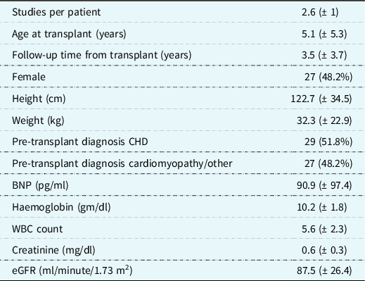
A summary of the population characteristics can be found in Table 1. A total of 56 patients and 143 observations were included in the analysis with a mean of 2.6 studies (+ 1) per patient. The mean age at the time of transplant was 5.1 (+ 5.3) years and mean follow-up time from transplant was 3.5 (+ 3.7) years. Forty-eight percent of the patients were female, 29 (51.8%) had a pre-transplant diagnosis of CHD, and 27 (48.2%) of cardiomyopathy. The mean B-type natriuretic peptide was 90.9 (+ 97.4) pg/ml, and the population was noted to have normal kidney function with a mean creatinine of 0.6 mg/dl (± 0.3).
Summary of patient (n = 56) demographics and baseline laboratory values

Data are presented as counts (percentage) or mean values (± standard deviation)
BNP = B-type natriuretic peptide; eGFR = estimated glomerular filtration rate; n = number of patients; WBC = white blood cell
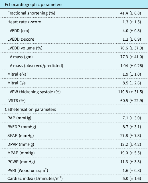
A summary of baseline echocardiographic and haemodynamic data is presented in Table 2. The majority of patients had normal systolic function as shown by a mean fractional shortening of 41.4% (+ 6.8%). Reference Tissot, Singh and Sekarski30 The mean right atrial and pulmonary capillary wedge pressures were also within normal limits with a mean right atrial pressure of 7.1 (+ 3.0) mmHg and a mean pulmonary capillary wedge pressure of 11.3 (+ 3.3) mmHg. Reference Nagueh, Smiseth and Dokainish31
Summary of paired studies (n = 143): echocardiographic and haemodynamic data

Data are presented as mean values (± standard deviation)
DPAP = pulmonary artery pressure in diastole; IVSTS = percent interventricular septal thickening in systole; LV mass (observed/predicted) = LV mass/(2.0574*
 $${\rm{h}}{{\rm{t}}^{0.3324}}{\rm{*w}}{{\rm{t}}^{0.5540}}$$
); LVEDD = left ventricular end-diastolic dimension; LVPW = left ventricular posterior wall; MPAP = mean pulmonary artery pressure; n = number of paired echocardiograms and cardiac catheterisations; PCWP = pulmonary capillary wedge pressure; PVRI = pulmonary vascular resistance index; RAP = right atrial pressure; RVEDP = right ventricular end-diastolic pressure; SPAP = pulmonary artery pressure in systole
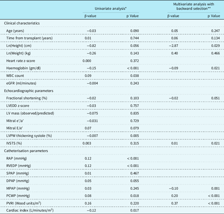
Based on the scatterplots, linear regression analyses were used to examine potential clinical characteristics associated with B-type natriuretic peptide including age, stature, laboratory values, echocardiographic, and haemodynamic indices and are summarised in Table 3. Among the haemodynamic parameters, mean right atrial pressure, right ventricular end-diastolic pressure, mean pulmonary capillary wedge pressure, and cardiac index were associated with B-type natriuretic peptide in the univariate analysis. However, only mean pulmonary capillary wedge pressure remained statistically significant in the multivariate analysis and mean pulmonary artery pressure and pulmonary vascular resistance index became significant in the multivariate analysis. Height was a statistically significant covariate of B-type natriuretic peptide in the multivariate analysis; however, age and weight were not. Haemoglobin was inversely related to B-type natriuretic peptide in both univariate and multivariate analyses, such that the lower the haemoglobin, the higher the B-type natriuretic peptide.
Linear regression analyses: relationships to B-type natriuretic peptide (LnBNP)

Data presented as β values (direction of correlation) and p values
BNP = B-type natriuretic peptide; DPAP = pulmonary artery pressure in diastole; eGFR = estimated glomerular filtration rate; IVSTS = percent interventricular septal thickening in systole; LV mass (observed/predicted) = LV Mass/(2.0574*
 $${\rm{h}}{{\rm{t}}^{0.3324}}{\rm{*w}}{{\rm{t}}^{0.5540}}$$
); LVEDD = left ventricular end-diastolic dimension; LVPW = left ventricular posterior wall; MPAP = mean pulmonary artery pressure; PCWP = pulmonary capillary wedge pressure; PVRI = pulmonary vascular resistance index; RAP = right atrial pressure; RVEDP = right ventricular end-diastolic pressure; SPAP = pulmonary artery pressure in systole; WBC = white blood cell count
* Univariate analysis adjusted for time
** Variables included simultaneously if p value <0.10 after adjusting for time, age, and weight
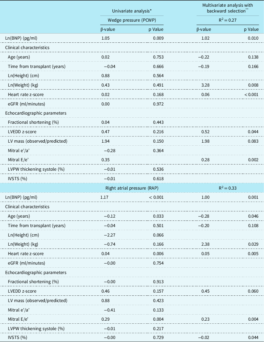
With univariate linear regression analysis, Ln(B-type natriuretic peptide) and mitral E/e’ were associated with mean pulmonary capillary wedge pressure (Table 4). In the multivariate analysis for covariates Ln(B-type natriuretic peptide), Ln(Weight), heart rate z-score, left ventricular end-diastolic dimension z-score, and mitral E/e’ were independently associated with mean pulmonary capillary wedge pressure. With linear regression analysis, Ln (B-type natriuretic peptide), age, heart rate z-score, left ventricular mass (observed/predicted), and mitral E/e’ were associated with right atrial pressure in the univariate analysis. In the multivariate analysis for covariates independently associated with mean right atrial pressure, Ln(B-type natriuretic peptide), age, Ln(weight), heart rate z-score, left ventricular end-diastolic dimension z-score, mitral E/e’, and percent interventricular septal thickening in systole were independently associated with mean right atrial pressure (Table 4).
Multivariate linear regression analyses: relationships of global right and left heart catheterisation derived filling pressures to B-type natriuretic peptide levels, demographic and echocardiographic parameters

Data presented as β values (direction of correlation) and p values
BNP = B-type natriuretic peptide; eGFR = estimated glomerular filtration rate; IVSTS = percent interventricular septal thickening in systole; LV mass (observed/predicted) = LV Mass/(2.0574*
 $${\rm{h}}{{\rm{t}}^{0.3324}}{\rm{*w}}{{\rm{t}}^{0.5540}}$$
); LVEDD = left ventricular end-diastolic dimension; LVPW = left ventricular posterior wall; WBC = white blood cell count
* Univariate analysis adjusted for time
** Variables included simultaneously if p value <0.10 after adjusting for time, age, and weight
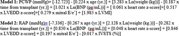
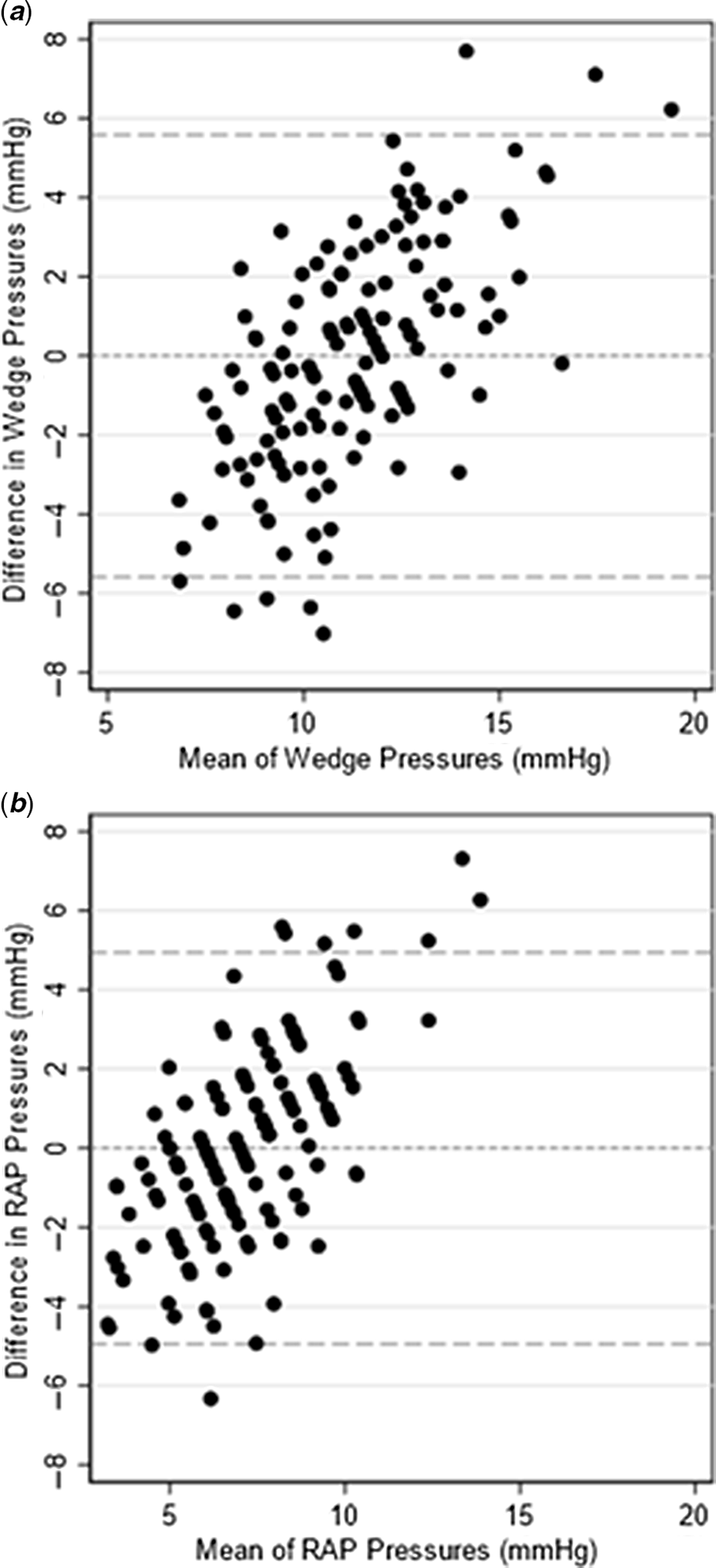
Within the limitations of predominantly normal filling pressures in the study cohort and based on the independent relationships identified in the multivariate analyses (Table 4), we derived predictive algorithms from the multi-variable models to estimate mean right atrial and pulmonary capillary wedge pressures in an equation format (Fig 1). Bland–Altman analyses comparing the variability between the measured and the equation estimated pulmonary capillary wedge and right atrial mean pressures was also performed (Fig 2). Note that most of the algorithm-predicted pressures were within 2 standard deviations, and there was a trend for the algorithms to overestimate the higher measured mean pulmonary capillary wedge, with 3 values greater than 2 standard deviations (Fig 2a) and right atrial pressures, with 6 values greater than 2 standard deviations (Fig 2b).
Equations created using univariate and multivariate regression analyses of non-invasive parameters and measured haemodynamic data. BNP = B-type natriuretic peptide; IVSTS = interventricular septal thickening in systole; LVEDD = left ventricular end-diastolic dimension; LVMI = left ventricular mass indexed; PCWP = pulmonary capillary wedge pressure; RAP = right atrial pressure.

Bland–Altman analyses comparing the variability between the measured and the equation estimated pulmonary capillary wedge ( a ) and right atrial ( b ) mean pressures. The difference between algorithm-predicted mean pressures and the respective measured pressures at cardiac catheterisation (y-axis) is plotted as a function of the sum of the algorithm-predicted and measured pressures/2 (x-axis) for each paired determination. The dashed lines are the zero line of identity and two standard deviations lower and higher than the line of identity. The majority of comparisons were less than two standard deviations of identity.

Discussion
In this study, we identified significant interrelationships between echocardiographic parameters, including multiple diastolic function parameters, and plasma B-type natriuretic peptide levels to directly measure right atrial and/or pulmonary capillary wedge pressures in paediatric heart recipients. In regression analyses, pulmonary capillary wedge pressure (multivariate analysis) and right atrial pressure (univariate analysis) were identified to be related to the Ln(B-type natriuretic peptide) levels (Table 3). Of the echocardiographic parameters studied, including estimates of diastolic function, only fractional shortening and percent interventricular septal thickening in systole were found to be related to Ln(B-type natriuretic peptide) levels (Table 3). In regression analyses, Ln(B-type natriuretic peptide) levels and several echocardiographic parameters, including the diastolic function parameter E/e’, were found to be related to pulmonary capillary wedge pressure and right atrial pressure (Table 4). These observations suggest that echocardiographic parameters of diastolic function and B-type natriuretic peptide levels are independently related to these pressures and provided the rationale for developing multi-parametric equations for estimating pulmonary capillary wedge pressure and right atrial pressure that include both echocardiographic parameters and B-type natriuretic peptide (Fig 1).
Diagnosing and tracking diastolic function in the cardiac allograft are clinically meaningful. Early detection of increased right atrial pressure and pulmonary capillary wedge pressure is particularly important in the surveillance of heart transplant patients as it potentially can detect the onset of late graft dysfunction and coronary allograft vasculopathy before the development of end-stage systolic dysfunction. Reference Lu, Magdo and Yu32,Reference Daud, Xu and Revelo33 However, in paediatric heart transplant recipients, direct measurement of right atrial pressure and pulmonary capillary wedge pressure requires sedation and vascular access and is typically reserved until the time of endomyocardial biopsy or coronary angiography which are only performed annually, or even less frequently after the first few years post-transplant. Reference Godown, Harris and Burger34–Reference Zhorne, Petit and Ing36
Cardiac filling pressures have been difficult to accurately ascertain by echocardiography alone, Reference Lunze, Colan and Gauvreau11–Reference Oh, Appleton and Hatle14 and the determinants of B-type natriuretic peptide levels have not been well characterised in paediatric heart transplant recipients. Reference Mehra, Uber and Walther25 Yet currently echocardiography and B-type natriuretic peptide testing are routinely used in serial outpatient monitoring for graft dysfunction in paediatric heart recipients. Echocardiography has been disappointing, particularly in paediatric heart recipients, in part because no single echocardiographic parameter has had acceptable predictive accuracy for rejection and, in part because the correlation between standard echocardiographic parameters of diastolic function and severity of diastolic dysfunction has not been rigorously validated. Reference Badano, Miglioranza and Edvardsen37–Reference Stengel, Allemann and Zimmerli40 Evidence from this study suggests that multiple echocardiographic parameters in conjunction with other non-invasive parameters could provide improved predictive equations for filling pressures. On the other hand, B-type natriuretic peptide levels can be elevated by factors in addition to wall stress. Reference Ationu, Sorensen and Whitehead20–Reference Maisel, Clopton and Krishnaswamy23 The trend is for B-type natriuretic peptide to increase in paediatric transplant recipients over time, and the relationship of B-type natriuretic peptide levels to rejection, graft failure, or cardiac allograft vasculopathy remains controversial. Reference Shaw and Williams41,Reference Lan, Chang and Alejos42 Therefore, it is important to understand common determinants of B-type natriuretic peptide levels which in this study were a shorter stature and lower haemoglobin. Evidence from this study suggests that including B-type natriuretic peptide along with multiple echocardiographic parameters could further improve predictive algorithms for filling pressures.
In the future, with a larger sample size, we would validate whether these algorithms can provide a robust prediction of these filling pressures as an important surrogate of diastolic function. We would also determine whether the estimation of filling pressures in this manner, including its change in individual patients, can aid in the diagnosis of rejection, development of cardiac allograft vasculopathy, and new idiopathic allograft dysfunction. With a larger sample size, a sub-analysis of patients with and without systolic dysfunction can also be performed.
Limitations
The limitations to this study include the retrospective study design with variable number of samples contributed by each patient at a variable duration after transplantation. These variations were adjusted for by selecting only routine annual surveillance cardiac catheterisation data. The small sample size also limited the Bland–Altman analyses as it could not be performed on a separate cohort to validate the predictive equations. We included this in the current cohort as a proof of concept and to support the predictive algorithms. All catheterisation data were measured while the recipient was under anaesthesia, and the echocardiographic and laboratory data were obtained on the same day but were not simultaneous with the cardiac catheterisation. The potential effect of anaesthetics and intravenous fluids, routinely used for paediatric cardiac catheterisation, could affect the haemodynamic measurements of right atrial pressure and pulmonary capillary wedge pressure. This design, however, best fits the intent of the study to develop a non-invasive algorithm in awake children useful in predicting catheterisation-derived left and right ventricular filling pressures. In addition, data collected at the time of rejection episodes were excluded so that this “normative” dataset and predictive algorithms could be developed. The algorithms describing the relationships between the selected covariates and filling pressures were generated over the range of predominantly normal post-transplant filling pressures. A larger sample size would also allow for inclusion of more extreme filling pressures as well as to include clinical events such as rejection, overt systolic graft dysfunction, and cardiac allograft vasculopathy as covariates as well as events predicted by the equations to estimate filling pressures. For the estimate of right atrial pressure, it would be advantageous to include echocardiographic parameters of right ventricular diastolic function in the future. Additionally, the equations generated from the models in this study included B-type natriuretic peptide, routinely followed at our institution, and would not be valid for NT-pro B-type natriuretic peptide.
Conclusions
In this study, we have identified important haemodynamic, echocardiographic, and clinical covariates such as haemoglobin that can affect the level of B-type natriuretic peptide in paediatric heart transplant recipients. This finding will help the clinician interpret B-type natriuretic peptide in a more accurate context. We incorporated B-type natriuretic peptide into novel multi-modal and multi-variable equations that included echocardiographic parameters referable to diastolic function to predict invasively acquired mean right atrial pressure and mean pulmonary capillary wedge pressure as the most acceptable measures of diastolic function in children. Given the difficulty of obtaining right atrial pressure and pulmonary capillary wedge pressure, this multi-modal approach warrants prospective testing to better follow global allograft diastolic function and help identify the need for additional evaluations, including cardiac catheterisation, especially when systolic function is normal. Based on these findings, this study identified candidate parameters for future prospective testing in a multi-parametric approach of their predictive value in screening for elevated left and right ventricular filling pressures.
Acknowledgements
None.
Financial support
This work was supported by NICHD K99 HD099387 and Stanford Maternal Child Research Institute MCHRI pilot grant to BH.
Conflicts of interest
None.
Ethical standards
The authors assert that all procedures contributing to this work comply with the ethical standards of the relevant national guidelines on human experimentation and with the Helsinki Declaration of 1975, as revised in 2008, and have been approved by the Institutional Review Board of Seattle Children’s Hospital.