Introduction
The birth of the first ‘test-tube’ baby, Louise Brown, created via the in vitro fertilization and embryo transfer (IVF-ET) technique pioneered by Robert Edwards, was a major advance in medical science. Since then, human-assisted reproduction technology has been rapidly developed and IVF babies account for ∼0.1% of the world’s total population. With the opening of the universal two-child policy in recent years, many couples have turned to the IVF-ET technique and there is an increasing demand for elective single-embryo transfer (SET). The rate of miscarriage in older pregnant women is generally more than that in young pregnant women; assisted reproductive technology (ART) faces greater challenges due to decreased ovarian function and fewer embryo transfers. Therefore, choosing high-quality embryos for implantation into the uterus is a challenge for human ART.
The retrieval of some high-quality mature oocytes and embryos is the key to IVF success. The increasing demands for SET highlight the need to identify biomarkers for the evaluation of embryo quality more accurately and objectively. At present, embryo selection is mainly based upon classic standard morphological evaluation; however, such morphological criteria remain poorly predictive of development or implantation. Cumulus cells (CCs) are the first cells to be directly contacted by oocytes and share the same microenvironment with the oocyte. Follicular development, oocyte maturation, cumulus expansion and ovulation rely on continuing crosstalk between the oocyte and the surrounding CCs (Carou et al., Reference Carou, Cruzans, Maruri, Farina, Fiorito, Olea and Lombardo2017; Kordus and LaVoie, Reference Kordus and LaVoie2017). Apoptosis is closely involved in most reproductive processes, including oocyte maturation, fertilization and subsequent developmental competence (Worku et al., Reference Worku, Rehman, Talpur, Bhattarai, Ullah, Malobi, Kebede and Yang2017; Rimon-Dahari et al., Reference Rimon-Dahari, Heinemann-Yerushalmi, Hadas, Kalich-Philosoph, Ketter, Nevo, Galiani and Dekel2018). An increase in CC apoptosis has also been associated with the immaturity of oocytes and with impaired fertilization and pregnancy rates (Lin, D. et al., Reference Lin, Ran, Zhu, Quan, Ye, Yu, Kang and Lin2017; Almeida et al., Reference Almeida, Ferreira, Silveira, Campos, Borges, Baeta, Silva, Reis and Del Puerto2018). Gene expression of CCs, including that of apoptosis-related genes, could predict oocyte developmental quality (Lin, L. T. et al., Reference Lin, Wang, Wen, Li, Chen, Tsai, Cheng and Tsui2017; Bouckenheimer et al., Reference Bouckenheimer, Fauque, Lecellier, Bruno, Commes, Lemaître, De Vos and Assou2018). To date, the precise mechanisms governing oocyte quality are still unclear. The tumour suppressor gene PTEN (encoding phosphatase and tensin homolog) has been shown to be crucial for mammalian reproduction, functions as a mediator of apoptosis in CCs and specifically suppresses primordial follicle activation in oocytes (Reddy et al., Reference Reddy, Liu, Adhikari, Jagarlamudi, Rajareddy, Shen, Du, Tang, Hämäläinen, Peng, Lan, Cooney, Huhtaniemi and Liu2008; Chang et al., Reference Chang, Lim, Yoon, Jeong, Bae, Lee, Yoon, Choi and Lee2015). Our previous studies found that increased PTEN expression promoted the apoptosis of granulosa cells (Yao et al., Reference Yao, Huang, Li, Jiang, Wu, Li, Peng, Hua, Huang, You, Chen, Lin and Yang2021). We speculated that PTEN might affect follicular development and predict oocyte/embryo developmental quality. In our study, we aimed to investigate the relationship between PTEN expression in human CCs and embryo developmental competence. We attempted to develop novel embryo assessment strategies that could be used alone or in combination with morphologic criteria.
Materials and methods
Patient and stimulation
In total, 52 intracytoplasmic sperm injection (ICSI) cycles performed at the Quanzhou Maternity and Child Healthcare Hospital between July 2014 and March 2016 were enrolled in the study. The patients with the following characteristics were included for analysis: basal follicle-stimulating hormone (Day 3 FSH) ≤10 IU/ml, a long protocol for the use of a gonadotropin-releasing hormone (GnRH) agonist, first stimulation cycle for IVF-ET, the presence of bilateral ovaries and the absence of ovulatory dysfunction, endometriosis, endocrine disorders such as polycystic ovarian syndrome or hyperprolactinemia. Primary infertility, immune infertility and women taking special drugs or undergoing ovarian surgery were also excluded to minimize etiological influences that could potentially alter the gene expression in cumulus granulosa cells.
Controlled ovarian hyperstimulation (COH) was performed using a long standard protocol with GnRH agonist (triptorelin acetate; Ipson; France)/recombinant human follicle-stimulating hormone (rFSH; Gonal-F; Seron; Merck)/human menopausal gonadotropin (hMG; Organon; The Netherlands). Briefly, triptorelin acetate was given at a dose ranging from 1.0 to 1.3 mg in the middle of luteal phase of menstrual cycle; ∼2 weeks later the adequate suppression of ovulation was assessed by monitoring serum estradiol (E2) levels and by ovarian ultrasonography. When the serum E2 level was 50 pg/ml or lower (conversion factor to SI unit, 3.671), a daily starting dosage of 150–300 IU of rFSH (75 IU/ampule) were given intramuscularly (IM) in the morning; then rFSH dose was adjusted or hMG was added according to the ovarian response. When the serum E2 levels reached the maximum peak and at the same time, two or more dominant follicles were 18 mm diameter or larger, 5000–1000 IU of human chorionic gonadotropin (hCG; Organon, Netherlands) was administered to trigger oocyte maturation. Ultrasound-guided transvaginal oocyte retrieval (TVOR) was performed ∼34–36 h after the hCG administration. The ovarian response was measured by number of oocytes retrieved. Cumulus cells were collected from infertile women undergoing ICSI treatment.
Evaluation parameters
In total, 52 ICSI cycles (caused by male infertility) were included in this study. In addition, in total, 187 individual CCs was included in this part of study. Oocyte maturation was initially evaluated based on the morphological characteristics of cumulus complex at the time of oocyte retrieval. At the time of ICSI (Day 0), the oocytes were classified into two categories based on nuclear status. If the first polar body was observed in the oocyte cytoplasm, the oocyte was regarded as being at the metaphase II (MII) stage and then was included in the matured group. conversely, the oocyte at the germinal vesicle (GV) stage and without first polar body (arbitrarily called metaphase I (MI)) was then included in the immatured group. The oocyte at the MII stage was fertilized with the help of the ICSI (intracytoplasmic sperm injection) procedure. ICSI was performed as described previously (Esteves et al., Reference Esteves, Roque, Bedoschi, Haahr and Humaidan2018). Oocyte fertilization was observed 18–19 h after ICSI. Normal fertilization was confirmed when two pronuclei (2PN) were found in the cytoplasm. Oocytes exhibiting abnormal fertilization include no pronuclei (0PN), 1 pronuclei (1PN) and multiple pronuclei (≥3PN). Embryo cleavage was examined 43–45 h after ICSI. Embryo evaluation was conducted 67–69 h after oocyte fertilization. A high-quality embryo on Day 3 should consist of 6–10 blastomeres with a uniform size and the fragment proportion should be less than 20% (Carrasco et al., Reference Carrasco, Arroyo, Gil, Gómez, Rodríguez, Barri, Veiga and Boada2017). Both the outcome of extended embryo culture and day of blastocyst development (Day 5/6) were recorded for each individually cultured embryo. The blastocyst assessment score was based on the expansion of the blastocoele cavity and the number and cohesiveness of the inner cell mass and trophectodermal cells (Gardner and Schoolcraft, Reference Gardner and Schoolcraft1999). Blastocysts scored ≥3BB were classified as ‘high-quality’ blastocysts.
Collection of human cumulus cells
Cumulus–oocyte complexes (COCs) were retrieved 34–36 h post hCG. COCs were incubated in fertilization medium covered with paraffin oil until denudation. At ∼0.5–1.0 h before ICSI, oocytes were denuded individually in a 50-μl droplet of G-MOPS plus containing 1 mg/ml hyaluronidase for a maximum of 40 s and washed sequentially in three droplets without enzyme. The CCs of individual oocytes were snap frozen by plunging in liquid nitrogen immediately after denudation until RNA extraction and MII oocytes were used for ICSI and were individually cultured after ICSI. All the collected CCs samples were well marked with the corresponding oocyte and subsequent embryo information.
RNA extraction and cDNA synthesis
Poly(A) RNA from pooled CCs from individual oocytes was extracted using the Real-Time Cell Lysis Kit (Roche) according to the manufacturer’s instructions. Details of cDNA synthesis are described in our previous paper (Yao et al., Reference Yao, Geng, Huang, Peng, Chen, Jiang, Yu, Li, Huang and Yang2017). The Transcriptor First Strand cDNA Synthesis Kit (Roche) was used to synthesize cDNA with a final volume of 20 μl containing 1 μl oligo(dT) primer, 4 μl reverse transcription buffer, 0.5 μl protector RNase inhibitor and 0.5 μl transcription reverse transcriptase. The reverse transcription was carried out at 65°C for 10 min, 55°C for 30 min and 85°C for 5 min to inactivate reverse transcriptase.
Quantitative real-time polymerase chain reaction
Quantitative analysis of each CCs sample was carried out using quantitative real-time PCR (qRT-PCR) of cDNA, with SYBR green and an ABI Prism 7500 system (Applied Biosystems), according to manufacturer’s protocol. Briefly, two-step qRT-PCR of the double-curve standard was performed to control for PCR variations. The quantity of each measured cDNA sample was normalized to the reference gene β-ACTIN (Chen et al., Reference Chen, Yao, Huang, Zheng, Jiang, Chen, Chen, Li, Huang, Jiang, Huang and Yang2015).
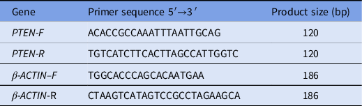
Primers for amplification of PTEN and β-ACTIN transcripts were designed by TaKaRa and are listed in Table 1. The PCR efficiency for each single primer pair was determined using serial five-fold dilutions of cDNA transcripts. The linear correlation coefficient (R2), an indicator of fit for the standard curve, was plotted to the standard data points of all genes, which ranged from 0.995 to 0.997. Cycling conditions were 95°C for 30 s, followed by 43 cycles at 95°C for 5 s, then 60°C for 34 s. Melting curves were generated to distinguish specific from non-specific amplicons under the following conditions: 95°C for 15s, 60°C for 1 min and 95°C for 15s. Results were collected in triplicate for each sample and threshold cycle values for PTEN were normalized relative to that of β-ACTIN. Finally, the 2−ΔCT method was used to quantify relative mRNA levels (Livak and Schmittgen, Reference Livak and Schmittgen2001).
Primer sequences for quantitative real-time polymerase chain reaction

Statistical analysis
All the results are presented as mean ± standard error (SE). The PTEN mRNA levels among different groups were analyzed using Student’s t-test and SPSS v.13.0 software. P-values < 0.05 were considered to be statistically significant. Data points determined to be outliers were excluded from graphic representation, but they were, however, included in statistical analysis.
Results
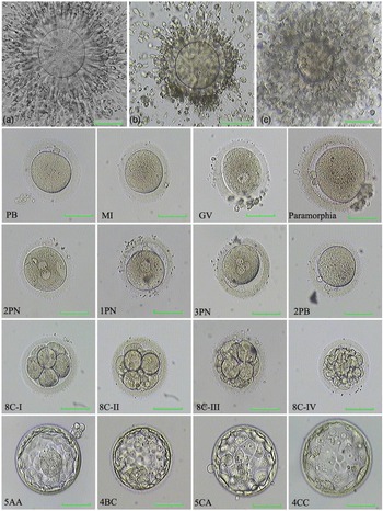
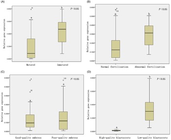
In total, 198 cumulus cell samples were obtained from the 52 women. Successful mRNA extraction was achieved and semiquantitative RT-PCR analysis was performed in 187 samples (based on β-ACTIN expression). We did not directly assay gene expression levels in the embryos; we measured PTEN expression levels in CCs retrieved from the corresponding oocyte that developed into an embryo. Upon nuclear maturation, 153 matured oocytes (MII) and 34 immatured oocytes (MI/GV) were analyzed. In addition, 11 abnormally shaped oocytes were discarded. At the time of fertilization observation, 125 oocytes showed normal fertilization and 28 oocytes showed abnormal fertilization or failed to be fertilized. Cleavage embryos were divided into ‘good-quality’ (Grade 1–2) and ‘poor-quality’ (Grade 3–4) embryos on Day 3 and Day 5/6 embryos were divided into ‘high-quality’ or ‘low-quality’ blastocysts. Upon normal fertilization, 53 oocytes developed into good-quality embryos and 72 developed into poor-quality embryos. During the blastocyst culture process, 46 oocytes developed into low-quality blastocysts and 26 into high-quality blastocysts (Fig. 1). Results in this study showed that a five-fold decreased PTEN mRNA level was observed in CCs isolated from MII oocytes compared with those isolated from MI/GV oocytes (relative mRNA abundance 0.084 ± 0.1843 vs. 0.4582 ± 0.1967, P < 0.001). Analysis using fertilization revealed differences in PTEN expression, with higher expression in CCs from oocytes that showed abnormal fertilization compared with those from oocytes with normal fertilization (0.3122 ± 0.1374 vs 0.1223 ± 0.1422, P = 0.015). Expression of PTEN in CCs from oocytes that developed into good-quality and poor-quality Day 3 embryos was only slightly different and did not reach statistical significance (0.0937 ± 0.1115 vs 0.1165 ± 0.1372, P = 0.653). PTEN was expressed at significant higher levels in CCs from oocytes that developed into low-quality blastocysts (0.2886 ± 0.1744 vs 0.0125 ± 0.0070, P < 0.001) than in those from oocytes that developed into high-quality blastocysts (Fig. 2).
Morphological observation of oocytes and embryos. (A) Mature cumulus–oocyte complex. (B) Over-mature cumulus–oocyte. (C) Immature cumulus–oocyte complex. At the time of ICSI (Day 0), the oocytes were first classified into two categories based on nuclear status: (i) immature oocytes exhibiting no polar bodies (MI) or immature oocytes at the germinal vesicle stage (GV) and (ii) mature MII oocytes that extruded a clearly visible polar body (PB), oocytes with paramorphia were discarded. On Day 1, oocyte fertilization was observed 18–19 h after ICSI and zygotes were classified into two groups: (i) oocyte exhibiting normal fertilization (2 pronuclei, 2PN) and (ii) oocyte exhibiting abnormal fertilization (including no pronuclei (0PN), 1 pronuclei (1PN) and multiple pronuclei (≥3PN). On Day 3 (67–69 h after oocyte fertilization), cleavage embryos were evaluated and divided into ‘good-quality’ (Grade 1–2, including 8C-I and 8C-II in this figure) and ‘poor-quality’ (Grade 3–4, including 8C-III and 8C-IV in this figure) embryos. During the blastocyst culture process, Day 5/6 embryos were divided into ‘high-quality’ (scored ≥3BB, including 5AA in this figure) or ‘low-quality’ blastocysts (scored ≥3BB or failed to formed blastocysts, including 4BC, 5CA and 4CC in this figure). Scale bars, 50 μm.

Relative expression levels of PTEN in cumulus cells isolated from human oocytes according to nuclear maturity, fertilization, cleavage embryo blastocyst and quality. (A) Cumulus cells from mature oocytes (MII) and immature oocyte (MI/GV). (B) Cumulus cells from oocytes that showed normal fertilization (two pronuclei, 2PN) and abnormal fertilization (including 0PN, 1PN and ≥3PN). (C) Cumulus cells from oocytes yielding good-quality embryos and poor-quality embryos at Day 3. (D) Cumulus cells from oocytes yielding good-quality and low-quality blastocysts after 5/6 days of in vitro culture. Results are expressed as means ± standard error of the mean (SEM) of relative expression to the reference gene β-ACTIN. The upper adjacent value and lower adjacent value (vertical lines) represent maximum and minimum data points. Standard errors of the means are indicated by bars. Outliers are indicated by closed dots. Values with different superscripts (a, b) refer to significant differences (P < 0.05).

Discussion
In addition to tumours and cardiovascular and cerebrovascular diseases, infertility will become the third major disease that threatens human health and family happiness in the 21st century, according to the World Health Organization (WHO). The incidence of infertility increased in the past 20 years and affects 15–20% of couples in China, reaching the level found in developed countries (Europe and America). Infertile couples have increased five-fold and the total number has reached 50 million. Under the guidance of social medicine and reproductive ethics, IVF-ET technology has developed rapidly and is a new milestone in the development of human science and technology. With the development of ovarian stimulation programmes and the improvement of the embryo culture system, the quality of the oocytes obtained is improved, which plays a key role in subsequent embryo development and clinical pregnancy (Xu et al., Reference Xu, Chen, Zhang, Wei and Cao2015; Dyer et al., Reference Dyer, Chambers, de Mouzon, Nygren, Zegers-Hochschild, Mansour, Ishihara, Banker and Adamson2016; Li et al., Reference Li, Wang, Lv, Huang, Wang, Pan and Zhang2017); however, ∼50% of female women still face pregnancy failure with ART. Currently, the criteria for oocyte and embryo selection are morphological at fixed times, which is simple and fast but strongly subjective.
There is growing evidence that the subjective morphological characteristics of the embryo are not completely consistent with its internal physiological state (Kurosawa et al., Reference Kurosawa, Utsunomiya, Shiga, Takahashi, Ihara, Ishibashi, Nishimoto, Watanabe, Abe, Kumagai, Terada, Igarashi, Takahashi, Fukui, Suganuma, Tachibana and Yaegashi2016; Sanchez et al., Reference Sanchez, Venturas, Aghvami, Yang, Fraden, Sakkas and Needleman2019; Fishel et al., Reference Fishel, Campbell, Foad, Davies, Best, Davis, Smith, Duffy, Wheat, Montgomery, Wachter and Beccles2020). Morphological assessment alone does not accurately predict oocyte developmental potential, so it is generally acknowledged that its reliability requires further improvement. It has been proposed that biochemical markers of embryos may be used as a parameter for selecting high-quality embryos but, so far, no powerful biochemical markers have been found to objectively and accurately reflect the developmental potential of embryos (Artini et al., Reference Artini, Tatone, Sperduti, D’Aurora, Franchi, Di Emidio, Ciriminna, Vento, Di Pietro, Stuppia and Gatta2017; Dhali et al., Reference Dhali, Javvaji, Kolte, Francis, Roy and Sejian2017; Biase and Kimble, Reference Biase and Kimble2018). Therefore, noninvasive, objective and reliable markers are needed that could identify the most quality oocytes and embryos with the highest implantation potential and would not compromise the success of IVF procedures in elective single-embryo transfer (SET). Optimization of the superovulation programme to ensure the accurate quality of follicle development in infertile women is a challenge. More recently, it has been reported that several genomic biomarkers expressed in CCs could accurately and noninvasively predict oocyte developmental competence (Assou et al., Reference Assou, Haouzi, De Vos and Hamamah2010; Huang et al., Reference Huang, Hao, Shen, Zhang and Liu2013; Rajabpour-Niknam et al., Reference Rajabpour-Niknam, Totonchi, Shahhosseini, Farrokhi, Alipour and Eftekhari-Yazdi2013; Artini et al., Reference Artini, Tatone, Sperduti, D’Aurora, Franchi, Di Emidio, Ciriminna, Vento, Di Pietro, Stuppia and Gatta2017). The mechanisms of early follicle development and oocyte growth are largely unknown. Follicular development, oocyte maturation, cumulus expansion and ovulation rely on continuing crosstalk between the oocyte and somatic follicular cells. Oocyte maturation and competence are acquired during follicular development. Follicular development determines the oocyte quality and oocyte quality is a key limiting factor in female fertility, yet we have a poor understanding of what constitutes oocyte quality or the mechanisms governing it. Mammalian oocytes grow and develop in the same follicle microenvironment as adjacent somatic cells. According to their positional relationship and functional characteristics with oocytes, granular cells are the first body cells to be directly contacted by oocytes and are divided into mural granular cells (MGCs) and cumulus granulosa cells (cumulus cells, CCs) (McLaughlin et al., Reference McLaughlin, Kinnell, anderson and Telfer2014; Biase and Kimble, Reference Biase and Kimble2018). The ovarian follicular microenvironment and maternal signals are mediated primarily through MGC and CCs. Oocytes and granular cells grow and develop in a highly coordinated and mutually dependent manner. The dysfunction of granulosa cells, especially cumulus granulosa cells, affects the follicular microenvironment, possibly resulting in oocyte meiotic chromosome abnormalities (Merhi et al., Reference Merhi, Polotsky, Bradford, Buyuk, Chosich, Phang, Jindal and Santoro2015). Therefore, CCs can more reflect the function of the follicular microenvironment and the changed ovarian function related to embryo development competence (Kordus and LaVoie, Reference Kordus and LaVoie2017). The oocyte plays a dominant role in regulating granular cell functions during folliculogenesis (Worku et al., Reference Worku, Rehman, Talpur, Bhattarai, Ullah, Malobi, Kebede and Yang2017); granulosa cells act as differentiation signals initiating the growth of primordial follicles (Appeltant et al., Reference Appeltant, Somfai, Nakai, Bodó, Maes, Kikuchi and Van Soom2015; Biase and Kimble, Reference Biase and Kimble2018). Therefore, it is believed that the functions of cumulus granular cells can indirectly reflect oocyte competence. CCs are a good tool for studying follicle development and the follicular microenvironment; moreover, functional studies of CCs do not affect the reproductive process.
Single-embryo transfer performs as a means of reducing multiple pregnancy rates. To achieve higher embryo implantation and successful pregnancy outcomes, the choice of the best embryos to transfer is important in ART. Using qRT-PCR technology, a multitude of studies have correlated changes in cumulus or granulosa cell gene expression with clinically relevant outcome parameters, including in vitro embryo development and pregnancy (Wathlet et al., Reference Wathlet, Adriaenssens, Segers, Verheyen, Van de Velde, Coucke, Ron El, Devroey and Smitz2011; Uyar et al., Reference Uyar, Torrealday and Seli2013; Pourret et al., Reference Pourret, Hamamah and Aït-Ahmed2016; Shen et al., Reference Shen, Chen, Zhao, Liu, Ren and Zhang2020; De Conto et al., Reference De Conto, Matte and Cunha-Filho2021). Although some studies have provided evidence for the genes expressed in CCs as potential biomarkers to predict oocyte quality/or embryo competence/or IVF pregnancy, biomarkers of CC gene expression require validation (Burnik Papler et al., Reference Burnik Papler, Vrtačnik Bokal, Maver and Lovrečić2015; Dhali et al., Reference Dhali, Javvaji, Kolte, Francis, Roy and Sejian2017; Kordus and LaVoie, Reference Kordus and LaVoie2017). Limited data are available regarding the close dialogue between human oocytes and their associated CCs. There is a growing awareness that apoptosis is closely involved in most reproductive processes; the incidence of apoptosis in CCs could influence oocyte quality (Jain et al., Reference Jain, Jain, Kumar, Kumar, De, Gohain, Kumar and Datta2016; Bosco et al., Reference Bosco, Ruvolo, Luparello, Ferrari, Valerio, Santi, Piomboni, Sarcina, Lispi and Roccheri2017). The tumour suppressor gene PTEN (phosphatase and tensin homolog deleted on chromosome 10), located at chromosome 10q23, is frequently mutated or inactivated in a variety of human cancers (Leslie and den Hertog, Reference Leslie and den Hertog2014; Papa et al., Reference Papa, Wan, Bonora, Salmena, Song, Hobbs, Lunardi, Webster, Ng, Newton, Knoblauch, Guarnerio, Ito, Turka, Beck, Pinton, Bronson, Wei and Pandolfi2014). The overexpression of PTEN leads to the suppression of cell proliferation, migration and invasion and the enhancement of apoptosis. Recent advances have demonstrated that the normal function of PTEN may also play an important role in the female reproductive system (McLaughlin et al., Reference McLaughlin, Kinnell, anderson and Telfer2014; Pulido, Reference Pulido2018). The PTEN/PI3K (phosphatidylinositol 3-kinase)–Akt (protein kinase B) signalling pathway plays a key role in cell apoptosis, growth and proliferation. Impaired PTEN signalling might cause ovarian dysfunction, resulting in premature ovarian failure (POF) (Shimizu et al., Reference Shimizu, Kimura, Takebayashi, Fujiwara, Takakura and Takahashi2009). Therefore, the present study aimed to identify and characterize the PTEN expression levels in CCs retrieved from the corresponding oocyte that developed into the embryo and then investigated the relationship between PTEN mRNA level and the oocyte quality/or embryo competence. However, cell samples from one individual oocyte are limited. The CC samples of individual oocytes should be collected independently and be well marked with the corresponding oocyte and subsequent embryo information. To truly reflect the correlation between gene expression and embryo quality, individual CC samples should not be mixed. It is difficult to complete the PTEN mRNA and PTEN protein detection in the same CC sample. So, our present study aimed to investigate the relationship between PTEN mRNA expression in human CCs and oocyte/embryo developmental quality.
Our previous studies showed that PTEN transcripts in MGCs varied significantly among different responders, with the highest levels in poor responders and the lowest levels in high responders (Yao et al., Reference Yao, Huang, Li, Jiang, Wu, Li, Peng, Hua, Huang, You, Chen, Lin and Yang2021). We speculate that the expression of PTEN may play a vital role in normal follicle development, which is closely related to oocyte and embryo quality. Past studies have also shown that embryo apoptosis could be closely related to the early embryo morphology; the incidence of apoptosis in CCs could influence oocyte quality (Bakri et al., Reference Bakri, Ibrahim, Osman, Hasan, Jaffar, Rahman and Osman2016). Originally, PTEN was reported to be a tumour suppressor gene frequently inactivated in some human cancers and its role as a transcription factor is well known (Leslie and den Hertog, Reference Leslie and den Hertog2014). Recently, altered expression of PTEN and its abnormal activity have been proposed to play a key role in human reproduction other than cancer, including oocyte growth and early follicle development. In human ovaries, PTEN might be involved in the regulation of proliferation and differentiation of granulosa cells (Brandmaier et al., Reference Brandmaier, Hou and Shen2017). Overexpression of PTEN in granulosa cells induced the arrest of the cell cycle and/or apoptosis. We also found that the apoptosis rate of granulosa cells declined significantly after 24 h transfection of the PTEN shRNA lentivirus (Yao et al., Reference Yao, Huang, Li, Jiang, Wu, Li, Peng, Hua, Huang, You, Chen, Lin and Yang2021). Considering that the function of CCs can better reflect the function of the follicular microenvironment and the embryo development competence than MGCs, our present study aimed to further investigate the relationship between the PTEN expression level in CCs and the oocyte quality and/or embryo competence. To our knowledge, this is the first study to investigate the PTEN gene in human CCs associated with the corresponding oocyte quality and their subsequent embryo competence. Our results showed that the PTEN mRNA level was five-fold lower in CCs that were surrounding mature oocytes than in those surrounding immature oocytes. The PTEN mRNA level was 2.5-fold lower in CCs from oocytes that showed normal fertilization than in those from oocytes with abnormal fertilization. The PTEN mRNA level was 23-fold lower in CCs from oocytes that developed into high-quality blastocysts than in CCs from oocytes that developed into low-quality blastocysts. However, the expression of PTEN in CCs from oocytes that developed into good-quality and poor-quality Day 3 embryos was only slightly different, without statistical significance. Therefore, we speculated that PTEN mRNA levels in human CCs that are surrounding oocytes negatively affect the follicular development, maturation, fertilization and embryo quality. However, the morphological criteria for grading and screening of D3 cleavage embryos are subjective and controversial and seem not to be related to the intrinsic competence of the cleavage embryos. Therefore, it is necessary to investigate new and noninvasive molecular markers in gamete and embryo selection; the measurement of PTEN mRNA levels in human CCs surrounding oocytes would reliably complement the morphological evaluation, providing a useful tool for selecting oocytes with greater chances to be fertilized and transplanted into the uterus, although this conclusion needs to be further verified in the future.
Why is the overexpression of PTEN not beneficial to normal follicle development? Apoptosis is a type of cell death and is closely involved in most of the reproductive processes, including abnormal ovarian function and follicular atresia. It has been reported that the apoptosis of granular cells may be a possible explanation for POF (Hong et al., Reference Hong, Peng, Li, Fang, Wang, Klausen, Yin, Wang, Leung and Yang2018). The overexpression of PTEN increases the apoptosis of CCs. The direct apoptosis of granular cells possibly leads to decreased oocyte quality and failed implantation. Several studies have reported that apoptosis may be the mechanism that causes female ovarian function to decline (Hussein, Reference Hussein2005; Chang et al., Reference Chang, Lim, Yoon, Jeong, Bae, Lee, Yoon, Choi and Lee2015; Worku et al., Reference Worku, Rehman, Talpur, Bhattarai, Ullah, Malobi, Kebede and Yang2017). Studies by Reddy showed that impaired PTEN function might result in POF in mice (Reddy et al., Reference Reddy, Liu, Adhikari, Jagarlamudi, Rajareddy, Shen, Du, Tang, Hämäläinen, Peng, Lan, Cooney, Huhtaniemi and Liu2008; Chang et al., Reference Chang, Lim, Yoon, Jeong, Bae, Lee, Yoon, Choi and Lee2015). Phosphatidylinositol 3,4,5-trisphosphate (PI(3,4,5)P3), a major substrate of PTEN, is an important second messenger molecule (Chang et al., Reference Chang, Lim, Yoon, Jeong, Bae, Lee, Yoon, Choi and Lee2015; Quan et al., Reference Quan, Wang, Gong, Peng, Richard, Zhang, Fornage, Alcorn and Wang2017). The PTEN protein dephosphorylates inositol phospholipids and converts phosphatidylinositol-3,4,5-trisphosphate (PIP3) to phosphatidylinositol-4,5-biphosphate (PIP2), through the PTEN/PI3K/Akt signalling pathway (Jagarlamudi et al., Reference Jagarlamudi, Liu, Adhikari, Reddy, Idahl, Ottander, Lundin and Liu2009; McLaughlin et al., Reference McLaughlin, Kinnell, anderson and Telfer2014). The proliferation and differentiation of granulosa cells are critical for follicular development (Jain et al., Reference Jain, Jain, Kumar, Kumar, De, Gohain, Kumar and Datta2016). In human ovaries, the phosphatidylinositol 3-kinase (PI3K) pathway plays a key role in the regulation of cell proliferation, migration, survival and metabolism (Reddy et al., Reference Reddy, Liu, Adhikari, Jagarlamudi, Rajareddy, Shen, Du, Tang, Hämäläinen, Peng, Lan, Cooney, Huhtaniemi and Liu2008). PTEN functions as a major negative regulator of PI3K/AKT signalling (Reddy et al., Reference Reddy, Liu, Adhikari, Jagarlamudi, Rajareddy, Shen, Du, Tang, Hämäläinen, Peng, Lan, Cooney, Huhtaniemi and Liu2008; McLaughlin et al., Reference McLaughlin, Kinnell, anderson and Telfer2014). PTEN dephosphorylates PIP3 to PIP2, followed by a decline in PIP3 levels and inhibition of the PI3K/Akt signalling pathway, resulting in cell apoptosis. PIP3 is an important second messenger that affects a variety of intracellular signals. The balance between PI3K and PTEN determines the PIP3 levels. The various activities involved in PIP2 are enhanced due to the overactivation of PTEN and, finally, apoptosis is induced. The occurrence of apoptosis in granulosa cells was closely associated with abnormal follicular development (Jain et al., Reference Jain, Jain, Kumar, Kumar, De, Gohain, Kumar and Datta2016). In contrast, loss of PTEN function might promote PI3K/AKT-mediated cell growth and survival. Good-quality oocytes and success of IVF treatments may be promoted by inhibition of PTEN expression. Although previous studies also showed that elevated apoptosis rates in granulosa cells are associated with lower pregnancy rates (Lin, D. et al., Reference Lin, Ran, Zhu, Quan, Ye, Yu, Kang and Lin2017), the exact intracellular signalling pathways leading to apoptosis within granulosa cells are largely unknown. The exact nature and diversity of oocyte–CCs signalling molecules are complex and dynamic. The differential expression of PTEN in human granulosa cells used as a molecular biomarker to predict embryonic development potential needs to be further studied. In our future research, we will use the PTEN inhibitor bpV (HOpic) and GC cell lines to investigate the underlying molecular mechanism by which PTEN affects ovarian follicular development and embryo developmental competence.
In conclusion, PTEN may play a vital role in human reproduction, especially in follicular development, but its exact functional importance and the mechanism of its involvement remain largely unknown. In the present study, our data suggest the expression of PTEN in human CCs and that it negatively affects follicular maturation, fertilization and embryo quality. Studying the PTEN signalling pathway may provide an effective treatment for infertility and the proapoptotic mechanism of PTEN in human reproduction needs to be further studied.
Data availability statement
All datasets generated for this study are included in the manuscript.
Author contributions
JY and ML collected samples, conducted experiments, analyzed data and wrote the manuscript. YL collected samples and designed the primers for qRT-PCR. JZ, YH WP analyzed data and conducted qRT-PCR. JL checked the draft. RH, XY designed experiment and revised the manuscript. JY, ML and LL revised the manuscript. All authors read and accepted the final version of the manuscript.
Financial support
This work was supported by the National Natural Science Foundation of China (grant number 81972714); the Young and Middle-aged Talent Training Programme of Fujian Province of China (grant number 2019-ZQN-91); the Natural Science Foundation of Fujian Province of China (grant number 2018J01148); the Natural Science Foundation of Fujian Province of China (grant number 2019J01140); the Hunan Provincial Natural Science Foundation of China (grant number 2021JJ30482) and the Quanzhou Science and Technology Plan Project (Type B, grant number [2004]0010).
Conflict of interest
The authors declare none.
Ethical approval
This study protocol was reviewed and approved by the Ethics Committee of Quanzhou Maternity and Child Healthcare Hospital and the registration number is Ethics 2018: No. 10. All the studied participants had consented in writing that their medical records could be used for research purposes, as long as the patients’ anonymities were protected. The authors assert that all procedures contributing to this work complied with the ethical standards of the relevant national and institutional committees on human experimentation and with the Helsinki Declaration of 1975, as revised in 2008.