Trypanosomatids are flagellated protozoan parasites belonging to the order Kinetoplastida. They can infect diverse hosts, ranging from plants through to higher mammals. In humans, Trypanosoma brucei and Trypanosoma cruzi are responsible for two major tropical diseases: human African trypanosomiasis (HAT) and Chagas disease, respectively. There are 11 million people infected and more than 150 million at risk. These diseases represent a major public health problem in regions of the world least able to deal with the associated economic burden. With no immediate prospect of vaccines, and no satisfactory drug treatments, the requirement for new therapies is a priority. However, drug discovery is high risk and expensive, and the development of agents designed specifically to target trypanosomal diseases is not perceived to be commercially attractive. Until recently, interest from large pharmaceutical corporations has been minimal, with most research and development occurring in academic settings. As a consequence, trypanosome infections are now referred to as ‘most neglected diseases’. This review focuses on how existing trypanocidal therapies mediate their activity, the problems associated with their usage and current areas of research that offer hope of new treatments.
Human African trypanosomiasis
The problem
Insect-transmitted parasites of the T. brucei species complex are the causative agents of HAT (also known as African sleeping sickness) and nagana, a disease common in domesticated animals in Africa. The parasite was first identified in 1895 by David Bruce while studying cattle (Ref. Reference Steverding1), and later from human blood by Forde and Dutton (Ref. Reference Steverding1). HAT is considered endemic in 36 countries of sub-Saharan Africa, with over 50 million people at risk (Refs Reference Barrett2, Reference Stuart3). Localised epidemics can arise readily following political and socioeconomic disruption, killing tens of thousands of people. As a result of coordinated surveillance and treatment programmes, the number of people dying from HAT has recently fallen to ~70 000 annually (Ref. Reference Barrett4). However, in regions of Angola, the Democratic Republic of Congo and southern Sudan, the mortality rate exceeds that of malaria and HIV/AIDS (human immunodeficiency virus and acquired immune deficiency syndrome).
HAT pathology has two distinct phases: the early (or haemolymphatic) stage and the late (or encephalitic) stage (Refs Reference Barrett2, Reference Stuart3). In the early stage, parasites are found in both the blood and the lymph systems. Symptoms begin with irregular fevers, headaches and joint pains, but can develop to generate other complications including enlarged lymph glands and spleen, local oedema and cardiac abnormalities. When parasites cross the blood–brain barrier, the disease then enters the late stage. Here, a series of neurologically related symptoms are observed, including severe headaches, alterations to the sleeping pattern, personality change, mental function impairment and weight loss. Without treatment, patients eventually fall into a coma and die.
HAT is caused by two distinct subspecies of T. brucei, each having separate geographical foci and differing pathologies (Ref. Reference Barrett2). In West and Central Africa, T. b. gambiense is prevalent, while elsewhere in sub-Saharan Africa, T. b. rhodesiense predominates. These subspecies are responsible for the West and East African forms of the disease, respectively. The main difference between the two infections is the rate of progression from the blood/lymphatic stage to the cerebral stage. In West African trypanosomiasis, this takes months and hence the infection is termed chronic. By contrast, with East African trypanosomiasis the infection is more acute, with progression occurring in as little as 1–3 weeks. In both cases, the disease is zoonotic, although with T. b. gambiense, humans are the main reservoir. Wild and domestic animals, especially cattle, are the major reservoirs with T. b. rhodesiense. A third closely related subspecies, T. b. brucei, is nonpathogenic to humans, but is responsible for many cases of nagana in cattle. This infection is of huge veterinary and economic importance and has had a profound effect on agricultural development in Africa.
T. brucei is an extracellular pathogen, with a surface coat composed almost entirely of a single antigen, the variant surface glycoprotein (VSG). The parasite avoids immune destruction by a process of antigenic variation, which involves the periodic switching of VSG expression to another of the antigenically distinct surface glycoproteins encoded by the large repertoire of VSG genes. As a result of this constant switching, vaccine development against this parasite is not thought to be a realistic prospect.
Treatment of HAT
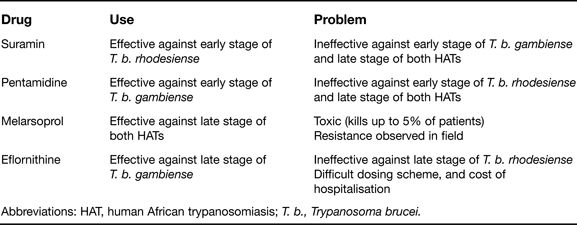
Treatment of African sleeping sickness is dependent on the subspecies and the disease stage. When parasites are restricted to the blood/lymphatic system, pentamidine is used against T. b. gambiense, and suramin against T. b. rhodesiense (Table 1). Neither compound can cross the blood–brain barrier effectively, and these drugs are therefore of little use against cerebral disease. The treatments available for this lethal stage are restricted to melarsoprol and eflornithine. Melarsoprol is the only drug active against both T. brucei subspecies once the central nervous system (CNS) has been accessed (Table 1). However, it is a highly toxic arsenical, and drug resistance is a major issue (Ref. Reference Matovu5). Eflornithine, in contrast, is relatively safe, but this compound is effective only against West African trypanosomiasis and the cost of treatment is problematic in underdeveloped countries (Table 1). Currently, Phase III clinical trials are evaluating the efficacy of a combination therapy consisting of eflornithine with nifurtimox against the cerebral stages of West African trypanosomiasis (Refs Reference Checchi6, Reference Priotto7, Reference Priotto8). This has proved very effective and has recently been recommended by the World Health Organization (WHO) as a front-line treatment for infections with T. b. gambiense.
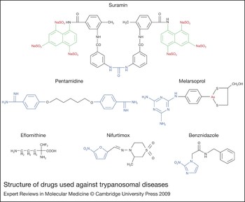
Structure of drugs used against trypanosomal diseases. The highlighted regions correspond to motifs described in the text. For suramin, urea (blue), napthalenes (green) and sulphonates (red) are shown. In pentamidine, the aromatic diamidine (blue) is outlined. For melarsoprol, the arsenic (red) and melamine ring (blue) are highlighted. The blue regions in nifurtimox and benznidazole correspond to the 5-nitrofuran or the 2-nitroimidazole group, respectively.

Problems associated with current drugs for human African trypanosomiasis
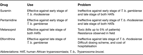
Abbreviations: HAT, human African trypanosomiasis; T. b., Trypanosoma brucei.
Suramin
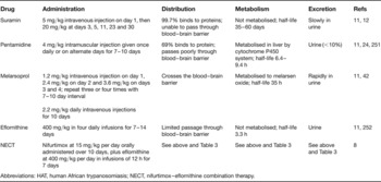
Suramin was first used against HAT in 1922 following observations that trypan dyes had trypanocidal activity (Refs Reference Docampo and Moreno9, Reference Nok10). This symmetrical polysulphonated naphthalene derivative of urea (Fig. 1) is negatively charged at physiological pH. The drug is typically administered to patients as an intravenous injection with a low dose on day 1 followed by higher amounts on days 3, 5, 11, 23 and 30 (Ref. Reference Legros11) (Table 2). Its anionic charge prevents it from freely crossing biological membranes, hinders transport across the blood–brain barrier and promotes binding to a variety of other molecules. Suramin is extremely stable in humans, shows no transformation in the liver, and is only slowly excreted in urine. These properties contribute to the relatively long half-life (Ref. Reference Spinks12) (Table 2).
Pharmacokinetic properties of drugs against human African trypanosomiasis

Abbreviations: HAT, human African trypanosomiasis; NECT, nifurtimox–eflornithine combination therapy.
Based on size and charge, it had been assumed that transport of suramin into trypanosomes occurs through bulk-flow uptake. However, in the presence of blood serum proteins, the drug transport rate exceeds the level attainable by this passive process alone (Ref. Reference Fairlamb and Bowman13). As suramin can readily complex with blood serum proteins, it has been suggested that the drug enters T. brucei through receptor-mediated endocytosis, possibly bound to low-density lipoprotein (LDL) (Refs Reference Coppens14, Reference Vansterkenburg15). However, analysis of endocytosis components indicates that suramin and LDL uptake are not directly linked, with the drug entering the parasite via an unidentified mechanism (Ref. Reference Pal, Hall and Field16).
Little is known about how suramin is active against trypanosomes. Suramin-resistant strains of T. brucei and Trypanosoma evansi, a blood-borne animal parasite, have been selected in vitro, but the mechanisms underlying this phenotype have not been established (Refs Reference Scott, Tait and Turner17, Reference Mutugi, Boid and Luckins18). By virtue of its negative charge, suramin can hinder uptake of serum proteins, or inhibit endocytosis and key enzymes in metabolic pathways such as glycolysis (Refs Reference Fairlamb and Bowman13, Reference Misset and Opperdoes19, Reference Willson20). Since suramin affects multiple targets this, coupled with its low toxicity, may explain why it has been used to treat early-stage East African trypanosomiasis for nearly 90 years.
Pentamidine
Pentamidine belongs to the aromatic diamidine class of antiprotozoal agents and was first used against T. b. gambiense in 1937, following observations that the related compound synthalin had potent antitrypanosomal activity (Refs Reference Docampo and Moreno9, Reference Nok10). Treatment involves daily intramuscular injections for 7–10 days (Ref. Reference Legros11) (Table 2). It is readily metabolised by the mammalian cytochrome P450 system (but not by the trypanosomal system) and excreted in urine, and therefore has a relatively short half-life (Refs Reference Conte, Upton and Lin21, Reference Berger22, Reference Berger and Fairlamb23) (Table 2).
Pentamidine is positively charged. As a result, it binds easily to blood serum proteins, does not readily diffuse across biological membranes, and passes into the cerebrospinal fluid very slowly (Ref. Reference Sanderson24). Because the drug must accumulate to high intracellular levels before having a trypanocidal effect (>1 mm), it is probable that active transport mechanisms are necessary for pentamidine activity. Uptake of diamidines by bloodstream T. brucei involves the P2 aminopurine permease (TbAT1), a transporter that also mediates melarsoprol uptake (Refs Reference Carter, Berger and Fairlamb25, Reference de Koning and Jarvis26, Reference Berger, Carter and Fairlamb27). However, T. brucei TbAT1-null mutants were found to retain sensitivity to pentamidine, whereas they were resistant to other diamidines such as berenil (Ref. Reference Matovu28). These results can now be explained following the characterisation of other uptake activities, including a high-affinity pentamidine transporter (HAPT) and low-affinity pentamidine transporters (LAPTs) (Refs Reference de Koning and Jarvis26, Reference De Koning29). As pentamidine uptake by T. brucei occurs by several routes, it has been speculated that resistance by reduced drug uptake is unlikely to arise in a clinical context. However, parasites deficient in two of the pentamidine transporters (TbAT1 and HAPT) have been generated in the laboratory, demonstrating that such events can occur (Ref. Reference Bridges30). In addition, recombination between resistant strains in the field is a possibility.
The mode of action for pentamidine is not fully understood. It can interact with anionic molecules and readily bind to DNA (Ref. Reference Wilson31). Analogues of pentamidine accumulate in the nucleus and kinetoplast, a disc-shaped structure containing the mitochondrial genome (Ref. Reference Mathis32). It has been proposed that pentamidine inhibits mitochondrial topoisomerase II activity, leading to the linearisation of the kinetoplast DNA and the generation of dyskinetoplastic trypanosomes (Ref. Reference Shapiro and Englund33). Exposure of parasites to pentamidine analogues appears to confirm this observation, as prolonged treatment leads to breakdown of the kinetoplast and its eventual disappearance (Refs Reference Wilson31, Reference Mathis32). However, it has been argued that formation of dyskinetoplastic parasites may not be sufficient to cure an infected mammalian host, and the mechanism of pentamidine transport into the mitochondrion has yet to be determined (Ref. Reference Wang34). Thus, diamidines may exert their trypanocidal activities through other mechanisms, possibly via inhibition of T. brucei S-adenosylmethionine decarboxylase or Ca2+-ATPase activities, by affecting mitochondrial membrane potential, or by acting to uncouple oxidative phosphorylation (Refs Reference Bitonti, Dumont and McCann35, Reference Benaim36, Reference Vercesi and Docampo37, Reference Moreno38). Intriguingly, pentamidine analogues accumulate in organelles lacking DNA, possibly acidocalcisomes, and this possible mode of action warrants further investigation (Ref. Reference Mathis32).
Melarsoprol
Melarsoprol (MelB) was introduced in 1949 and is currently the only treatment used against the late stage of both forms of HAT (Ref. Reference Friedheim39). This trivalent melaminophenyl arsenical (Fig. 1) functions as a prodrug, with activation leading to the formation of melarsen oxide (MelOx). MelB is highly toxic and causes reactive encephalopathy in approximately a fifth of patients, and has a mortality rate of 5% (Ref. Reference Enanga40). Additionally, it is responsible for other side effects including convulsions, fever, loss of consciousness, rashes, bloody stools, nausea and vomiting. The drug is administered as an intravenous injection with a number of different schedules (Ref. Reference Blum and Burri41) (Table 2). For T. b. rhodesiense infections, a regime consists of one injection daily over 3–4 days, followed by a rest period of 7–10 days; this is then repeated twice more. Based on pharmacokinetic properties, an alternative scheme is now routinely used for T. b. gambiense infections, consisting of a daily injection for 10 days (Refs Reference Legros11, Reference Burri42). Although this regime can lead to adverse skin reactions, it is as effective as the previous schedules and significantly cheaper: less drug is used and hospitalisation is shorter. Additional problems are also encountered because MelB has poor solubility in water, alcohol or ether, and the drug is normally dissolved in propylene glycol. As a result, MelB can only be given using glass syringes and the injections are very painful, with patients frequently reporting a burning sensation during administration.
Accumulation of melaminophenyl arsenical drugs by trypanosomes occurs through the P2 aminopurine permease TbAT1, and loss of this activity has been implicated in drug resistance in both laboratory and field isolates (Refs Reference Matovu5, Reference Matovu28, Reference Carter and Fairlamb43, Reference Maser44). The transporter was identified using competition studies, which demonstrated that uptake of purine nucleosides occurs by two distinct activities (Ref. Reference Carter and Fairlamb43). One transport system (designated P1) functions as a general purine nucleoside transporter, and a second (TbAT1) carries adenosine and its nucleobase adenine. MelB and MelOx hinder adenine uptake through the latter mechanism. Comparison of TbAT1 substrates has identified a common amidinium-like motif (Refs Reference Barrett and Fairlamb45, Reference Tye46, Reference de Koning and Jarvis47, Reference de Koning48). This pattern has now been incorporated into several novel chemical structures, some of which display considerable trypanocidal activity (Refs Reference Tye46, Reference Baliani49, Reference Stewart50, Reference Baliani51, Reference Chollet52). Intriguingly, TbAT1-null mutants are only threefold more resistant to MelB and MelOx than controls, significantly lower than the levels of resistance reported in laboratory-selected cells adapted to grow in the presence of arsenical drugs (Refs Reference Matovu5, Reference Fairlamb53). This has led to the proposal of alternative uptake routes, with HAPT being a likely candidate (Refs Reference Matovu28, Reference Bridges30, Reference de Koning54). The involvement of this pentamidine transporter in arsenical uptake stems from observations that sequential loss of TbAT1 followed by HAPT generates parasites with a greater resistance to MelOx than the TbAT1-null mutant (Ref. Reference Bridges30).
Arsenical drugs lyse T. brucei rapidly, but their modes of action are not fully understood. MelB inhibits a range of glycolytic and pentose phosphate pathway enzymes (Refs Reference Van Schaftingen, Opperdoes and Hers55, Reference Hanau56), and can readily form stable adducts with trypanothione, the glutathione–spermidine conjugate that is the major thiol in trypanosomes (Refs Reference Fairlamb57, Reference Fairlamb, Henderson and Cerami58). These adducts can block activity of trypanothione reductase (TR), the parasite-specific enzyme that maintains trypanothione in its reduced form (Refs Reference Fairlamb, Henderson and Cerami58, Reference Cunningham, Zvelebil and Fairlamb59). However, in all cases the rapidity with which the drug kills parasites cannot be explained by the systems so far implicated in its mode of action. For example, in the time taken for MelB-treated cells to lyse, ATP levels have not been sufficiently depleted to kill the parasite (Ref. Reference Fairlamb, Henderson and Cerami58). In addition, only a small fraction of trypanothione is actually conjugated to the arsenical, and T. brucei with reduced TR levels are as sensitive to MelOx as controls (Ref. Reference Krieger60).
In some regions, the relapse rate to MelB is up to 30% (Ref. Reference Legros11). The emergence of resistance is of concern, given that the drug is the only cheap treatment available against the late stage of both forms of HAT. In a significant number of cases, MelB-nonresponsive parasites retrieved from HAT patients were found to contain mutations in TbAT1, suggesting that resistance correlates with reduced drug uptake (Ref. Reference Matovu5). However, TbAT1-null mutants do not display a high level of resistance (Ref. Reference Matovu28), although it has not yet been established whether other transporters such as HAPT contribute to this phenotype. In other organisms, multidrug resistance is often mediated through active efflux mechanisms involving ABC transporters. Analysis of the T. brucei genome data suggests that the parasite possesses 22 potential members of this protein class (Ref. Reference Sauvage61). Overexpression of one of these, TbMRPA, results in tenfold resistance to MelB (Ref. Reference Shahi, Krauth-Siegel and Clayton62). This protein is a putative thiol-conjugate transporter and it has been postulated that TbMPRA functions by removing arsenical/trypanothione moieties from the cell. However, its role in mediating resistance in a clinical context is open to debate (Ref. Reference Alibu63).
Eflornithine
Originally developed as an anticancer therapy, eflornithine (dl-α-difluoromethylornithine) was first used to treat late-stage West African trypanosomiasis in the early 1980s (Ref. Reference Bacchi64) (Fig. 1). It quickly gained the nickname ‘resurrection drug’ for its ability to cure and revive comatose patients. However, despite being relatively safe, usage is restricted as a result of the difficult dosing scheme (Ref. Reference Robays65): eflornithine is administered under medical supervision by intravenous infusion in four large doses daily for 7–14 days (Ref. Reference Legros11) (Table 2). One explanation for the high concentrations of drug needed to cure the disease is poor transport across the blood–brain barrier (Ref. Reference Sanderson66). Despite these problems, eflornithine is now recommended as the front-line treatment for late-stage West African trypanosomiasis (Refs Reference Robays65, Reference Balasegaram67, Reference Politi68, Reference Chappuis69).
In mammals, eflornithine enters cells by passive diffusion, a mechanism that may operate in bloodstream T. brucei (Refs Reference Bellofatto70, Reference Erwin and Pegg71, Reference Bitonti72). However, as uptake by trypanosomes is temperature sensitive and follows Michaelis–Menten-type kinetics, it could be carrier-mediated, possibly by amino acid transporters (Ref. Reference Phillips and Wang73; M. Barrett, pers. commun.). Once in the parasite, the drug functions as an irreversible inhibitor of ornithine decarboxylase (ODC), the rate-controlling enzyme in polyamine biosynthesis (Ref. Reference Willert and Phillips74). The interaction between T. brucei ODC and eflornithine has been extensively studied and the mechanism of drug selectivity has been determined (Refs Reference Phillips, Coffino and Wang75, Reference Ghoda76). It stems from the differential stability of the parasite ODC (half-life ~18 h) and mammalian ODC (half-life ~20 min) (Refs Reference Phillips, Coffino and Wang75, Reference Tabor and Tabor77, Reference Iten78). When eflornithine irreversibly binds to the mammalian enzyme, the resultant complex is rapidly degraded and replaced with newly synthesised ODC. By contrast, the T. brucei eflornithine–ODC molecule is relatively stable and the enzyme slowly replaced. Thus, the level of active ODC is effectively decreased, leading to a cessation of putrescine formation. Eventually, parasites stop growing and the nondividing cells are cleared by the immune system.
Chagas disease
The problem
Chagas Disease (or American trypanosomiasis) is caused by T. cruzi. This is a zoonotic infection spread by blood-feeding triatomid (or kissing) bugs. The parasite can infect a wide range of mammals including dogs, cats, monkeys, rodents, ground squirrels, opossums and armadillos. It is found throughout Latin America, where as many as 8–11 million people are infected, resulting in over 15 000 deaths per year (Refs Reference Barrett2, Reference Stuart3, Reference Schofield, Jannin and Salvatella79). Normally, the disease occurs in rural areas where triatomid bugs transmit parasites from animals to humans. Recently, as a result of human migration, the disease has also been found in urban areas. This, in combination with secondary routes of infection, such as through blood and organ transplantation, has resulted in Chagas disease becoming a problem in the USA, which has an estimated 100 000 infected people (Ref. Reference Bern80).
Chagas disease has three distinct phases: acute, indeterminate and chronic (Refs Reference Barrett2, Reference Stuart3). The acute stage commonly affects children, with up to 5% of diagnosed cases resulting in death. During this phase, parasites multiply as intracellular amastigotes in macrophages and tissue cells at the site of the insect bite. Although generally asymptomatic, the acute stage can present clinical signs in the form of fever, skin lesions (chagoma), oedema, enlarged lymph nodes and conjunctivitis (Romana's sign). These can appear 1–6 weeks after infection. In the indeterminate stage, patients are asymptomatic and parasites largely disappear from the bloodstream, although they can be detected in cardiac and smooth muscle. In this phase, patients remain a reservoir of infection. In most cases, the disease does not advance further. However, 30% of individuals eventually progress to the chronic stage, often 10–20 years after the initial infection. The long-lasting, symptomatic chronic phase frequently presents with extensive cardiac and digestive tract pathologies and in these instances prognosis is poor. Reactivation of latent Chagas disease in HIV/AIDS patients has also been observed, sometimes with unusual clinical manifestations, including CNS involvement (Ref. Reference Diazgranados81).
In Latin America, the economic impact of Chagas disease outweighs the combined effects of other parasitic diseases, including malaria and leishmaniasis. Although there is no effective cure for the chronic disease, it is possible to halt transmission through the elimination of the insect vector. In 1991, governments of the six Southern Cone countries launched an initiative to control Chagas disease, which has had remarkable success, with transmission halted in previously endemic regions (Ref. Reference Dias, Silveira and Schofield82). New programmes, aimed at repeating this accomplishment in other areas of Latin America, are now under way (Ref. Reference Schofield, Jannin and Salvatella79). However, even if all transmission could be blocked, Chagas disease would remain a public health problem for many years.
Chagas disease chemotherapy
Treatment of Chagas disease is controversial because the two available drugs, nifurtimox and benznidazole, are toxic, may be carcinogenic, and have poor efficacy against the chronic stage (Refs Reference Rodriques Coura and de Castro83, Reference Gorla84, Reference Ferreira, Schwarz and Ferreira85). Additionally, the proposition that antiparasitic drugs are appropriate for treating the chronic stage has been questioned, given the belief that there is a large autoimmune component associated with this phase of the disease (Ref. Reference Kalil and Cunha-Neto86). Recently, a series of reports using murine models has shown that parasite persistence is both necessary and essential for development of Chagasic heart disease, that drugs prevent development of chronic cardiomyopathy, and that drug-induced pathogen clearance results in a stable protective T cell memory (Refs Reference Tarleton, Zhang and Downs87, Reference Tarleton and Zhang88, Reference Tarleton89, Reference Garcia90, Reference Bustamante, Bixby and Tarleton91). These studies demonstrate that chemotherapeutic intervention against any stage of Chagas disease is an appropriate course of action.
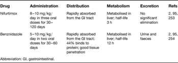
Nifurtimox and benznidazole have been used to treat the acute phase of Chagas disease for more than 40 years. Both compounds are orally administered, and because they are metabolised by the cytochrome P450 system (Refs Reference Walton and Workman92, Reference Montalto de Mecca, Diaz and Castro93, Reference Mecca94), multiple doses have to be given daily (Refs Reference Barrett2, Reference Raether and Hanel95) (Table 3). A course of treatment lasts 1–4 months for nifurtimox and 1–2 months for benznidazole. In many cases the recommended drug schedules are not completed, often as a result of side effects, resulting in considerable scope for the development of resistance.
Pharmacokinetic properties of drugs against Chagas disease

Abbreviation: GI, gastrointestinal.
Both nifurtimox and benznidazole are nitroheterocyclic compounds. They contain a nitro group linked to a furan or imidazole ring, respectively (Fig. 1). These agents function as prodrugs and must undergo enzyme-mediated activation to have cytotoxic effects, reactions catalysed by nitroreductases (NTRs). The precise mode of action of nitroheterocyclic drugs in trypanosomes was originally unclear, with two hypotheses proposed.
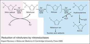
The first stemmed from observations that activation of either drug can lead to the formation of reactive oxygen species (ROS) (Refs Reference Docampo96, Reference Docampo97, Reference Viode98) (Fig. 2), a process that involves a one-electron reduction of the drug by a type II NTR activity. In the presence of oxygen, this promotes superoxide anion production and drug regeneration, a process known as futile cycling (Fig. 2). To date, several trypanosomal flavin adenine dinucleotide (FAD)-containing enzymes, including TR, lipoamide dehydrogenase and cytochrome P450 reductase have been implicated in nifurtimox reduction (Refs Reference Viode98, Reference Henderson99, Reference Blumenstiel100). This mode of action was considered attractive because trypanosomes were thought to lack many of the ‘classical’ eukaryotic enzymes responsible for ROS detoxification (Refs Reference Boveris101, Reference Fairlamb and Cerami102). However, far from being deficient in such activities, trypanosomatids actually possess a series of novel oxidative defence pathways (Refs Reference Flohe, Hecht and Steinert103, Reference Wilkinson104, Reference Wilkinson and Kelly105, Reference Irigoín106). The only direct link between drug-induced ROS formation and trypanocidal activity stems from gene deletion experiments on T. brucei SODB1, which encodes a superoxide dismutase. Parasites lacking SODB1 are hypersensitive to nifurtimox and benznidazole (Ref. Reference Prathalingham107). Functional analysis of other oxidative defence pathways has failed to find a link with the trypanocidal activity of nitroheterocyclic drugs (Refs Reference Wilkinson104, Reference Wilkinson108, Reference Wilkinson109, Reference Wilkinson110, Reference Wilkinson111, Reference Wilkinson112, Reference Kelly113).
Reduction of nitrofurans by nitroreductases. Type I nitroreductases (NTRs) (blue background) mediate a two-electron reduction of the conserved nitro group on the nitrofuran drug to generate the unstable nitroso derivative. This then undergoes further reduction to the amine form via a hydroxylamine intermediate. The hydroxylamine can also react to form a nitrenium ion, which then promotes DNA damage. Type II NTRs (lilac background) mediate a one-electron reduction of the conserved nitro group, leading to the formation of an unstable nitro radical. In the presence of oxygen, this can react to form superoxide anions (O2−). This process, known as futile cycling, also results in the regeneration of the drug.

The second hypothetical mechanism was based on the demonstrated antimicrobial activity of nitrofurans (Refs Reference Peterson114, Reference Roldan115). Here, flavin mononucleotide (FMN)-containing, oxygen-insensitive type I NTRs mediate a series of two-electron reductions of the conserved nitro group, through nitroso, to a hydroxylamine derivative, using reduced nicotinamide adenine dinucleotide (phosphate) [NAD(P)H] as the source of reducing equivalents. The hydroxylamine can react to generate nitrenium cations, which promote DNA breakage (Refs Reference McCalla, Reuvers and Kaiser116, Reference Streeter and Hoener117) (Fig. 2). In addition to this, the highly electrophilic intermediaries may affect other molecules in the cell. The decrease in free thiol content observed in nifurtimox- and benznidazole-treated T. cruzi could be due to conjugation of the thiol with the nitroso form of the drugs, thus affecting the redox status of the cell (Ref. Reference Maya118). Two trypanosomal enzymes with this type I activity have been reported. The first is a prostaglandin F2α synthase (PGFS), although this can only mediate two-electron reduction of nifurtimox under anaerobic conditions (Ref. Reference Kubata119). The second, for which there is now strong experimental evidence, is a type I NTR (Ref. Reference Wilkinson120). This class of enzyme had been regarded as specific to bacteria, and absent from eukaryotes; trypanosomes are now a major exception.
T. cruzi resistance to nifurtimox and benznidazole is encountered throughout Latin America and can be readily mimicked through continuous culturing of the parasite in the presence of drug (Refs Reference Wilkinson120, Reference Nozaki, Engel and Dvorak121, Reference Murta and Romanha122). Until recently though, the molecular basis for resistance had not been elucidated and appeared complex. Parasites selected for nitroheterocyclic drug resistance often displayed significant karyotypic alterations (Refs Reference Wilkinson120, Reference Nozaki, Engel and Dvorak121), with the resulting effects on global gene expression masking the precise mechanism(s) (Refs Reference Nogueira123, Reference Murta124, Reference Murta125, Reference Andrade126, Reference Rego127, Reference Campos128, Reference Nogueira129). A link between the activity of a trypanosome type I NTR and cross-resistance to nifurtimox and benznidazole has now been established in T. cruzi and T. brucei. In both cases, deletion of the corresponding gene generates parasites resistant to nitroaromatic compounds, while overexpression confers hypersensitivity (Ref. Reference Wilkinson120). Furthermore, laboratory selection of nifurtimox-resistant T. cruzi gives rise to parasites that lack one of the chromosomes containing the NTR gene. This phenotype can be complemented by reintroduction of the gene. These experiments clearly implicate NTR as a key player in the activation of both drugs and demonstrate the potential for resistance by a simple mechanism (Ref. Reference Wilkinson120). The extent to which this occurs in the field remains to be established. Another possible resistance mechanism involving drug detoxification has also been proposed (Ref. Reference Portal130). Overexpression of a trypanosomal cytochrome P450 reductase (CPR) confers resistance to benznidazole. However, given that nitroheterocyclic drugs are readily metabolised by hepatic CPRs to generate toxic products, it is unclear how this mechanism would mediate resistance within the parasite. This warrants further attention to dissect the precise details underlying the pathway.
Future perspectives in chemotherapy
The research landscape
As outlined, new treatments for HAT and Chagas disease are urgently needed in the respective regions. This will require a major international effort since important specifications must be fulfilled by any new drugs targeted at these diseases. In the case of HAT, the priority is a safe, oral, short-course therapy for late-stage T. b. gambiense CNS infection. The requirement for active compound to reach parasites within the brain is a major issue in drug design, since the blood–brain barrier impedes the passage of most small molecules. For Chagas disease, a drug able to arrest or alleviate the symptoms of the chronic stage of the infection is the challenging goal. Progress in both areas has been limited, in part, by the lack of good animal models that accurately mirror the course of disease in humans. However, the outlook for research is improving with schemes such as the Drugs for Neglected Diseases Initiative (DNDi) and the Consortium for Parasitic Drug Development (CPDD), as well as the engagement of organisations such as the Bill & Melinda Gates Foundation and the Wellcome Trust. In addition, academic and biotechnology/pharmaceutical organisations, facilitated by public–private partnerships, are building portfolios of projects based on validated targets and high-throughput screening of chemical libraries. These approaches build on significant advances that have been made in our understanding of trypanosome biology and genetics, including the completion of the T. brucei and T. cruzi genome projects in 2005 (Refs Reference El-Sayed131, Reference Berriman132). The development of sophisticated and flexible genetic manipulation systems, particularly for T. brucei (Refs Reference Meissner, Agop-Nersesian and Sullivan133, Reference Motyka and Englund134), has also been an important factor, in that it facilitates rapid analysis of gene function and target validation.
New targets for chemotherapy
There are now a large number of trypanosomatid enzymes and/or biochemical pathways that have been identified as targets for drug development. Below are listed some examples that are currently under investigation in this context.
Sterol biosynthesis
Analysis of T. cruzi sterol composition revealed that the parasite contains ergosterol, a molecule previously identified as a component of fungal cell membranes (Ref. Reference Furlong135). Candidate genes encoding enzymes in the ergosterol biosynthetic pathway have been identified in T. cruzi, with many of these having now been characterised and chemically validated as drug targets (Refs Reference Buckner136, Reference Buckner137, Reference Buckner138, Reference Urbina139, Reference Lepesheva140, Reference Chen141, Reference Konkle142). The pathway is present in T. brucei, but this parasite can also scavenge cholesterol from the host bloodstream (Ref. Reference Coppens and Courtoy143). Several inhibitors of the fungal sterol biosynthetic pathway have been found to have activity against T. cruzi. These include compounds that block sterol 14α-demethylase (CYP51), such as triazoles (Refs Reference Urbina and Docampo144, Reference Roberts145, Reference Urbina146), imidazoles (Refs Reference Buckner136, Reference Lepesheva140, Reference Hucke147, Reference Suryadevara148, Reference Kraus149), indomethacin amides (Ref. Reference Konkle142) and a novel structure (C155-0123) based on the moiety N-[4-pyridyl]-formamide (Refs Reference Chen141, Reference McKerrow150). A number of these are reported to display curative activity against acute/chronic Chagas disease, with some able to eradicate nifurtimox- and benznidazole-resistant T. cruzi from mice, even in immunosuppressed hosts (Refs Reference Urbina146, Reference Molina151, Reference Urbina152).
The allylamine derivative terbinafin, and heteroallyl 5-nitrofuranes, which are both inhibitors of squalene epoxidase, are also active against T. cruzi (Refs Reference Urbina153, Reference Gerpe154). Terbinafin is of particular interest, as it potentiates the activity of the triazole CYP51 inhibitors (Refs Reference Urbina153, Reference Maldonado155). Inhibitors of lanosterol synthase (oxidosqualene cyclase) have been reported to have good in vitro activity against T. cruzi, but in vivo effects have yet to be identified (Refs Reference Buckner138, Reference Hinshaw156). Some azasterols have been shown to target sterol methyltransferase activity, whereas others do not (Refs Reference Urbina157, Reference Urbina158, Reference Magaraci159, Reference Lorente160, Reference Gros161, Reference Gigante162). These latter molecules do however still display trypanocidal activity, even against T. b. rhodesiense, an organism able to scavenge sterols from its surrounding environment, indicating that these compounds may target other essential parasitic enzymes (Refs Reference Lorente160, Reference Gros161, Reference Gigante162, Reference Gros163).
Cysteine proteases
Several trypanosomal cysteine proteases (CPs) have been identified and biochemically characterised, including the cathepsin L-like enzymes TcCATL (cruzipain) and TbCATL (rhodesain or brucipain) (Refs Reference Cazzulo164, Reference Murta165, Reference Eakin166, Reference Caffrey167), and the cathepsin B-like CPs TcCATB and TbCATB (Refs Reference Lima168, Reference Garcia169, Reference Nobrega170, Reference Mackey171) (nomenclature in accordance with Ref. Reference Caffrey and Steverding172). Validation of these enzymes as drug targets is under way. Chemical validation of both cathepsin L-like CPs and TbCATB has resulted in the identification of inhibitors that block enzymatic function, with several compounds displaying trypanocidal activity in vivo and parasitological cure in animal models (Refs Reference McKerrow150, Reference McKerrow, McGrath and Engel173, Reference Engel174, Reference Du175, Reference Roush176, Reference Du177, Reference Greenbaum178, Reference Nkemgu179, Reference Doyle180, Reference Chen181, Reference Mallari182, Reference Mallari183). For TcCATL and TbCATL, structural data have informed the molecular mechanisms underlying some of these enzyme–inhibitor interactions (Refs Reference McGrath184, Reference Eakin185, Reference Huang, Brinen and Ellman186, Reference Choe187, Reference Kerr188). Genetic validation of the T. cruzi CPs has not been reported, presumably because of their essential nature, although overexpression of TcCATL does enhance metacyclogenesis (Ref. Reference Tomas, Miles and Kelly189). Intriguingly, RNA interference (RNAi) experiments in T. brucei have demonstrated that TbCATB, but not TbCATL, is essential for parasite viability (Refs Reference Mackey171, Reference Abdulla190). However, in murine models, trypanosomes with reduced TbCATL are less efficient at crossing the blood–brain barrier and parasitaemia is suppressed, leading to the suggestion that anti-TbCATL inhibitors may function by preventing the onset of late-stage HAT (Ref. Reference Abdulla190).
Thiol metabolism
Trypanothione is a low molecular weight thiol unique to trypanosomes (Refs Reference Fairlamb57, Reference Fairlamb and Cerami102, Reference Krauth-Siegel and Comini191). Therefore, enzymes involved in its metabolism, especially TR and trypanothione synthetase, are good candidates for drug design (Refs Reference Fairlamb and Cerami102, Reference Krauth-Siegel and Inhoff192, Reference Jaeger and Flohe193). Many of these systems have been genetically validated as drug targets (Refs Reference Krieger60, Reference Irigoín106, Reference Wilkinson111, Reference Krauth-Siegel and Comini191, Reference Comini, Krauth-Siegel and Flohe194, Reference Comini195, Reference Ariyanayagam196, Reference Wyllie197), whereas chemical inhibitors studies have focused primarily on TR (Refs Reference Henderson99, Reference Jockers-Scherubl, Schirmer and Krauth-Siegel198, Reference Fournet199, Reference Zani200, Reference Bond201, Reference Gallwitz202, Reference Fournet203, Reference Salmon-Chemin204, Reference Salmon-Chemin205, Reference Zani and Fairlamb206, Reference Hamilton207, Reference Hamilton208, Reference Hamilton209, Reference Stump210, Reference Martyn211, Reference Czechowicz212, Reference Stump213, Reference Stump214, Reference Holloway215, Reference Holloway216, Reference Cavalli217, Reference Richardson218, Reference Patterson219); several compounds blocking TR activity have been identified, with some displaying trypanocidal activity. A significant aid to such TR inhibitor studies is the availability of a three-dimensional structure (Refs Reference Bond201, Reference Zhang220, Reference Lantwin221, Reference Jacoby222, Reference Zhang223). This has facilitated high-throughput virtual enzyme–ligand screens that then guide the synthesis of candidate compounds (Refs Reference Saravanamuthu224, Reference Meiering225, Reference Ogungbe and Setzer226). The structures of several other enzymes involved in trypanothione metabolism have now been elucidated and it is envisaged that this resource should assist in the identification of inhibitors (Refs Reference Alphey227, Reference Alphey, Konig and Fairlamb228, Reference Melchers229). However, despite considerable efforts, no inhibitor targeting trypanothione metabolism has yet entered clinical trials.
Polyamine biosynthesis
The use of eflornithine against HAT has prompted the characterisation of other components of polyamine biosynthesis (Ref. Reference Bacchi64). In T. brucei, several of these enzymes have been biochemically characterised (Refs Reference Bitonti, Dumont and McCann35, Reference Bitonti, Kelly and McCann230, Reference Tekwani, Bacchi and Pegg231, Reference Yarlett232, Reference Taylor233) and shown to be essential for parasite viability (Refs Reference Willert and Phillips74, Reference Taylor233, Reference Huynh234, Reference Xiao, McCloskey and Phillips235). Compounds targeting these activities have been identified (Refs Reference Bitonti, Dumont and McCann35, Reference Tekwani236, Reference Guo237, Reference Brun238, Reference Marasco239, Reference Hirth240), some of which cure T. brucei-infected mice (Refs Reference Bitonti241, Reference Bacchi242, Reference Bacchi243). Of these, the S-adenosylmethionine decarboxylase irreversible inhibitor MDL 73811 is the most potent, although it is rapidly cleared in rodent models (Refs Reference Bitonti241, Reference Byers244). Recently, a stable derivative, Genz-644131, has been developed that overcomes these problems and shows considerable promise in the treatment of East African trypanosomiasis (Refs Reference Bacchi245, Reference Barker246).
Bridging the gap until the development of novel drugs
There are currently no novel classes of chemotherapeutic agents undergoing trials against late-stage HAT or chronic Chagas disease. As noted by Bernard Pécoul, Executive Director of DNDi, ‘No new drugs for stage 2 [late] sleeping sickness are expected in the next five years, so there is an urgent need to develop new treatments based on currently available drugs, especially through combinations’ (http://www.msfaccess.org/media-room/press-releases). Given this scenario, the evaluation of combinatorial therapies is important, especially in the short term. Evaluation of cotherapies using existing drugs has several other advantages: it could delay emergence of drug resistance to the parental compounds, it may allow a reduction in drug dosage (thereby minimising toxicity while maintaining efficacy), and it could streamline drug administration. Different combinations of three trypanocidal agents (melarsoprol, eflornithine and nifurtimox) have recently been trialled against West African trypanosomiasis, with one cotherapy (nifurtimox–eflornithine) showing particular promise in terms of safety and efficacy (Refs Reference Checchi6, Reference Priotto7, Reference Priotto8, Reference Priotto247). Follow-up studies at different geographical locations have confirmed the potential of this combination, resulting in NECT (nifurtimox–eflornithine combination therapy) being added to the Essential Medicines List of the WHO (Refs Reference Checchi6, Reference Priotto7, Reference Priotto8, Reference Priotto247).
Another well-tried approach to improving chemotherapy is to investigate derivatives related to drugs already in use. An example of this is parfuramide, an orally available prodrug based on furamidine, a diamidine with trypanocidal activity (Refs Reference Rane, Rane and Kinnamon248, Reference Das and Boykin249, Reference Ansede250). This compound did reach Phase III clinical trials against early-stage HAT and was shown to have good efficacy. However, a retrospective Phase I trial in South Africa required by the US Food and Drug Administration revealed nephrotoxicity in a small number of the volunteers. Currently, these unexpected findings are being investigated. In other studies, several nitroheterocyclic derivatives have been shown to have considerable trypanocidal activity (Refs Reference Baliani49, Reference Stewart50, Reference Baliani51, Reference Chollet52). Notable among these is fexinidazole, an orally administered 5-nitroimidazole that displays trypanocidal activity against both HAT subspecies, has little toxicity in animal studies and can cross the blood–brain barrier. This has resulted in fexinidazole entering clinical development with the ultimate goal of using it to target both stages of HAT. The precise mode of action of nitroaromatic compounds is currently unknown, but they may undergo activation involving a type I NTR (Ref. Reference Wilkinson120). Further dissection of this activity in terms of substrate specificity will provide a valuable resource for designing the next generation of NTR-activatable nitroheterocyclic drugs.
Conclusion
Although several drugs are available for use against HAT and Chagas disease, none is satisfactory. Each has limited efficacy, resistance is an increasing problem, dosage regimes can be complex and drug administration requires medical supervision. There has been significant progress in resolving the mechanisms by which these trypanocidal drugs operate and in identifying how resistance might arise. In addition, advances in our understanding of basic parasite biochemistry and genetics have led to identification of a large number of potential drug targets. New funding initiatives, prompted by the recognition that research on these debilitating neglected diseases has long been under resourced, now provide an opportunity to exploit these findings and improve the range of treatments.
Acknowledgements and funding
The authors acknowledge The Wellcome Trust for their financial support and thank Martin Taylor, Belinda Hall and the reviewers for their constructive comments on the manuscript.