INTRODUCTION
Parasites are usually given the opportunity to choose between a variety of unique sites on or in their hosts. Indeed, the current knowledge allows us to say that site selection is universal among them, though varying between the species and groups (Rohde, Reference Rohde1979). Each given site constitutes a unique microhabitat – due to its very own abiotic and biotic features – determining, to some extent, the parasite's choice.
Site selection is well documented in the literature for the parasitic copepods of fish (see for example, Geets et al. Reference Geets, Coene and Ollevier1997; Lo and Morand, Reference Lo and Morand2001; Scott-Holland et al. Reference Scott-Holland, Bennett and Bennett2006; Timi et al. Reference Timi, Lanfranchi and Poulin2010). In particular, among the chondracanthid copepods, the members of the genus Acanthochondria were described to infect, specifically, the branchial chambers of flatfish (see for example, Kabata, Reference Kabata1959, Reference Kabata1979, Reference Kabata1992). These latter constitute a unique and protected environment that offers the parasites, a number of different possible sites for attachment. Also, they allow blood-feeding species to feed abundantly, particularly when they infect the filaments of the holobranchs and pseudobranchs. In accordance with Llewellyn (Reference Llewellyn1956), variability in the volumes of water that pass by the 4 holobranchs might lead to different infection levels, once the larval specimens are given different opportunities to attach on each of them. Such variability was demonstrated, for instance, for the brown trout (Salmo trutta forma fario L.), with the volume of water passing over holobranchs II and III being significantly greater than that passing over holobranchs I and IV (Paling, Reference Paling1968). Besides this, several intrinsic factors of the parasites may also determine how they distribute among the different holobranchs (Ramasamy et al. Reference Ramasamy, Ramalingam, Hanna and Halton1985; Geets et al. Reference Geets, Coene and Ollevier1997).
In recent years, several studies have documented high infection levels of a species of Acanthochondria, i.e., A. cornuta, in European flounder, Platichthys flesus (Linnaeus, 1758) (Teleostei: Pleuronectidae), of North Sea (Schmidt et al. Reference Schmidt, Zander, Körting and Steinhagen2003) and Atlantic (Marques et al. Reference Marques, Teixeira and Cabral2006; Cavaleiro and Santos, Reference Cavaleiro and Santos2007, Reference Cavaleiro and Santos2009) waters and considered issues like the spatial and seasonal occurrence of infection. Notwithstanding, several aspects of the species ‘niche volume’ (see Rohde, Reference Rohde1994), including, among others, parasite spatial distribution on the host and food, remain still to be elucidated.
The main aim of this paper is to describe in both qualitative and quantitative terms, the pattern of site selection of A. cornuta in the European flounder. The possible driving forces of the observed pattern of spatial distribution are discussed.
MATERIALS AND METHODS
Host sampling
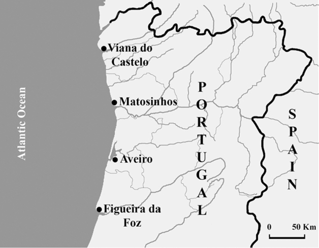
In total, 210 European flounder were collected: in September 2005, off 4 localities (Viana do Castelo, Matosinhos, Aveiro and Figueira da Foz) of north-central Portugal (41°40′N, 8°50′W to 40°8′N, 8°52′W), Eastern North Atlantic (Fig. 1); and during 1 year (once per season) in the geographical locality where flounders were most infected, i.e., Matosinhos, between September 2005 and May 2006. Additional details on sampling are provided in the papers by Cavaleiro and Santos (Reference Cavaleiro and Santos2007, Reference Cavaleiro and Santos2009).
Geographical location of the 4 sampling areas in the north-central Portuguese coast, Eastern North Atlantic.

Parasite survey
Ocular and blind branchial chambers were examined for Acanthochondria parasites. In each of them, 17 possible infection sites were considered for analysis. They were: (i) the operculum (internal surface); (ii) the branchiostegal pocket (delimited by the operculum and the branchiostegal membrane); (iii) the urohyal; (iv) the internal wall of the chamber; (v-xvi) the 4 holobranchs, in each of which, 3 sites were considered for analysis – i.e., the bony part (raker) and the inner and outer hemibranchs (filaments); and (xvii) the pseudobranch (filaments). The opercula and holobranchs were first dissected out of the fish and only then examined for parasites. Both the ocular and blind holobranchs were numbered from I to IV. The one nearest the operculum was assigned as holobranch I, whereas the innermost was assigned as holobranch IV. Besides the branchial chambers, examined sites on the fish's body surface included also the skin, fins, eyes, nostrils and mouth cavity. Observations were carried out under a stereo- dissecting microscope at 30× magnification. Chondracanthids did not detach from the tissues to which they were attached during the host manipulation. Consequently, they could be localized (and counted) with precision on the host's body. Their identification to the species level, that is, as representatives of A. cornuta, was made following the description of Kabata (Reference Kabata1979, Reference Kabata1992). As for the life-cycle stages i.e. copepodite (I-V), pre-adult female or adult female/male, they were identified in accordance with Heegaard (Reference Heegaard1947). Different stages of sexual maturity were considered for the adult female: (i) non-gravid, i.e. female without egg sacs; (ii) gravid, i.e. female with intact egg sacs; and (iii) post-gravid, i.e. female with remnants of egg sacs.
Analysis of parasite site selection
Infection levels, that is, prevalence, intensity and abundance, were estimated in accordance with Bush et al. (Reference Bush, Lafferty, Lotz and Shostak1997). Also, the mean, standard deviation (s.d.) and range of variation were calculated (for intensity and abundance) and recorded whenever necessary. Statistical analyses were conducted on SPSS for Windows, version 17.0 (SPSS Inc., Chicago, Illinois), with the significance level set at P<0·05. The different levels of analysis considered were as described below.
General site selection
To characterize the spatial distribution of A. cornuta on the host's body, the mean intensity was computed for each class of fish including specimens with the same number of infection sites (sites on the ocular and blind sides of the fish's body were considered separately). The existence of a significant relationship between the 2 variables, that is, intensity and number of infection sites, was evaluated by the Spearman's rank correlation test. Also, the existence of a significant difference in infection occurrence among the ocular and blind sides was investigated. Moreover, the infection prevalence in the 2 sides was compared using the McNemar's test for matched pairs of dichotomic variables. The abundance levels recorded for the 2 sides were compared using the Wilcoxon matched-pairs signed ranks test. Non-parametric tests were preferred over parametric ones owing to the non-normal distribution of both intensity and abundance data (Kolmogorov-Smirnov test: 1·598 ⩽ Z ⩽2·494; 0·000 ⩽ P ⩽0·012).
Site selection within the branchial chambers
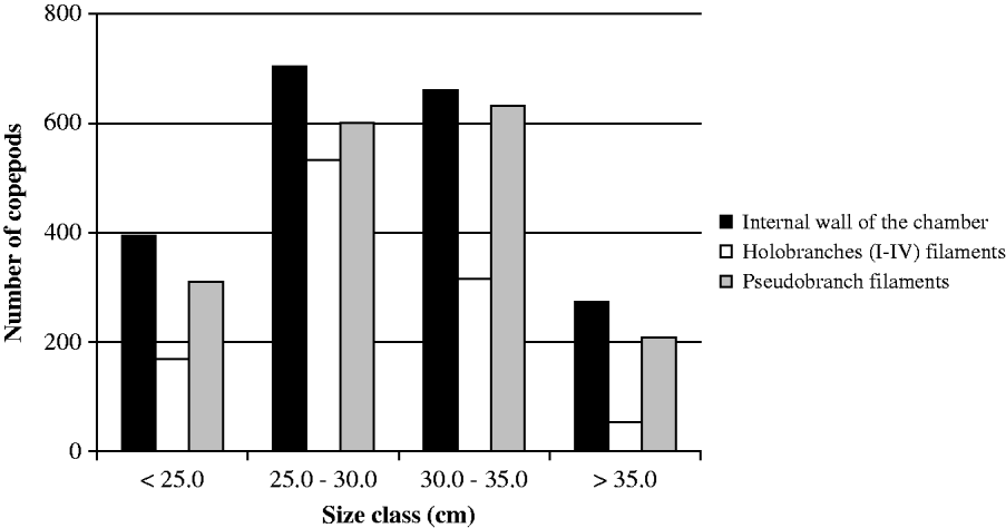
Prevalence and intensity (mean±s.d.) levels were assessed for each infection site in the ocular and blind branchial chambers. Next, the existence of particular spatial distributions inside them was investigated. Moreover, significant differences in parasite abundance among the filaments of the inner and outer hemibranchs of a given holobranch were evaluated by the Wilcoxon matched-pairs signed ranks test, whereas differences in abundance among the 4 holobranchs and among the internal wall of the chamber, the holobranch (I-IV) filaments and the pseudobranch filaments were tested by the Friedman rank sum test. All these analyses were conducted separately for the ocular and blind chambers. The number of specimens in different stages of development and sexual maturity i.e., copepodites (I-V), pre-adult females, non-gravid adult females, gravid adult females, post-gravid adult females and adult males, was quantified for each site of attachment in the branchial chamber. In particular, the distribution of copepodites (I-V), pre-adult females, non-gravid adult females and gravid adult females was assessed for the 4 holobranchs. Finally, an analysis of the number of copepods at different infection sites in the branchial chamber was performed for fish of different size. The classes considered in this analysis were as follows: class 1: <25·0 cm (N=43); class 2: [25·0–30·0 [cm (N=107); class 3: [30·0–35·0 [cm (N=39); and class 4: ⩾35·0 cm (N=21).
RESULTS
The overall infection levels of a total of 4841 specimens of A. cornuta were: prevalence=80·0%; intensity (mean±s.d. [range])=28·8±24·0; (1–110) parasites were isolated from the examined fish.
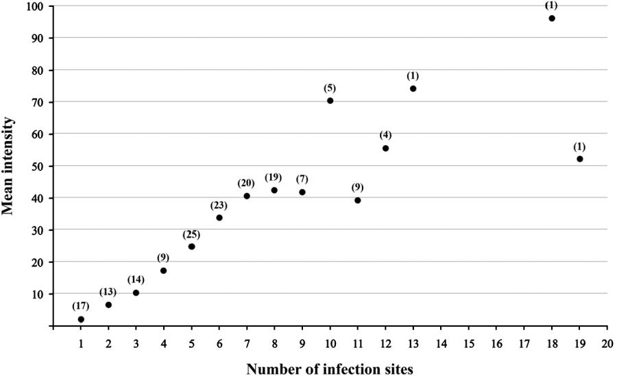
The branchial chamber constituted, as expected, the preferred site of attachment. However on rare occasions, i.e. in 3 fish recording several infection sites, a few parasites were also isolated from the skin (N=2) and fins (N=5). Internally, no parasite was found attached to the bony parts of the holobranchs, urohyal and operculum, and within the branchiostegal pocket. Also, no specimen was found infecting the eyes, nostrils and mouth cavity. The recorded data support the existence of a positive correlation between the intensity and the number of infection sites (Spearman's rank correlation test: r s=0·795, N=168, P=0·000) (Fig. 2). Besides this, the existence of a side bias in infection was partially supported by the results of the performed statistical analyses, i.e., for abundance but not for prevalence (Wilcoxon matched-pairs signed ranks test: Z=−5·099, N= 210, P=0·000; McNemar's test: N=210, P=0·134; respectively), with the parasites preferring the ocular side (mean±s.d. abundance: 13·1±14·4 parasites) to the blind side (mean±s.d. abundance: 10·0±11·2 parasites) of the fish's body.
Relationship between the mean intensity of Acanthochondria cornuta and the number of infection sites on the host's body (the numbers of fish are given in parentheses).

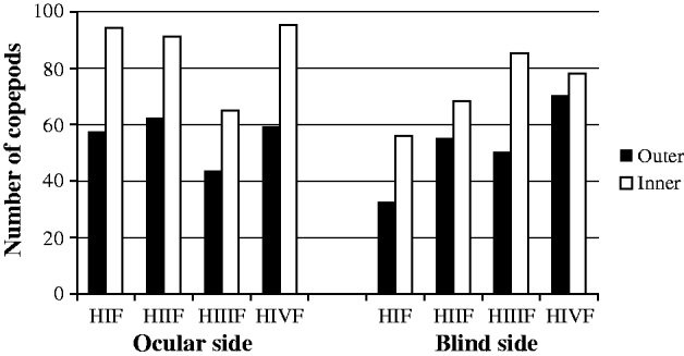
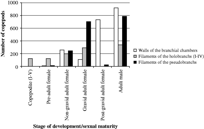
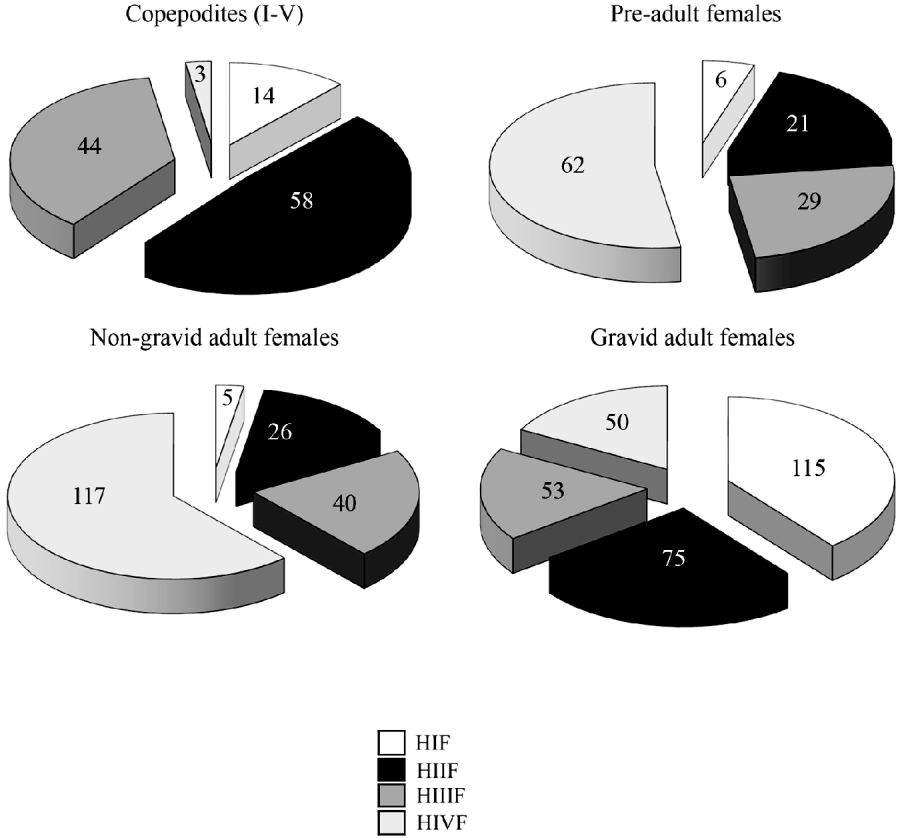
Similar infection trends were found when the sites of attachment on the ocular and blind branchial chambers were considered separately. Moreover, the internal wall was the site most frequently/heavily parasitized, followed closely by the filaments of the pseudobranchs. The infection levels recorded for the filaments of the holobranchs (I-IV) were comparatively lower (Table 1). It is worth noting that, when infecting the internal wall, the parasites were frequently crowded in the posterior region, moreover, in a restricted and narrow space between the end of holobranch I and the pseudobranch. As for the variability in parasite distribution among the inner and outer hemibranchs, significant differences were found only for the ocular side, that is, for holobranchs I, II and IV, with the parasites accumulating, to a greater extent, on the inner hemibranch (Fig. 3 and Table 2). The internal wall (mean±s.d. abundance [ocular and blind together]: 9·7±13·2 parasites) was preferred over the pseudobranch (mean±s.d. abundance [ocular and blind together]: 8·3±11·3 parasites), and this over the holobranchs (I-IV) together (mean±s.d. abundance [ocular and blind together]: 5·1±6·9 parasites), with significant differences in infection abundance recorded between the 3 sites, both for the ocular and blind sides (Friedman rank sum test: χ2=15·356, d.f.=2, P=0·000 [ocular side] and χ2=11·548, d.f.=2, P=0·003 [blind side]). The parasite exhibited no preference for any given holobranch, for either the ocular or blind sides (Friedman rank sum test: χ2=4·429, d.f.=3, P=0·219 [ocular side] and χ2=3·482, d.f.=3, P=0·323 [blind side]). Some interesting trends on the spatial distribution of the different stages of development and sexual maturity inside the chamber were noticed. Moreover, the younger stages i.e., the copepodites (I-V) and pre-adult females, dominated on the filaments of the holobranchs (I-IV); instead, the adults were mainly found infecting the filaments of the pseudobranchs – in the case of the gravid adult females – and the internal walls of the chambers – in the case of the post-gravid females and adult males. As for the non-gravid adult females, they distributed fairly equally among the internal wall, the filaments of the holobranchs (I-IV) and the filaments of the pseudobranchs. Their distribution is, however, remarkable in that it resembles that of the adult males (Fig. 4). Concerning the distribution among the 4 holobranchs, the copepodites (I-V) were dominant on holobranchs II and III, the pre-adult and non-gravid adult females on holobranch IV and the gravid adult females on holobranch I (Fig. 5). Site selection seems not to depend on host size (Fig. 6).
Spatial distribution of Acanthochondria cornuta among the inner and outer hemibranchs of the ocular and blind holobranchs (I-IV) of the European flounder, Platichthys flesus (L.). (Abbreviations: HIF, holobranch I filaments; HIIF, holobranch II filaments; HIIIF, holobranch III filaments; HIVF, holobranch IV filaments).

Infection levels – prevalence (%) and intensity (mean±s.d.) – of Acanthochondria cornuta recorded for the different sites of attachment in the ocular and blind branchial chambers of the European flounder, Platichthys flesus (L.)

Site distribution of the different stages of development and sexual maturity of Acanthochondria cornuta inside the branchial chamber of the European flounder, Platichthys flesus (L.).

Site distribution of Acanthochondria cornuta (copepodites (I-V), pre-adult females, non-gravid adult females and gravid adult females) on the holobranchs (I-IV) of the European flounder, Platichthys flesus (L.). (Abbreviations: HIF, holobranch I filaments; HIIF, holobranch II filaments; HIIIF, holobranch III filaments; HIVF, holobranch IV filaments).

Site distribution of Acanthochondria cornuta recorded for 4 size classes of European flounder, Platichthys flesus (L.).

Results for the Wilcoxon matched-pairs signed ranks test, which compared between parasite abundance on the inner and outer hemibranchs of holobranchs (I-IV)

The site of attachment of the male parasites was always the female genito-abdomen, as indeed typical for the male chondracanthids. However, it was found that 3 of the isolated females were bigamous, that is, were coupled with 2 males, each of which attached to a different nuptial organ.
DISCUSSION
Overall, the evidence found in this work suggests that A. cornuta distributes according to a particular pattern in the European flounder's branchial chambers. The intensity is indicated as an important regulator of its spatial distribution, with high levels resulting, sometimes, in the infection of ‘atypical’ sites, such as the skin and fins. This might be due to increased intraspecific competition, once A. cornuta is a large body-size species, compared to the amount of free space under the operculum, that tends to occur in high numbers in the European flounder, as reported in this study and others (Schmidt et al. Reference Schmidt, Zander, Körting and Steinhagen2003; Marques et al. Reference Marques, Teixeira and Cabral2006). The side bias in infection abundance might reflect the fact that the fish spends most of its lifetime buried in the bottom sediment. This means that the ocular side offers less resistance to the respiratory water flow, being therefore, comparatively better ventilated. As a consequence, most of the infective stages carried by the ventilating current will be directed to it. Despite the previous efforts to characterize the ecology of Acanthochondria infections in flatfish (see Kabata, Reference Kabata1959), no similar bias in parasite abundance was recorded. This latter author reported, however, the occurrence of parasite crowding at the same region of the internal wall of the plaice, Pleuronectes platessa Linnaeus, 1758. As for the differences found in other host-parasite systems e.g., sites of attachment and overall infection levels, they should relate, at the least in part, to differences in the anatomy of the respiratory apparatus, that of the European flounder being of the ‘open’ type (Kabata, Reference Kabata1959). As A. cornuta develops from one stage into another, it seems to migrate to different sites within the chamber, with the copepodites (I-V) located mostly in the central holobranchs, and the remaining stages in sites at some distance from the chamber's centre. Moreover, the intensity trend found for the copepodites (I-V), that is II-III-I-IV, along with the preferential infection of the inner hemibranchs and the crowding in the posterior region of the internal wall suggest that the ventilating water current is a major driving force of spatial distribution. Another possible constraint of site selection is the blood supply (probably a main food source) since (i) the parasite was absent from the operculum and branchiostegal membrane, both of which are poorly vascularized and (ii) the filaments of the holobranchs (I-IV) and pseudobranch together accounted for most of the parasite records. Less easy to explain is the variability in the main sites of attachment of the gravid and post-gravid females. On the one hand, the pseudobranchs seem to constitute an adequate place for the egg sac development, since they are not under the direct effect of the main direction of the ventilating current. On the other hand, while in the internal wall, the ovigerous females can liberate their eggs easily to the surrounding environment, therefore becoming post-gravid. Competition (for space and nourishment) with other branchial parasites present in the examined fish i.e., Nerocila orbignyi (Guérin-Méneville, 1832) (Isopoda: Cymothoidae) and gnathiid pranizae (Isopoda: Gnathiidae) (see Cavaleiro and Santos, Reference Cavaleiro and Santos2007, Reference Cavaleiro and Santos2009), should have been a less important factor in shaping A. cornuta spatial distribution. This is because the infection levels recorded for those 2 taxa were very low.
In conclusion, the results found in this work seem valuable in providing new insights on the behaviour of the chondracanthid copepods. Moreover, in the parasitic copepods, site selection has been suggested as being highly dependent on the species ability to move freely over the host's body surface e.g., Lepeophtheirus pectoralis (Copepoda: Caligidae) on the European flounder (see Scott, Reference Scott1901) and, to a lesser extent, on both morphological and physiological factors (Lo and Morand, Reference Lo and Morand2001). However, in the case of A. cornuta (and all other gill copepods), the parasite was expected to remain stationary, that is, attached to the initial infection site. This is because it lives in a perpetual waterfall needing, therefore, to be securely fastened, a situation that argues against mobility (Kabata, Reference Kabata, Metrick and Desser1982). Such an expectation is not supported by our data, which suggest instead, a displacement of the parasite along its successive stages of development and sexual maturity. This behaviour might be advantageous since it will reduce the competition among the different stages. The latter might become particularly problematic in the summer season, when the number of adult females increases approximately 3–7 times, as compared with other seasons of the year (Cavaleiro and Santos, Reference Cavaleiro and Santos2009). The condition of having more than 1 male attached is not new to female chondracanthids. Indeed, it was previously reported for Rhynchochondria longa Ho, Reference Ho1967 by Ho (Reference Ho1967) and for Juanettia cornifera Wilson, 1921 by Ho (Reference Ho1970). To the best of our knowledge, however, no report on such a condition exists for A. cornuta. In this species, the female is provided with paired nuptial organs (Østergaard and Boxshall, Reference Østergaard and Boxshall2004), which suggests that coupling with more than 1 male may constitute a relevant aspect of its reproductive biology. Notwithstanding, bigamous females were rare in this study.
ACKNOWLEDGEMENTS
The authors would like to thank the Portuguese Foundation for Science and Technology and the European Social Fund for the grants awarded to Francisca I. Cavaleiro (SFRH/BM/23063/2005; SFRH/BD/65258/2009), and also two anonymous referees for their helpful comments and suggestions on a previous version of the manuscript.