P-wave dispersion is a parameter determined by non-invasive surface electrocardiographic leads. The first clinical use of the increase in P-wave dispersion was described as an indicator for diagnosis of atrial arrhythmias.Reference Li, Hertervig, Carlson, Johansson, Olsson and Yuan1, Reference Li, Hertervig, Yuan, Yang, Lin and Olsson2 Following this incident, an increase in P-wave dispersion caused by drugs and in patients with autonomic dysfunction had been reported.Reference Cheema, Ahmed, Kadish and Goldberger3–Reference Dogangun, Guzeltas and Emul5 In the adult patients, who had diastolic dysfunction, an increased value of P-wave dispersion had been found.Reference Gunduz, Binak and Arinc6, Reference Yazici, Ozdemir and Altunkeser7 Different studies showed both systolic and diastolic dysfunction in the diabetic patients.Reference Cosson and Kevorkian8–Reference Karamitsos, Karvounis and Dalamanga10 In the patients with type 1 diabetics, diastolic dysfunction without any cardiac disorder as an early finding of myocardial disease was reported.Reference Freire, Moura, Barbosa Mde, Machado, Nogueira and Ribeiro-Oliveira11 The increase in the P-wave dispersion in the diabetic patients was shown in a few studies.Reference Yazici, Ozdemir and Altunkeser7, Reference Vranka, Penz and Dukat12 Regarding the current literature, there is only one study that displayed such an increase in diabetic children.Reference Imamoglu, Oztunc, Eroglu, Onal and Guzeltas13 This study aimed to show the relations between the P-wave dispersion and diastolic function in type 1 diabetic children.
Methods
A total of 33 diabetic patients without cardiovascular diseases or hypertension, with a mean age of 12 plus or minus 4.2 years, and 29 healthy controls, with a mean age of 10.4; plus or minus 3.9 years, were enrolled for this study. The diabetic patients were on insulin medication only. After a resting period of 10 minutes, 12-lead electrocardiograms were obtained from all the patients. The cases of right bundle-branch block, left bundle-branch block, Wolff–Parkinson–White syndrome, and intraventricular conduction defect in resting electrocardiogram were excluded. The patients with a thyroid disorder were also eliminated from the study. All echocardiographic evaluations were performed in left lateral decubitus position by using a Sonos 5500 device. For the measurement of the diastolic function belonging to the left ventricular, transmitral diastolic flow, doppler tracing was imaged in the apical four-chamber view by using pulsed Doppler echocardiography with sample volume sited at the tip of the mitral leaflets. The mitral early diastolic velocity, mitral late diastolic velocity, mitral early diastolic velocity/mitral late diastolic velocity, E-wave deceleration time, and isovolumetric relaxation time were measured accordingly. The isovolumetric contraction time was also measured. The right ventricular diastolic function was measured by the method that is described as left ventricular regarding the sample volume of Doppler but was symmetrically set at the tricuspid valve instead of mitral valve. The study protocol was approved by the local Ethics Committee; and the informed consent was approved by the parents of children.
Electrocardiography and P-wave measurements
The 12-lead surface electrocardiograms of all cases were obtained in a supine position with a Nihon Kohden Cardiofax GEM electrocardiography device. Electrocardiogram was recorded at a paper speed of 50 millimetres per second and 2 millivolts per centimetre standardisation. During the recording period, all the cases were allowed to breathe without speaking or holding their breath. Electrocardiograms of the cases were evaluated by the same cardiologist who did not know the clinical status of the patients. The analysis calibers and magnifying glass had been used in order to decrease the errors in measurements of P-wave. P-waves measured in less than 9-leads in electrocardiogram were not analysed and excluded. The onset of P-wave was defined as the junction between the isoelectric line and the start of P-wave deflection while the offset of the P-wave was defined as the junction between the end of the P-wave deflection and the isoelectric line. Pmax and PminP-waves were calculated on a simultaneously recorded 12-lead surface electrocardiogram; and the difference was defined as P-wave dispersion = Pmax − Pmin. The intraobserver and interobserver coefficients of variation, standard deviation of differences between two observations divided by the mean value and expressed as a percentage, for the P dispersion were found to be 3.2% and 4.0%, respectively.
Statistical analysis
For the statistical analysis of the collected data, SPSS software was used. All of the data was expressed as mean plus or minus standard deviation. Student’s t-test was used for the comparison of the means of independent groups. The differences between the groups were assessed by χ2 test, while Pearson correlation analysis was used for the linear variables; p < 0.05 was considered as statistically significant in all of the analyses.
Results
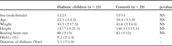
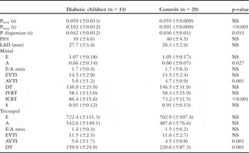
The clinical and demographical aspects of the diabetic children and healthy controls were demonstrated in Table 1. No significant differences were found for the values of resting heart rates, sexes, ages, weights, and lengths of children in the comparison of both groups. The systolic and diastolic blood pressures of the diabetics and control group were within normal limits determined for Turkish children.Reference Tumer, Yalcinkaya and Ince14 The comparison of P-wave dispersions and diastolic functions of the diabetics and control group can be seen in Table 2. Pmax and P-wave dispersion significantly increased in the diabetics group. Mitral A-wave and A-wave velocity time integral showing diastolic functions were also found to be significantly increased in diabetics. Likewise, the value of isovolumic contraction time was found to be significantly higher for the diabetics. When the tricuspid valve measurements were compared, A-wave velocity time integral was found significantly higher, whereas E-wave deceleration time was significantly lower in diabetics. No relation was found between the left ventricular diastolic functions and the duration of diabetes, HbA1c levels, and P-wave dispersion for the diabetic children. Likewise, no correlation was found between the diastolic functions and Pmin, Pmax, and P-wave dispersion for all of the participants; and no correlation was found between left atrial dimensions and Pmin, Pmax, and P-wave dispersion. There was a weak positive correlation between left atrial dimensions and mitral E-wave velocity time integral, A-wave velocity time integral, and E-wave deceleration time (r = 0.328, r = 0.308, r = 0.267, respectively).
Demographical aspects of diabetic children and healthy controls.

NS, non-significant (p > 0.05)
P-wave measurements and echocardiographic results in diabetic children and controls.

A, A velocity (m/s); AVTI, A wave velocity time integral (cm); DT, E wave deceleration time (ms); E, E velocity (m/s); EVTI, E wave velocity time integral (cm); FS, fractional shortening (%); ICRT, izovolemic contraction time (ms); IVRT, isovolumic relaxation time (ms); LAD, left atrial dimension; Pmin, minimum P-wave duration; S, peak systolic myocardial velocity at mitral annulus (m/s); s, second; cm, centimetre; mm, millimetre ms, millisecond; NS, non-significant (p > 0.05); Pmax, maximum P-wave duration
Discussion
The early sign of diabetic heart muscle disease is diastolic dysfunction preceding the systolic injury. The pathophysiological mechanisms leading to ventricular dysfunction are not understood well and are still controversial.Reference Francis15
The diastolic dysfunction, without cardiovascular diseases or other diabetic complications, has been put forward as an earlier functional change detected in specific diabetic cardiomyopathy.Reference Raev16 The characteristic findings of diastolic dysfunction are prolonged isovolumic relaxation period, delayed mitral valve opening, and impairment in rapid diastolic filling, increased atrial contribution of left ventricular filling, and reduced mitral early diastolic velocity/mitral late diastolic velocity ratio.Reference Cosson and Kevorkian8
The left ventricular diastolic dysfunction without systolic dysfunction in diabetics has been displayed in many similar studies. An intact systolic function with a higher prevalence of diastolic dysfunction has been determined in a study made by Raev et alReference Raev16 in asymptomatic type-1 diabetic children. The diastolic dysfunction has been reported 8 years after the onset of diabetes; which is earlier than systolic dysfunction occurring after 18 years of diabetes duration.Reference Cosson and Kevorkian8
In this study, the mean duration of diabetes was 5.1 years. A defect was only detected in the late diastolic filling. The other components of left ventricular diastolic dysfunction were not present in the patients; and this may be attributed to a relatively shorter duration of diabetes. In the studies conducted on the diabetic adults, the diastolic dysfunction of the right ventricle has been rarely reported. Since the data about the right ventricular performance in diabetics were insufficient, left ventricular dysfunction, compared with right ventricular dysfunction. has been a more recognised and common complication seen in the patients with type 1 diabetics.Reference Kosmala, Przewlocka-Kosmala and Mazurek17 By using either conventional or tissue Doppler echocardiography, an impaired diastolic function in both of the ventricles has been found in adult diabetics.Reference Karamitsos, Karvounis and Dalamanga10 No study, as far as is known, has ever demonstrated a diastolic dysfunction in both of the ventricles of type-1 diabetic children. In this study, the diastolic functions of both the ventricles were observed to be affected negatively together. No correlation was found between the duration of diabetes and ventricular functions. The isovolemic contraction time, however, was the first systolic parameter to be affected.
P-wave dispersion is a measure of heterogenicity in atrial refractoriness and its prolongation shows intraatrial and interatrial non-uniform conduction.Reference Perzanowski, Ho and Jacobson18 P-wave dispersion has been examined in various diseases of adults. In the paediatric age group, P-wave dispersion has been studied in healthy children, diabetic children, children with congenital cardiac diseases, atrial septal defect, tetralogy of Fallot, and in children using some drugs.Reference Imamoglu, Oztunc, Eroglu, Onal and Guzeltas13, Reference Kose, Kilic, Iyisoy, Kursaklioglu and Lenk19–Reference Yavuz, Nisli and Oner21 There was only one study regarding P-wave dispersion in type 1 diabetic children.Reference Imamoglu, Oztunc, Eroglu, Onal and Guzeltas13 In that study, similar to the results of this study, P-wave dispersion and Pmax duration was found longer in diabetics group compared with healthy control group; and no correlation was found between the P-wave dispersion duration and the duration of diabetes as well as the levels of HbA1c.
P-wave duration depends upon some factors such as age, autonomic tone, left atrial dimension, and left atrial pressure.Reference Vranka, Penz and Dukat12 Besides, the left ventricular diastolic dysfunction has cardinal effects on the left atrial properties. The left atrial volume is also a predictor for the severity of LV diastolic dysfunction.Reference Tsang, Barnes, Gersh, Bailey and Seward22 There are a few studies in the current literature regarding diastolic dysfunction and P-wave dispersion in adults.Reference Gunduz, Binak and Arinc6, Reference Yazici, Ozdemir and Altunkeser7 Yazici et alReference Yazici, Ozdemir and Altunkeser7 showed that the values of P-wave dispersion and Pmax has been prolonged, compared with the healthy control group, in diabetic adults but they have not conducted a correlation analysis between diastolic dysfunction and these parameters. Gunduz et alReference Gunduz, Binak and Arinc6 found that P-wave dispersion has been increased in patients with diastolic dysfunction unrelated to the severity or cause of diastolic dysfunction. No correlation was found between P-wave dispersion and age, sex, heart rate, left atrial diameter, early diastolic velocity/late diastolic velocity ratio, isovolumic relaxation time, and E-wave deceleration time in the abovementioned study. Likewise, no relation was found between the diastolic functions and P-wave dispersion, Pmax, and Pmin in this study. Moreover, no difference has been detected for the left atrial dimensions between the groups; and no correlation has been found between the left atrial dimensions and P-wave dispersion. Those results might be secondary to the relatively shorter duration of diabetes which has not yet prominently affected diastolic functions. The difference in the P-wave dispersion value was considered to be possibly due to the autonomous neuropathy of diabetes.
As far as is known, this study is the first one that concerns the relations between the P-wave dispersion and both left and right ventricular diastolic function in the diabetic children.
In conclusion, both ventricular diastolic functions deteriorate in type-1 diabetic children; and diabetes might cause a prolongation of P-wave dispersion. No relation seems to exist between the diastolic dysfunction and P-wave dispersion in type-1 diabetic children with ventricular dysfunction.