




No CrossRef data available.
Article contents
Immunization with cytoplasmic repetitive antigen and flagellar repetitive antigen of Trypanosoma cruzi stimulates a cellular immune response in mice
Published online by Cambridge University Press: 05 October 2004
Abstract
In previous studies, we demonstrated that CRA and FRA recombinant proteins, used for diagnosis of Chagas' disease, elicited a humoral immune response in susceptible and resistant mice. To understand better the immune response to these proteins, we have evaluated, the cellular immune response in CRA- and in FRA-immunized BALB/c and C57BL/6 mice. A specific cellular lymphoproliferative response was observed in both strains of mice. Spleen cell cultures mainly from CRA-immunized C57BL/6 and FRA-immunized BALB/c mice produced high levels of IFN-γ, indicating the induction of a Type 1 immune response. Regarding the T cell subsets, CD4+ T cells were the major source of IFN-γ in CRA- and FRA-immunized mice. These results suggest that CRA and FRA are important immunogens in inducing a Type 1 immune response and that they may be considered as potential vaccine antigens.
Information
- Type
- Research Article
- Information
- Copyright
- © 2004 Cambridge University Press
INTRODUCTION
Chagas' disease, caused by Trypanosoma cruzi, is still a major health problem in Latin America, where 11–18 million people are infected (WHO, 2002). Human infection is primarily transmitted by way of an insect vector; transmission by blood transfusion also occurs but on a much smaller scale. Better vector control measures and blood bank screening have practically eradicated transmission in previously endemic areas of several countries including Brazil and Chile. However, vector transmission is still high in several regions of Latin America. No vaccine is currently available for prevention of this disease and the available chemotherapy is often accompanied by severe side-effects.
In the mouse model of the infection, especially in the acute phase, several abnormalities of the immune response can be observed, such as non-specific polyclonal activation (Minoprio et al. 1986; D'Imperio-Lima et al. 1986; Cordeiro da Silva et al. 1998; Montes et al. 1999) or immunosuppresive effects of the parasite (Abrahamsohn & Coffman, 1995). In addition, T. cruzi and normal mammalian cells share common epitopes, that can be a possible cause of autoimmunity manifestations (Ribeiro-dos-Santos et al. 2001; Minoprio et al. 1989; Van Voorhis, Barret & Koelling, 1993; Cunha-Neto et al. 1996; Soares, Pontes-de-Carvalho & Ribeiro-dos-Santos, 2001).
The cloning and characterization of two T. cruzi genes that encode proteins bearing repetitive epitopes were described by Lafaille et al. (1989). One of the antigens, the flagellar repetitive antigen (FRA), is located in the region of the flagellum that faces the body of the parasite and displays a 68-amino acid repeat, and is found in both epimastigote and trypomastigote forms. The other, cytoplasmic repetitive antigen (CRA), is distributed throughout the cytoplasm of the parasite and has a 14-amino acid repeat and is detected in amastigotes and epimastigotes (Lafaille et al. 1989). Previous studies have demonstrated that these recombinant antigens are highly polymorphic in T. cruzi (Krieger et al. 1990). This polymorphism can be seen at the genomic level, where distinct restriction fingerprints are obtained for CRA and FRA in different T. cruzi strains (Goldenberg et al. 1991). However, these antigens were recognized by chagasic sera from different endemic regions of the country, and are thus being used for diagnosis (Lafaille et al. 1989; Krieger et al. 1992; Gomes et al. 2001). Although these antigens have been used with success in serodiagnosis for Chagas' disease (Krieger et al. 1992; Gomes et al. 2001), little is known about their immunogenic properties. According to Ibañez et al. (1988), proteins containing repeated structures, either secreted or associated with the surface of the parasite, are generally highly antigenic. We have previously reported that T. cruzi CRA recombinant protein is able to elicit humoral immune response in BALB/c and C57BL/6 mice by inducing the production of specific IgG immunoglobulins mainly of IgG1 and IgG3 isotypes. FRA-immunized BALB/c mice induced antibodies of the IgG1 isotype. No significant increase of specific immunoglobulin isotypes was detected in C57BL/6 mice immunized with FRA (Pereira et al. 2003a,b). In the present paper, we investigated whether immunization of mice with the recombinant CRA or FRA proteins would also induce cellular immune responses to these proteins.
MATERIALS AND METHODS
Mice
Male BALB/c and C57BL/6 mice (6 to 8-week-old) were raised at the animal facilities of the Oswaldo Cruz Foundation (Rio de Janeiro, Brazil) and maintained at the animal facilities of the Aggeu Magalhães Research Center of the Oswaldo Cruz Foundation in Recife, Brazil. All mice were sacrificed and treated in accordance with the Oswaldo Cruz Foundation Commission for Experiments with Laboratory Animals (Ministry of Health, Brazil).
Antigens
The CRA and FRA recombinant proteins were used as immunizing antigens. The genes encoding CRA and FRA antigens were obtained according to previously described protocols (Krieger et al. 1992) and cloned onto pQE 30 (Qiagen). Expression of the recombinant antigens by transfected Escherichia coli (TOP 10F′) was induced by the addition of isopropyl-β-D-thiogalactoside (IPTG) to a final concentration of 1 mM. After 4 h of induction, the protein lysates were harvested by centrifugation at 4000 g for 20 min and the recombinant proteins purified by nickel affinity-chromatography according to the supplier's instructions (Qiagen). The His-tagged recombinant proteins were recovered by elution with 0·5 M, pH 8·0 imidazole gradient, and the fractions analysed by electrophoresis after silver-staining and periodic acid-Schiff treatment.
Immunization of mice
Eighteen mice of each strain, BALB/c and C57BL/6, were injected 3 times subcutaneously at 20-day intervals with CRA (20 μg) or FRA (12 μg) (equimolar doses). The first immunizing dose was emulsified in a complete Freund's adjuvant and the others in an incomplete Freund's adjuvant (Sigma Chemical Co., St Louis, MO). Control mice (n=12) from each group were injected with phosphate-buffered saline (PBS) in adjuvant. The cellular immune response was evaluated 20 days after the final immunization.
Cell proliferation assay
Spleen and inguinal lymph node cell suspensions of 3 mice per group were pooled and cultured in 96-well plates at a density of 4×10⁵ cells/well, in RPMI-1640 containing 10% of fetal calf serum (FCS), 2 mM L-glutamine, 100 U/ml penicillin and 100 μg/ml streptomycin (Sigma). The cells were cultured for 72 h in the absence or in the presence of CRA (20 μg/ml) or FRA (12 μg/ml) antigens or concanavalin A (Con A; 2·5 μg/ml). Plates were pulsed with 0·5 μCi/well of methyl-[3H]thymidine (specific activity: 5·0 Ci mmol) (Amersham Pharmacia, Little Chalfont, UK) for 18 h. Radioactivity incorporation was measured by liquid scintillation counting and the results were expressed as the arithmetic mean counts per minute (cpm) of triplicate sample±standard deviation (S.D.).
Detection of cytokines in culture supernatant
Splenocytes were cultured in 24-well plates at a density of 107 cells/well. Cytokines were quantified in 24 h supernatants from cultures stimulated with Con A (2·5 μg/ml), CRA (20 μg/ml) or FRA (12 μg/ml) antigens, or maintained only in culture medium. The levels of IL-4 and IFN-γ were measured by sandwich ELISA, according to the manufacturer's suggested protocols. The antibody pairs used for the detection of IL-4 (detection limit 31·3 pg/ml) and IFN-γ (detection limit of 156 pg/ml) were purchased from R&D Systems (Minneapolis, MN), followed by streptavidin-peroxidase (Sigma). The reaction was developed with 2,2′-azino-bis(3-ethylbenzothiazoline-6-sulfonic acid) diammonium salt (ABTS) and read at 405 nm in an ELISA reader (Bio-Rad 3550; BioRad, Hercules, CA).
Flow cytometry analysis of intracellular cytokines
The spleens of 3 mice per group were pooled and the cell suspension double-stained for T cell markers (CD4 and CD8) and IFN-γ. The cells were cultured at a density of 107 cells/ml in polypropylene tubes (Falcon # 2059) stimulated with CRA (2 and 20 μg/ml) or FRA (2 and 12 μg/ml) for 1 or 6 h at 37 °C and 5% CO2. Cells of each group were also stimulated with 10 ng/ml of phorbol-12-myristate-13-acetate (PMA) and 1 μg/ml ionomycin for 4 h. Unstimulated cells were incubated in parallel to serve as negative controls. In order to inhibit cytokine secretion, all cultures received 10 μg/ml brefeldin A (Sigma Chemical Co, St Louis, MO) during the last 4 h of incubation. The cells were then harvested, washed in PBS and fixed in 1% formaldehyde at room temperature for 10 min. The cells were again washed in PBS, resuspended in 0·1% saponin in PBS, and stained (2 h, 4 °C, in the dark) simultaneously with anti-CD8-FITC and anti-CD4-FITC (P3067 and F7400, respectively; Sigma) and anti-IFN-γ-PE (18115) (Pharmingen, San Diego, CA, USA). After washing in 0·1% saponin in PBS, the cells were resuspended in 1% paraformaldehyde in 0·67% sodium chloride buffered with 1% sodium cacodylate. The fluorescence intensity of 10 000 cells was determined by flow cytometry (FACScan, Becton Dickinson, San Jose, CA, USA) and the data analysed using the Cell Quest software (Becton Dickinson). In flow cytometry analyses, cytokine-positive cells were expressed as a percentage of each analysed population, i.e. CD 4 or CD 8.
Histopathology
The spleens were removed post-mortem, and the organ/body weight ratio calculated. Samples of heart, spleen and liver of BALB/c and C57Bl/6 mice were fixed in 10% neutral buffered formalin, embedded in paraffin, sectioned (5 μm), and stained with haematoxylin-eosin for examination under light microscopy.
Statistical analysis
The significance of the observed differences was determined by Mann–Whitney's non-parametric test and Student's t-test was used to analyse the data from the lymphoproliferative assays. For the cytometry analysis the results of the positive cells were expressed as percentage of the respective population and the χ2 test was used for the comparision of proportions. Differences were considered to be significant when P<0·05.
RESULTS
Lymphoproliferative responses of CRA- or FRA-immunized mice
The purity of the CRA and FRA recombinant proteins was determined by SDS-polyacrylamyde gel electrophoresis. Bands of 50 and 30 kDa, corresponding to CRA and FRA, respectively, were visualized in the gel after silver staining (Fig. 1). To evaluate the cellular immune responses after immunization with CRA and FRA recombinant proteins, groups of BALB/c and C57BL/6 mice were immunized 3 times with equimolar doses of either recombinant protein. Proliferative responses in the cellular culture were measured and are presented in Fig. 2. Immunization with CRA induced an antigen-specific proliferative response when compared to the non-immunized group (Fig. 2A and C). Significant results were observed mainly in CRA-immunized C57BL/6 mice, with 8- and 3-fold higher incorporation of thymidine after stimulation with CRA, in spleen and lymph node cells respectively (Fig. 2C). In addition, spleen and lymph node cells of FRA-immunized BALB/c mice also showed a proliferative response higher than cell cultures of control mice on in vitro stimulation with FRA (Fig. 2B), indicating a successful immunization. In contrast, cellular proliferative response upon stimulation with FRA-immunized C57BL/6 was observed only in splenocytes (Fig. 2D). The proliferative responses to Con A by splenocytes or lymph node cells from CRA or FRA-immunized mice were similar to those of control-immunized groups (data not shown).

Fig. 1. SDS–PAGE analysis of CRA and FRA recombinant proteins purified by nickel affinity-chromatography. The lanes correspond to CRA (5 μg) and FRA (3 μg). The position of molecular weight markers is shown, corresponding to 94, 67, 43, 30, 20 and 14 kDa.

Fig. 2. Proliferative responses of cells from CRA and FRA-immunized BALB/c and C57BL/6 mice. Spleen and lymph node cells were stimulated in vitro with CRA (20 μg) and FRA (12 μg). Proliferation was assessed at 72 h of culture and the results are expressed as the average values±standard deviations obtained from triplicate cultures in each group. * P<0·05 immunized vs. non-immunized ([squf ]) spleen cells of CRA or FRA-immunized BALB/c and C57BL/6 mice; (□) spleen cells from control adjuvant-immunized BALB/c and C57BL/6 mice.
Immunization with CRA induces IFN-γ production in B6 mice
The levels of IL-4 and IFN-γ were determined in splenocyte cultures from BALB/c and C57BL/6 immunized mice. In BALB/c mice, spleen cells produced IFN-γ after stimulation with CRA and FRA in comparision with the control group (Fig. 3A and B). Immunization with CRA induced production of IFN-γ 3-fold higher than non-immunized mice, whereas immunization with FRA induced 10-fold higher IFN-γ levels. CRA-immunized C57BL/6 induced a potent IFN-γ response 13-fold higher than that observed in the control (Fig. 3C). The IFN-γ responses induced in these mice confirm that mainly CRA-immunized C57BL/6 mice developed antigen-specific Th1-like responses. In contrast, IFN-γ levels were undetectable in cultures of cells obtained from FRA-immunized C57BL/6 mice (Fig. 3D). Levels of IL-4 were undetectable on stimulation with CRA or FRA in both strains of mice, although this cytokine could be detected in Con A-stimulated cultures (data not shown).

Fig. 3. IFN-γ production by spleen cells from BALB/c and C57BL/6 mice stimulated in vitro with Con A, CRA or FRA. Unstimulated cells served as a negative control. The graphs A, B, C and D represent the results obtained by the ELISA assay. Bars represent average values±standard deviations obtained from duplicate cultures. ([squf ]) Spleen cells of CRA or FRA-immunized BALB/c and C57BL/6 mice; (□) spleen cells of control BALB/c and C57BL/6 mice.
Identification of IFN-γ-producing T cell subsets
To evaluate which T cell subsets produced IFN-γ upon immunization with CRA and FRA, splenocyte cultures were analysed using flow cytometry (Table 1). On stimulation with PMA and ionomycin, cultures of spleen cells from all the groups showed an increase in both CD4 and CD8 IFN-γ+ producing cells, compared to incubation in culture medium alone, except for CD8 IFN-γ+ cells in CRA-immunized C57BL/6 mice. IFN-γ+ cells were present in significant levels in splenocyte cultures from CRA or FRA-immunized BALB/c mice stimulated for 6 h with these antigens. However, CD4+ T cells were the main producers of IFN-γ in cultures of CRA-immunized mice showing a 2-fold higher level IFN-γ+ (12·03%), when compared to non-immunized mice (5·62%). Conversely, in FRA-immunized mice both CD4+ and CD8+ T cell subsets expressed IFN-γ after stimulation with FRA, at 5- and 4-fold higher levels than non-immunized mice respectively. Cells obtained from C57BL/6 mice were also stained for IFN-γ 1 h after stimulation with CRA or FRA and again significant percentages were obtained. Both CD4+ and CD8+ T cells were positive for IFN-γ in splenocyte cultures of C57BL/6 stimulated with CRA, whereas the major source of IFN-γ production in FRA-immunized C57BL/6 mice were the CD4+ T cells (2·89%) when compared with the control group (0·62%).

Gross and microscopical morphology
Differences in spleen/body weight ratios between immunized and non-immunized mice were not statistically significant. Histological examination of spleens from control mice showed a white pulp of normal appearance. In non-immunized mice, the red pulp contained small lymphocytes, granulocytes, some macrophages and a few plasmocytes. Megakaryocytes were rare. Immunization with CRA and FRA gave rise to a slight enlargement of the lymphoid follicles and a moderate hypercellularity of the red pulp, with an apparent increase in the number of reticular cells, macrophages, monocytes and megakaryocytes, besides the cell types detected in control animals. However, immature progenitor cells of either red or white blood cell lineages were not observed. These aspects were similar in both mouse strains. In order to determine if CRA and FRA induced tissue damage, hearts and livers were also examined, but they showed the same macroscopic and histological features as uninfected control mice.
DISCUSSION
In the current study, the immunogenicity of CRA and FRA recombinant proteins of T. cruzi was evaluated in BALB/c and C57BL/6 mice, which are considered susceptible and resistant strains to T. cruzi infection, respectively. These strains are widely used for the study of pathogenesis in Chagas' disease and the interplay between the host's and the parasite's genetic differences may influence the result of the murine model of T. cruzi infection (Roggero et al. 2002).
Available recombinant antigens used in vaccination are scarce and immunization protocols generally provide only partial protection in mice (Pereira-Chioccola et al. 1999; Schnapp et al. 2002). The current trend, therefore, is to focus on antigens that stimulate cell-mediated immune responses resulting in immune recognition of the parasite that is independent of the mouse strain (Miller, Wrightsman & Manning, 1996; Wizel, Nunes & Tarleton, 1997). In this paper it has been demonstrated that CRA and FRA antigens are capable of inducing specific cellular immune responses in both BALB/c and C57BL/6 mice, which display significantly distinct susceptibility to infection by T. cruzi. This response indicates that these antigens are highly immunogenic and elicit the same type of immune response which is independent of the genetic background of the animals. This was demonstrated by the lymphoproliferative response and IFN-γ production assays.
In the present study, it was observed that the splenic and lymph node cells, in both strains of mice, were stimulated in vitro in the presence of CRA and FRA recombinant proteins. In relation to the stimulus with CRA, significant proliferation was observed in splenic and lymph node cells of C57BL/6 mice. On the other hand, in BALB/c mice this antigen stimulated only the proliferation of splenic cells. When the stimulus was carried out with FRA, proliferation was observed in both splenic and lymph node cells of BALB/c but only in splenic cells of C57BL/6 mice. These results showed that the protocol of immunization used was good enough to generate immunological memory. According to Zinkernagel (2000), antigen localization-dose-time antigens determine immune reactivity: (i) antigen that does not reach secondary lymphoid organs in minimum doses or for sufficiently long time-periods, is immunologically ignored; (ii) antigen that either usually exists in the lymphoid system or reaches it and persists in excessive amounts for long periods deletes T cells. These arguments may explain the reason why proliferation in the lymph node cells did not occur.
A Th1-type immune response has been associated with protection against several infectious agents (Trinchieri, 1997). It is well established that resistance to T. cruzi infection is associated with IFN-γ production. Treatment of mice with neutralizing anti-IFN-γ antibodies or with recombinant IFN-γ confers susceptibility and resistance, respectively (Reed, 1988; Torrico et al. 1991). IFN-γ production was detected in cells from BALB/c mice stimulated with CRA and FRA, whereas in C57BL/6 mice only CRA induced significantly high levels of IFN-γ production. IFN-γ response induced in these mice confirms that mainly CRA-immunized C57BL/6 mice developed antigen-specific Th1-like response. Thus, immunization with just one of these 2 T. cruzi antigens induces a Type 1 immune response, and may protect against T. cruzi infection since IFN induces macrophages to express trypanocidal activity (Gazzinelli et al. 1992). The preferential activation of lymphocyte Th1 was confirmed when IL-4 was not detected in the cultures stimulated by antigens. On the other hand, spleen cells from BALB/c mice immunized with cruzipain, devoid of enzymatic activity, showed a Th2 cytokine profile (Giordanengo et al. 2002). Our results suggest that immunization with CRA and FRA produced a significant up-regulation of Th1-type cytokine whereas Th2-type cytokine was down-regulated.
In addition, spleen T CD4+ T culture cells of CRA-immunized BALB/c mice, were the main producers of IFN-γ, whereas in FRA-immunized BALB/c mice, both CD4+ and CD8+ T cells expressed IFN-γ. Similar results were obtained with FRA-immunized C57BL/6 mice and with CRA antigen (CD4+ and CD8+ T cells subsets produced this cytokine). With both the ELISA technique and flow cytometry the dose of each antigen used in the stimulation of the cultures was based on the equivalence of the molecule number. Using the same molecule number of CRA and FRA, does not necessarily mean the same number of antigenic sites will stimulate an immune response. In addition, differences observed between the ELISA results and single-cell intracytoplasmic cytokine staining may be due to the sensitivity of ELISA and to the cytoplasmic accumulation of the cytokine after addition of brefeldin A (Prussin & Metcalfe, 1995; Gomes et al. 2003). This could explain why CRA and FRA antigens stimulated different levels of IFN-γ and source of IFN-γ (CD4 and/or CD8) in spleen cells in both strains of mice, when data in Table 1 and Fig. 3 were compared.
Resistance to T. cruzi is associated with CD4+ and CD8+ T cells, antibodies and cytokine production (Abrahamson & Coffman, 1996; Cardillo, Voltarelli & Reed, 1996). It is well established that CD4 Th1 or TC1 cells stimulate strong trypanocidal activity by producing important mediators of resistance to T. cruzi such as IFN-γ and TNF-α (Abrahamson & Coffman, 1996; Cardillo et al. 1996; Silva et al. 1992; Tarleton, 1995). Immunization of mice with recombinant TolT generates a population of CD4+ T lymphocytes that recognize T. cruzi-infected macrophages resulting in the production of IFN-γ, which leads to nitric oxide production and a 50 to 60% reduction in parasite numbers compared to that seen with infected macrophages incubated with naive T cells (Quanquin et al. 1999). Depletion of CD4+ or CD8+ T cells increases the susceptibility of mice to T. cruzi infection (Russo et al. 1988; Rotemberg et al. 1996). Hoft et al. (2000) showed that the induction of a T. cruzi-specific Th1 response but not of a Th2 response in susceptible BALB/c mice, was associated with lower parasitaemias and increased survival after virulent parasite challenges. These facts show that both CD4+ and CD8+ T cells are effective against the causative agent of Chagas' disease.
Previous reports from our group demonstrated that immunization with CRA stimulates the production of antibodies mainly of the IgG1 and IgG3 isotypes (Pereira et al. 2003a,b) in both strains of mice. IFN-γ is the main factor regulating IgG2a switch and a minor regulator of the switch to IgG3 in mice, whereas IL-4 is one of the factors controlling the switch to IgG1 (Finkelman et al. 1990; Boehm et al. 1997). Although IL-4 production in cell cultures obtained from CRA-immunized mice was not detected, it is likely that this cytokine plays a role in the induction of anti-CRA IgG1 production. In contrast to the response to CRA, only IgG1 was detected in FRA-immunized BALB/c mice (Pereira et al. 2003a,b). The finding that mainly cells from FRA-immunized BALB/c and CRA-immunized C57BL/6 mice proliferate in vitro secreting IFN-γ- led us to suggest that a cell-mediated response is the effector mechanism activated by these proteins.
Taken as a whole, our results show that immunization with CRA or FRA antigens from T. cruzi stimulated cellular immune responses in BALB/c and C57BL/6 mice, suggesting that these antigens may be involved in protection against T. cruzi infection. The ability of the immune response generated against these two recombinant antigens in the protection against an infection challenge from live T. cruzi forms is currently under investigation.
We thank Mineo Nakazawa and Roni Evencio Araújo for their excellent technical assistance and Ulisses Montarroyos and Carlos Luna for assistance in statistical (computer) analysis. This work was supported by Biomanguinhos/Fundação Oswaldo Cruz (FIOCRUZ), Fundação de Amparo à Pesquisa do Estado do Rio de Janeiro (FAPERJ) and Conselho Nacional de Desenvolvimento Científico e Tecnológico (CNPq).
References
REFERENCES
ABRAHAMSOHN, I. A. &
COFFMAN, R. L. (
1995).
Cytokine and nitric oxide regulation of the immunosuppression in Trypanosoma cruzi infection.
Journal of Immunology
155,
3955–3963.Google Scholar
ABRAHAMSON, I. A. &
COFFMAN, R. L. (
1996).
Trypanosoma cruzi: IL-10, TNF, IFN-γ and IL-12 regulate innate and acquired immunity to infection.
Experimental Parasitology
84,
231–244.CrossRefGoogle Scholar
BOEHM, U.,
KLAMP, T.,
GROOT, M. &
HOWARD, J. C. (
1997).
Cellular responses to interferon-γ.
Annual Review of Immunology
15,
749–795.CrossRefGoogle Scholar
CARDILLO, F.,
VOLTARELLI, J. C. &
REED, S. G. (
1996).
Regulation of Trypanosoma cruzi infection in mice by gamma interferon and interleukin 10: role of NK cells.
Infection and Immunity
64,
128–134.Google Scholar
CORDEIRO DA SILVA, A.,
ESPINOZA, A. G.,
TAIBI, A.,
OUAISSI, P. &
MINOPRIO, P. (
1998).
A 24000 MW Trypanosoma cruzi antigen is a B-cell activator.
Immunology
94,
189–196.Google Scholar
CUNHA-NETO, E.,
COELHO, U. P. C.,
GUILHERME, L.,
FIORELLI, A.,
STOLF, N. &
KALIL, J. (
1996).
Autoimmunity in Chagas' disease: identification of cardiac miosin-B13 Trypanosoma cruzi protein crossreactive T cell clones in heart lesions of a chronic Chagas cardiomyopathy patient.
Journal of Clinical Investigation
98,
1709–1712.CrossRefGoogle Scholar
D'IMPERIO-LIMA, M. R.,
EISNEN, H.,
MINOPRIO, P.,
JOSKOWICZ, M. &
COUTINHO, A. (
1986).
Persistence of polyclonal B cell activation with undetectable parasitemia in the late stages of experimental Chagas' disease.
Journal of Immunology
137,
353–356.Google Scholar
FINKELMAN, F.,
HOMES, J.,
KATONA, I.,
URBAN, J.,
BECKMANN, M.,
PARK, L.,
SCHOOLEY, K.,
COFFMAN, R.,
MOSSMAN, T. &
PAUL, W. (
1990).
Lymphokine control of in vivo immunoglobulin isotype selection.
Annual Review of Immunology
8,
303–333.CrossRefGoogle Scholar
GAZZINELLI, R. T.,
OSWALD, I. P.,
JAMES, S. L. &
SHER, A. (
1992).
IL-10 inhibits parasite killing and nitrogen oxide production by IFN-γ-activated macrophages.
Journal of Immunology
148,
1792–1796.Google Scholar
GIORDANENGO, L.,
GUIÑAZÚ, N.,
STEMPIN, C.,
FRETES, R.,
CERBÁN, F. &
GEA, S. (
2002).
Cruzipain, a major Trypanosoma cruzi antigen, conditions the host immune response in favor of parasite.
European Journal of Immunology
32,
1003–1011.3.0.CO;2-P>CrossRefGoogle Scholar
GOLDENBERG, S.,
KRIEGER, M. A.,
LAFAILLE, J. J.,
ALMEIDA, E. &
OELEMANN, W. (
1991).
Use of Trypanosoma cruzi antigens in the immunological diagnosis of Chagas' disease.
Memórias do Instituto Butantan
53,
71–76.Google Scholar
GOMES, J. A. S.,
BAHIA-OLIVEIRA, L. M. G.,
ROCHA, M. O. C. &
MARTINS-FILHO, O. A. (
2003).
Evidence that development of severe cardiomyopathy in human Chagas' Disease is due to a Th1-specific immune response.
Infection and Immunity
71,
1185–1193.CrossRefGoogle Scholar
GOMES, Y. M.,
PEREIRA, V. R. A.,
NAKAZAWA, M.,
ROSA, D. S.,
FEREIRA, A. G. P.,
SILVA, E. D.,
KRIEGER, M. &
GOLDENBERG, S. (
2001).
Serodiagnosis of chronic Chagas' disease by using EIE-Recombinant-Chagas-Biomanguinhos.
Memórias do Instituto Oswaldo Cruz
96,
497–501.CrossRefGoogle Scholar
HOFT, D. F.,
SCHNAPP, A. R.,
EICKHOFF, C. S. &
ROODMAN, S. T. (
2000).
Involvement of CD4-Th1 cells in systemic immunity protective against primary and secondary challenges with Trypanosoma cruzi.
Infection and Immunity
68,
197–204.CrossRefGoogle Scholar
IBAÑEZ, C. F.,
AFFRANCHINO, J. L.,
MACINA, R. A.,
REYES, M. B.,
LEGUIZAMON, S.,
CAMARGO, M. E.,
ASLUND, L.,
PETERSSON, U. &
FRASCH, A. C. C. (
1988).
Multiple Trypanosoma cruzi antigens containing tandemly repeated amino acid sequence motifs.
Molecular and Biochemical Parasitology
30,
27–34.CrossRefGoogle Scholar
KRIEGER, M. A.,
SALLES, J. M.,
ALMEIDA, E.,
LINSS, J.,
BONALDO, M. C. &
GOLDENBERG, S. (
1990).
Expression and polymorphism of a Trypanosoma cruzi gene encoding a cytoplasmic repetitive antigen.
Experimental Parasitology
70,
247–254.CrossRefGoogle Scholar
KRIEGER, M. A.,
ALMEIDA, E.,
OELEMANN, W.,
LAFAILLE, J. J.,
PEREIRA, J. B.,
CARVALHO, M. R. &
GOLDENBERG, S. (
1992).
Use of recombinant antigens for the accurate immunodiagnosis of Chagas' disease.
American Journal Tropical Medicine Hygiene
46,
427–434.CrossRefGoogle Scholar
LAFAILLE, J. J.,
LINSS, J.,
KRIEGER, M. A.,
SOUTO-PADRON, T.,
DE SOUZA, W. &
GOLDENBERG, S. (
1989).
Structure and expression of two Trypanosoma cruzi genes encoding antigenic proteins bearing repetitive epitopes.
Molecular and Biochemical Parasitology
35,
127–136.CrossRefGoogle Scholar
MILLER, M. J.,
WRIGHTSMAN, R. A. &
MANNING, J. E. (
1996).
Trypanosoma cruzi: protective immunity in mice immunized with paraflagellar rod proteins is associated with a T-helper type 1 response.
Experimental Parasitology
84,
156–167.CrossRefGoogle Scholar
MINOPRIO, P.,
ITOHARA, S.,
HEUSSER, C.,
TONEGAWA, S. &
COUTINHO, A. (
1989).
Immunobiology of murine Trypanosoma cruzi infection: the predominance of parasite non-specific responses and the activation of TCRI T cells.
Immunological Reviews
112,
183–207.CrossRefGoogle Scholar
MINOPRIO, P. M.,
EISEN, H.,
FORNI, L.,
D'IMPERIO LIMA, M. R.,
JOSKOWICZ, M. &
COUTINHO, A. (
1986).
Polyclonal lymphocyte responses to murine Trypanosoma cruzi infection I – Quantitation of both T- and B-cell responses.
Scandinavian Journal of Immunology
24,
661–668.CrossRefGoogle Scholar
MONTES, C. L.,
ZUÑIGA, E.,
MINOPRIO, P.,
VOTTERO-CIMA, E. &
GRUPPI, A. A. (
1999).
Trypanosoma cruzi induces polyclonal B-cell activation of normal murine spleen cells by T-cell-independent, BCR-directed stimulation.
Scandinavian Journal of Immunology
50,
159–166.CrossRefGoogle Scholar
PEREIRA, V. R. A.,
LORENA, V. M. B.,
NAKAZAWA, M.,
GALVÃO DA SILVA, A. P.,
MONTARROYOS, U.,
CORREA-OLIVEIRA, R. &
GOMES, Y. M. (
2003 a).
Evaluation of immune response in C57Bl/6 mice immunized with CRA and FRA recombinant antigens of Trypanosoma cruzi.
Revista da Sociedade Brasileira de Medicina Tropical
36,
435–440.Google Scholar
PEREIRA, V. R. A.,
LORENA, V. M. B.,
VERÇOSA, A. F. A.,
GALVÃO DA SILVA, A. P.,
SILVA, E. D.,
FERREIRA, A. G. P.,
MONTARROYOS, U. &
GOMES, Y. M. (
2003 b).
Antibody isotype responses in BALB/c mice immunized with the CRA and FRA recombinant antigens of Trypanosoma cruzi.
Memórias do Instituto Oswaldo Cruz
98,
823–825.Google Scholar
PEREIRA-CHIOCCOLA, V. L.,
COSTA, F.,
RIBEIRÃO, M.,
SOARES, I. S.,
ARENA, F.,
SCHENKMAN, S. &
RODRIGUES, M. M. (
1999).
Comparison of antibody and protective immune responses against Trypanosoma cruzi infection elicited by immunization with a parasite antigen delivered as naked DNA or recombinant protein.
Parasite Immunology
21,
103–110.CrossRefGoogle Scholar
PRUSSIN, C. &
METCALFE, D. D. (
1995).
Detection of intracytoplasmic cytokine using flow cytometry and directly conjugated anti-cytokine antibodies.
Journal of Immunological Methods
188,
117–128.CrossRefGoogle Scholar
QUANQUIN, N. M.,
GALAVIZ, C.,
FOUTS, D. L.,
WRIGHTSMAN, R. A. &
MANNING, J. E. (
1999).
Immunization of mice with a TolA-like surface protein of Trypanosoma cruzi generates CD4+ T-cell-dependent parasiticidal activity.
Infection and Immunity
67,
4603–4612.Google Scholar
REED, S. (
1988).
In vivo administration of recombinant IFN-γ induces macrophage activation and prevents acute disease, immunosuppression and death in experimental Trypanosoma cruzi infection.
Journal of Immunology
140,
4342–4347.Google Scholar
RIBEIRO-DOS-SANTOS, R.,
MENGEL, J. O.,
POSTOL, E.,
SOARES, R. A. O.,
FERREIRA-FERNANDEZ, E.,
SOARES, M. B. P. &
PONTES DE CARVALHO, L. C. A. (
2001).
Heart-specific CD4+ T-cell line obtained from a chronic chagasic mouse induces carditis in heart-immunized mice and rejection of normal heart transplants in the absence of Trypanosoma cruzi.
Parasite Immunology
23,
93–101.CrossRefGoogle Scholar
ROGGERO, E.,
PEREZ, A.,
TAMAE-KAKAZU, M.,
PIAZZON, I.,
NEPOMNASCHY, I.,
WIETZERBIN, J.,
SERRA, E.,
REVELLI, S. &
BOTTASSO, O. (
2002).
Differential susceptibility to acute Trypanosoma cruzi infection in BALB/c and C57 BL/6 mice is not associated with a distinct parasite load but cytokine abnormalities.
Clinical and Experimental Immunology
128,
421–428.CrossRefGoogle Scholar
ROTEMBERG, M. E.,
CARDONI, R.,
ANDERSON, R.,
SEGURA, E. &
ORN, A. (
1996).
Resistance to Trypanosoma cruzi requires T helper inductor cells as well as natural killer cells.
Scandinavian Journal of Immunology
28,
573–582.Google Scholar
RUSSO, M.,
STARONBINAS, N.,
MINOPRIO, P.,
COUTINHO, A. &
HONTEBEYRIE-JOSKOWICZ, M. (
1988).
Parasite load increases and myocardial inflammation decreases in Trypanosma cruzi-infected mice after inactivation of T helper cells.
Annales du Institut Pasteur/Immunolgie
139,
225–236.CrossRefGoogle Scholar
SCHNAPP, A. R.,
EICKHOFF, C. S.,
SIZEMORE, D.,
CURTIS III, R. &
HOFT, D. F. (
2002).
Cruzipain induces both mucosal and systemic protection against Trypanosoma cruzi in mice.
Infection and Immunity
70,
5065–5074.CrossRefGoogle Scholar
SILVA, J. S.,
MORRYSSEY, P. J.,
GRABSTEIN, K. H.,
MOHLER, K. M.,
ANDERSON, D. &
REED, S. G. (
1992).
Interleukin 10 and interferon-γ regulation of experimental Trypanosoma cruzi infection.
Journal of Experimental Medicine
175,
169–174.CrossRefGoogle Scholar
SOARES, M. B. P.,
PONTES-DE-CARVALHO, L. &
RIBEIRO-DOS-SANTOS, R. (
2001).
The pathogenesis of Chagas' disease: when autoimmune and parasite-specific immune responses meet.
Anais da Academia Brasileira de Ciências
73,
547–559.CrossRefGoogle Scholar
TARLETON, R. L. (
1995).
The role of T cells in Trypanosoma cruzi infections.
Parasitology Today
11,
7–12.CrossRefGoogle Scholar
TORRICO, F.,
HEREMANS, H.,
RIVERA, M. T.,
VAN MARCK, E.,
BILLIAU, A. &
CARLIER, Y. (
1991).
Endogenous IFN-gamma is required for resistance to acute Trypanosoma cruzi infection in mice.
Journal of Immunology
146,
3626–3632.Google Scholar
TRINCHIERI, G. (
1997).
Cytokines acting on or secreted by macrophages during intracellular infection (Il-10, IL-12, IFN-γ).
Current Opinion in Immunology
9,
17–23.CrossRefGoogle Scholar
VAN VOORHIS, W. C.,
BARRET, L. &
KOELLING, R. (
1993).
FL-160 proteins of Trypanosoma cruzi are expressed from a multigene family and contain two distinct epitopes that mimic nervous tissues.
Journal of Experimental Medicine
178,
681–694.CrossRefGoogle Scholar
WIZEL, B.,
NUNES, M. &
TARLETON, R. L. (
1997).
Identification of Trypanosoma cruzi trans-sialidase family members as targets of protective CD8+ Tc1 responses.
Journal of Immunology
159,
6120–6130.Google Scholar
WORLD HEALTH ORGANIZATION (
2002).
The World Health Report,
Geneva.
ZINKERNAGEL, R. M. (
2000).
Localization dose and time antigens determine immune reactivity.
Seminars in Immunology
12,
163–171.CrossRefGoogle Scholar
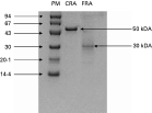
Fig. 1. SDS–PAGE analysis of CRA and FRA recombinant proteins purified by nickel affinity-chromatography. The lanes correspond to CRA (5 μg) and FRA (3 μg). The position of molecular weight markers is shown, corresponding to 94, 67, 43, 30, 20 and 14 kDa.
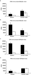
Fig. 2. Proliferative responses of cells from CRA and FRA-immunized BALB/c and C57BL/6 mice. Spleen and lymph node cells were stimulated in vitro with CRA (20 μg) and FRA (12 μg). Proliferation was assessed at 72 h of culture and the results are expressed as the average values±standard deviations obtained from triplicate cultures in each group. * P<0·05 immunized vs. non-immunized ([squf ]) spleen cells of CRA or FRA-immunized BALB/c and C57BL/6 mice; (□) spleen cells from control adjuvant-immunized BALB/c and C57BL/6 mice.
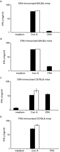
Fig. 3. IFN-γ production by spleen cells from BALB/c and C57BL/6 mice stimulated in vitro with Con A, CRA or FRA. Unstimulated cells served as a negative control. The graphs A, B, C and D represent the results obtained by the ELISA assay. Bars represent average values±standard deviations obtained from duplicate cultures. ([squf ]) Spleen cells of CRA or FRA-immunized BALB/c and C57BL/6 mice; (□) spleen cells of control BALB/c and C57BL/6 mice.
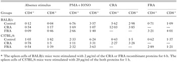
Table 1. Percentage of CD4+ and CD8+ cells positive for IFN-γ in splenocyte populations from BALB/c or C57BL/6 mice immunized with Trypanosoma cruzi recombinant proteins CRA or FRA*
 You have
Access
- 17
- Cited by