Introduction
Our understanding of the neural encoding and processing of sensory information and its relationship to the physiology of the molecular, synaptic, and cellular underpinnings is often limited by the lack of information regarding the wiring specificity of the underlying neural circuits. When one considers the spatial resolution necessary for synaptic identification (10’s of nanometer) and complete cellular reconstruction and identification (often ≥1 mm), it is not difficult to understand why a complete reconstruction of the 100 billion neurons and vast synaptic connectivity within the brain has remained elusive. Fortunately, for retinal physiologists, a complete assessment of the wiring specifics of the inner and outer synaptic/plexiform layers is likely on the horizon. As the details regarding the wiring specificity of retinal circuitry and the physiology of the underlying components (i.e., synaptic and cellular levels) continue to be revealed, a more complete picture of inner retinal encoding and processing of visual information and its relationship to the diversity of cellular and molecular mechanisms is becoming ever clearer.
Various aspects/features of the visual world are encoded within the parallel neural circuitries of the retina and transmitted onto the brain via the axons of retinal ganglion cells (i.e., the optic nerve). Within these parallel circuitries, it is the highly diverse class of amacrine cell interneurons and their extensive connectivity that are responsible for many higher order neural computations such as local edge detection and motion encoding (e.g., Caldwell et al., Reference Caldwell, Daw and Wyatt1978; Famiglietti, Reference Famiglietti2005; Baccus, Reference Baccus2007). On the cellular level, amacrine cells prove to be an extremely diverse class of interneurons with more than two dozen morphologically distinct subtypes (Kolb et al., Reference Kolb, Nelson and Mariani1981; MacNeil & Masland, Reference MacNeil and Masland1998; MacNeil et al., Reference MacNeil, Heussy, Dacheux, Raviola and Masland1999). On the molecular level, amacrine cells utilize a plethora of neurotransmitters, ligand-gated receptors, and hetero- and homotypic gap junctions to mediate communication to other neurons in the inner plexiform layer (IPL), including, but not limited to, neurotransmitters such as GABA, glycine, acetylcholine, and dopamine (described in detail elsewhere in this special issue). It is within this context of molecular and cellular diversity that we are now beginning to draw more direct connections between the physiology of individual neurons and synapses and the encoding of specific visual features and/or signal processing.
Classification according to connectivity
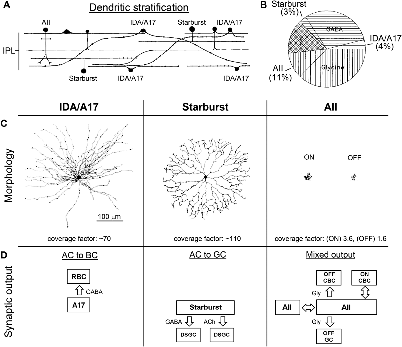
The great diversity of amacrine cell morphologies and physiologies (i.e., subtypes) and the general lack of information regarding their wiring specificity and circuit contributions have, not surprisingly, led to multiple approaches to classification (Fig. 1). Morphological classification systems (Fig. 1A and 1C) have grouped amacrine cell subtypes according to features of their neuritic arbors (e.g., wide/narrow field, bistratified/monostratified, stratification level) and cell density (MacNeil & Masland, Reference MacNeil and Masland1998), whereas molecular classification systems have typically grouped subtypes by their primary output transmitter (e.g., GABA vs. glycine; Pourcho, Reference Pourcho1980; Dick & Lowry, Reference Dick and Lowry1984; Fig. 1B). Although these approaches have been successful in providing evidence for the extreme diversity of morphologies and in estimating the divisions according to primary transmitter, the relationship between these anatomical features and the physiological contributions of individual subtypes to visual processing remains largely unclear.
To date, amacrine cell studies have revealed a diversity of circuit functions and cellular physiologies, but lack of information regarding many of the subtypes leaves classification schemes open to interpretation. The stratification (A), primary neurotransmitter (B), morphological features (C), and primary synaptic output targets (D) of the three best characterized amacrine cells (A17, starburst, and AII) exemplify the diverse nature of amacrine cell subtypes and physiologies. (A) All layers of the inner plexiform layer (IPL) are innervated by neurites of various amacrine cell subtypes. Adapted from Masland (Reference Masland1988). (B) Amacrine cells can be subdivided roughly in half according to molecular expression and identification of the inhibitory neurotransmitters, GABA or glycine (adapted from Strettoi & Masland, Reference Strettoi and Masland1996). Other than AII (11%), no single amacrine cell subtype accounts for more than 5% of the total population of amacrine cells found within mammalian species (MacNeil & Masland, Reference MacNeil and Masland1998). (C) Two-dimensional projections of A17 (left), starburst (center), and AII (right) amacrine cells, from a viewpoint parallel to incident light under physiological conditions (i.e., whole mount), reveal details of their unique morphological features and the extent of lateral dendritic spread. The coverage factor (i.e., dendritic area • cell density) represents the dendritic overlap of neurons of the same subtype, such analysis has been used to correctly predict (Masland, Reference Masland1988) that local processing exists within the dendrites of A17 (Grimes et al., Reference Grimes, Zhang, Graydon, Kachar and Diamond2010) and starburst (Euler et al., Reference Euler, Detwiler and Denk2002; Hausselt et al., Reference Hausselt, Euler, Detwiler and Denk2007) amacrine cells. A17 and starburst reconstructions are adapted from Oesch et al. (Reference Oesch, Kothmann and Diamond2011) and the AII reconstruction is courtesy of G.W. Schwartz. (D) A simplified representation of the primary synaptic outputs (target and type) where single-headed arrows represent chemical synapses and double-headed arrows represent electrical synapses. This representation highlights that amacrine cell outputs can be considered in terms of regulators of pre- and/or postsynaptic functions and mechanisms, with outputs to bipolar cells being considered presynaptic and outputs to ganglions cells considered postsynaptic. Amacrine cell to amacrine cell connections create higher-order regulation of the bipolar and ganglion cells connections.
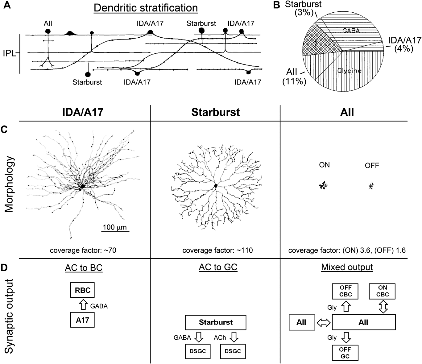
In this review, I classify amacrine cells according to their synaptic output targets (i.e., bipolar cells, BCs; ganglion cells, GCs; and/or amacrine cells, ACs; Fig. 1D). This approach, though clearly subjective, has several advantages: 1) Classification according to connectivity can be determined unambiguously via electron microscopy. 2) Despite molecular and wiring complexities and uncertainties (e.g., multiple output targets and lack of information regarding the inner retina wiring), basic physiological influence can be inferred on a synapse-by-synapse basis. For example, synaptic input to bipolar cell axon terminals has one primary function: to positively and/or negatively regulate the presynaptic membrane potential of the bipolar cell terminal, which subsequently regulates Cav activation and ribbon synapse (i.e., bipolar cell) output. Although this form of signal and/or noise processing can certainly be both spatially and temporally complex, its basic function can be described in a straightforward manner. Similarly, the primary function of amacrine cell outputs to ganglion cells is to directly influence postsynaptic integration, dendritic computation, and spike output. 3) Classification according to synaptic output target allows for higher-order classification of more complex wiring arrangements within individual subtypes without confusing basic features of functional connections. For example, the AII amacrine cell has been shown to play an essential role in the transmission of scotopic signals (Kolb & Nelson, Reference Kolb and Nelson1983; Nelson & Kolb, Reference Nelson and Kolb1984; Tsukamoto et al., Reference Tsukamoto, Morigiwa, Ueda and Sterling2001; Deans et al., Reference Deans, Volgyi, Goodenough, Bloomfield and Paul2002) but has also been shown to be involved in other forms of signal processing under photopic conditions (Manookin et al., Reference Manookin, Beaudoin, Ernst, Flagel and Demb2008; Munch et al., Reference Munch, da Silveira, Siegert, Viney, Awatramani and Roska2009), but, regardless, the basic classification remains the same: AII⇔AII, AII⇔ON cone bipolar cell (CBC), AII=>OFF CBC, AII=>OFF GC, where “⇔” represents potentially bidirectional gap junctions and “=>” indicates one-way conventional chemical synapses (Fig. 1D, right). Although this idea of a single interneuron subserving multiple functional circuitries is relatively new (Munch et al., Reference Munch, da Silveira, Siegert, Viney, Awatramani and Roska2009; Oesch & Diamond, Reference Oesch and Diamond2009; Oesch et al., Reference Oesch, Kothmann and Diamond2011), it could prove to be a common theme among amacrine cells and should be accounted for. Furthermore, albeit tempting to generally refer to amacrine cell synaptic output to bipolar cells, ganglion cells, and amacrine cells as feedback, feedforward, and serial connections, respectively, it is not clear whether such descriptions appropriately describe more complex retinal circuit features such as crossover inhibition between the ON and OFF parallel circuitries/pathways. For the remainder of this review, I will focus primarily on features of the synapses, cellular anatomy, and circuits that mediate inputs to bipolar cell axon terminals and their implications for inner retinal processing.
Chemical synapses (AC=>BC): Lateral inhibition versus local feedback
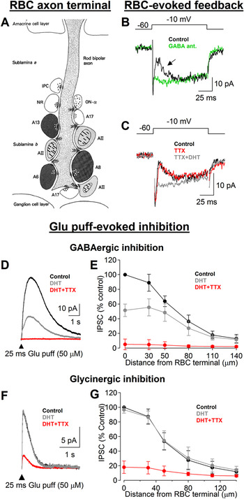
Retinal bipolar cell axon terminals have been shown to receive input from many different amacrine cell subtypes via both reciprocal and nonreciprocal synaptic connections (Fig. 2A). Furthermore, it is known that in the adult mammalian retina that the majority of amacrine cell subtypes are immunohistochemically positive for GABA or glycine markers suggesting that these are likely the two most common neurotransmitters underlying chemical synaptic transmission from amacrine cells (Davanger et al., Reference Davanger, Ottersen and Storm-Mathisen1991; Crooks & Kolb, Reference Crooks and Kolb1992; Fig. 1B). In accordance, intracellular recordings from bipolar cells have revealed that functional GABA- and glycine receptors are expressed on bipolar cell axon terminals (Suzuki et al., Reference Suzuki, Tachibana and Kaneko1990; Euler & Wassle, Reference Euler and Wassle1998; Morkve & Hartveit, Reference Morkve and Hartveit2009), and that these receptors types mediate direct synaptic input from costratified amacrine cells (Hartveit, Reference Hartveit1999; Eggers & Lukasiewicz, Reference Eggers and Lukasiewicz2006; Ivanova et al., Reference Ivanova, Muller and Wassle2006; Chavez & Diamond, Reference Chavez and Diamond2008; Chavez et al., Reference Chavez, Grimes and Diamond2010; Fig. 2).
Amacrine cell-mediated synaptic input to bipolar cell axon terminals is best understood at the rod bipolar cell (RBC). (A) Electron microscopy studies have identified numerous synaptic contacts between amacrine cell processes and RBC terminals in the IPL of the mammalian retina. Morphologically, these amacrine cell output connections can be readily identified as either making reciprocal feedback or nonreciprocal synapses onto the RBC axon terminal. Adopted from Sterling & Lampson (Reference Sterling and Lampson1986). (B–C) Reciprocal feedback can be elicited and observed in a single RBC using a somatic recording electrode (arrow in B). These recordings indicate that reciprocal feedback to the RBC terminal is GABAergic (B), and that the response to this particular stimulation does not rely upon tetrodotoxin (TTX)-sensitive Nav activity in the amacrine cells responsible (C). Furthermore, bath application of the toxic serotonin analog, 5,7-dihydroxytryptamine (5,7-DHT), selectively ablates indolamine-accumulating amacrine cells (i.e., A17) and abolishes reciprocal feedback. Adopted from Chavez et al. (Reference Chavez, Singer and Diamond2006). (D–G) Reciprocal feedback and other forms of both GABAergic and glycinergic inhibition to the RBC terminal can be elicited by puffing exogenous glutamate directly into the IPL. (D) Elicited in this manner, GABAergic inhibition (isolated in the presence glycine receptor antagonists; Chavez et al., Reference Chavez, Grimes and Diamond2010) is reduced by 5,7-DHT and abolished by further inclusion of TTX. (E) Experiments involving a lateral translation of the puffer pipette indicate that A17 amacrine cells mediate local inhibition, whereas longer-range inhibition is mediated by amacrine cells that require neuritic Nav channel activity to drive synaptic output. Adapted from Chavez et al. (Reference Chavez, Grimes and Diamond2010). (F–G) In comparison, glycinergic inhibition (isolated in the presence of GABA receptor antagonists; Chavez & Diamond, Reference Chavez and Diamond2008) is insensitive to 5,7-DHT and largely, but not completely, blocked by further inclusion of TTX. Adapted from Chavez and Diamond (Reference Chavez and Diamond2008). These results indicate that glycinergic inhibition to the RBC is not mediated by indolamine-accumulating amacrine cells and is instead largely mediated by amacrine cells that require Nav activity to drive synaptic output. However, it should be noted that in the presence of DHT and TTX, a small portion of the response remains thus suggesting that some glycinergic output synapses share Ca2+ microdomains with glutamate receptors within the same amacrine cells.

From an anatomical standpoint, it has generally been noted that the neuritic fields of GABAergic amacrine cells have wide-field morphologies compared to their glycinergic counterparts (Pourcho & Goebel, Reference Pourcho and Goebel1983, Reference Pourcho and Goebel1985), but an inference that lateral inhibition (i.e., nonreciprocal AC=>BC) might come strictly from GABAergic amacrine cells, whereas feedback inhibition stems exclusively from glycinergic amacrine cells is incorrect. For example, electron microscopy studies indicate that local reciprocal inhibitory feedback circuits are provided to bipolar cells on a synapse-by-synapse basis by wide-field GABAergic amacrine cells (Nelson & Kolb, Reference Nelson and Kolb1984, Reference Nelson and Kolb1985; Cuenca et al., Reference Cuenca, De Juan and Kolb1995; Cuenca & Kolb, Reference Cuenca and Kolb1998). Although this synaptic organization/arrangement (i.e., one cell containing many functional feedback circuits) does not exclude a functional role in lateral inhibition, recent physiological studies indicate that in some cases, synaptic feedback circuits within individual amacrine cells can function independently (Chavez et al., Reference Chavez, Singer and Diamond2006; Grimes et al., Reference Grimes, Zhang, Graydon, Kachar and Diamond2010). In contrast, the input and output synapses of narrow-field glycinergic amacrine cells (e.g., AII) are often segregated into distinct inner retina sublamina, therefore producing amacrine cell-mediated connections between bipolar cells of different subtypes (e.g., crossover inhibition), but, albeit direct or serial, longer range effects of glycinergic transmission on presynaptic inhibition likely exist (Roska et al., Reference Roska, Nemeth and Werblin1998; Hsueh et al., Reference Hsueh, Molnar and Werblin2008; Venkataramani & Taylor, Reference Venkataramani and Taylor2010). Because of these ambiguities and because ionotropic GABA- and glycine receptors differ primarily in kinetics but not general permeability (i.e., reversal potential), I will instead distinguish amacrine cell=>bipolar cell synapses/connections according to a single anatomical distinction for the remainder of this review: feedback synapses connect reciprocally to the same bipolar cell from which they receive input, whereas lateral synapses do not.
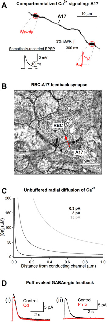
Classically, negative feedback helps improve the stability and control of a circuit’s output, and it is quite possible that this holds true for inner retinal inhibitory feedback. Although it is likely that many features of reciprocal feedback synapses are yet to be understood, several lines of evidence implicate reciprocal synapses in controlling bipolar cell release statistics (Freed et al., Reference Freed, Smith and Sterling2003), synaptic gain (Grimes et al., Reference Grimes, Li, Chavez and Diamond2009), and the time course of stimuli-evoked bipolar cell signaling in vivo (Dong & Hare, Reference Dong and Hare2003). Generally speaking, GABAergic reciprocal feedback synapses are conventional (i.e., non-ribbon); however, in the A17 amacrine cell feedback circuit (Fig. 3B), GABA release onto rod BC terminals can be triggered directly by Ca2+ influx through Ca2+-permeable glutamate receptors (Chavez et al., Reference Chavez, Singer and Diamond2006; Fig. 3D). This mechanistic feature eliminates the need for strong varicosity/bouton depolarization and Cav channel activation and allows feedback microcircuits to function in a synapse-specific manner if electrical and biochemical compartmentalization exists within and between neuritic branches of the same amacrine cell (Grimes et al., Reference Grimes, Zhang, Graydon, Kachar and Diamond2010; Fig. 3A). However, if input and output synapses are spatially segregated and do not share Ca2+ microdomains, as is typically the case for most neurons in the central nervous system (for review on presynaptic Ca2+ microdomains and transmitter release see Oheim et al., Reference Oheim, Kirchhoff and Stuhmer2006; Fig. 4A), then synaptic release will require propagation and innervation of electrical depolarization to drive Cav channel activation and local Ca2+ influx at the nerve terminal (Fig. 4B).
A diversity of molecular and cellular attributes supports a diversity of physiologies: Part 1. (A) Recent evidence indicates that synaptically evoked Ca2+ signaling can be highly compartmentalized within individual neurites of amacrine cells. In a single A17 amacrine cell, electrical and biochemical compartmentalization can allow many reciprocal feedback synapses to operate independently under certain conditions. Adapted from Grimes et al. (Reference Grimes, Zhang, Graydon, Kachar and Diamond2010). (B–D) Reciprocal feedback synapses between RBCs and A17 amacrine cells are highly colocalized and often share Ca2+ microdomains. (B) Most reciprocal synapses (input/black arrow and output/red arrow) are separated by no more than 1 μm. Image courtesy of J. Zhang. (C) Although information regarding the endogenous buffering mechanisms within individual amacrine cell neurites is lacking, simple modeling of steady-state [Ca]i near a static conducting source (modeled in terms of constant current) indicates that concentrations relevant to the triggering of transmitter release (~10–100’s of μM) can occur within a 1 μm proximity (for review, see Parekh, Reference Parekh2008). (D) Results from glutamate puff-evoked feedback experiments demonstrate that GABAergic feedback from A17 amacrine cells does not require Ca2+ influx through Cav channels (i) but instead utilizes Ca2+ influx mediated by calcium-permeable AMPARs (ii). Adopted from Chavez et al. (Reference Chavez, Singer and Diamond2006).

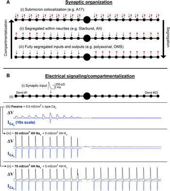
A diversity of molecular and cellular attributes supports a diversity of physiologies: Part 2. (A) Fundamentally, amacrine cells do not appear to obey a single rule when it comes to synaptic organization; input and output synapses can exhibit (i) submicron colocalization within individual neurites, (ii) coexpression within individual neurites without sharing microdomains, or (iii) full segregation to distinct neurites branching directly from the cell body. These various arrangements strongly influence the properties of neuronal integration and the dependencies on electrical and biochemical signaling for driving synaptic output. (B) A passive model of the A17 amacrine cell (adapted from Grimes et al., Reference Grimes, Zhang, Graydon, Kachar and Diamond2010) can be generalized to aid in thinking about the input–output relationships of amacrine cells of various synaptic organizations (ii). The voltage response to strong synaptic input (comparable to release of the entire readily releasable pool; i) can be restricted to individual neurites by morphology and passive properties alone (iii). This means that without significant nonlinear excitatory conductances, a neuron (such as the A17 amacrine cell) will respond to synaptic input with only local increases in intracellular calcium ([Ca2+]i). Such an arrangement can support local synaptic output from individual neurites (if input and output synapses are coexpressed) without triggering syanaptic output from other neurites. Active (e.g., Nav and Kv) membrane properties combine with morphology to shape the spatial extent of the neuronal response. With a simple uniform distribution of active properties, Nav conductance can be increased to produce spiking behavior in response to local stimulation (iv–v). However, the spatial extent of spike propagation can be limited to individual neurites under certain conditions (i.e., large leaky cell body) but such barriers can be overcome by sufficiently high Nav conductance. The reader should be reminded that the input–output relationship of any particular cell type is uniquely constrained by its specific morphology and biophysical membrane properties, and that the model presented here is only meant to provide a general conceptual framework for understanding amacrine cell physiologies. Details of the electrotonic model can be found in Grimes et al. (Reference Grimes, Zhang, Graydon, Kachar and Diamond2010). In brief, the following properties were used: R axial = 110 Ω cm, R mem = 3,571 Ω cm2, C mem = 1 μF/cm.

Seemingly complimentary to reciprocal inhibition, lateral inhibitory (AC=>BC) connections in the inner retina underlie aspects of space and motion encoding (e.g., Baccus et al., Reference Baccus, Olveczky, Manu and Meister2008) and have also been shown to extend or change the operating range of bipolar cell outputs (e.g., Zaghloul et al., Reference Zaghloul, Manookin, Borghuis, Boahen and Demb2007; Manookin et al., Reference Manookin, Beaudoin, Ernst, Flagel and Demb2008). For example, in the object motion sensitivity (OMS) circuit, amacrine cells need to integrate inputs over a relatively large spatial scale and utilize spatially offset inputs and outputs to allow the circuit (via bipolar cell regulation) to distinguish between head or eye movements and object movements (Baccus et al., Reference Baccus, Olveczky, Manu and Meister2008). Notably, the amacrine cells underlying OMS have polyaxonal morphologies (an indication of segregated input and output; Fig. 4A) and utilize action potential signaling in order to perform their computations.
Morphology, intrinsic biophysical properties, and the connection to amacrine cell physiology
All-or-none action potentials are employed by the polyaxonal amacrine cells in the OMS circuit, but many amacrine cells do not employ action potential signaling. While action potentials are often critical to the reliability and propagation of long-range neural signals, all-or-none neural transmission over long distances is not always a requirement of the functional circuit. To support a variety of retinal processes, amacrine cells have diverse morphological and biophysical properties that seem tailored to the unique computations performed by each subtype, and it has been clearly demonstrated that various subtypes rely on action potential signaling, Nav activity, and membrane excitability to varying extents (Shields & Lukasiewicz, Reference Shields and Lukasiewicz2003; Bloomfield & Volgyi, Reference Bloomfield and Volgyi2007; Chavez & Diamond, Reference Chavez and Diamond2008; Chavez et al., Reference Chavez, Grimes and Diamond2010; Grimes et al., Reference Grimes, Zhang, Graydon, Kachar and Diamond2010; Tian et al., Reference Tian, Jarsky, Murphy, Rieke and Singer2010).
As expected, Nav expression and morphology seem to play a critical role in the spatial extent of inhibition from amacrine cells. As a null example, the A17 amacrine cell has a wide-field morphology, but recent physiology and electrotonic modeling data indicate that the density of Nav channels in these interneurons is approximately 50-fold too low for action potential signaling (Grimes et al., Reference Grimes, Zhang, Graydon, Kachar and Diamond2010). It is this fact in combination with other morphological and biophysical features that may allow more than 100 feedback microcircuits to function independently within a single A17 amacrine cell. Intermediates to the full propagation of actions potentials in polyaxonal amacrine cells and the independence of microcircuits in the A17 also exist. For example, a recent study indicates that Nav channels in AII amacrine cells can speed signaling in a voltage-dependent manner without the need for action potential signaling (Tian et al., Reference Tian, Jarsky, Murphy, Rieke and Singer2010).
In accordance with our understanding of synaptic organization and amacrine cell physiology, direct recordings of inhibition to rod bipolar cells indicate that feedback and lateral pathways can function independently (Chavez et al., Reference Chavez, Grimes and Diamond2010; Eggers & Lukasiewicz, Reference Eggers and Lukasiewicz2010), and that signaling from the lateral pathways is heavily dependent on Nav channel activation (Shields & Lukasiewicz, Reference Shields and Lukasiewicz2003; Chavez & Diamond, Reference Chavez and Diamond2008; Chavez et al., Reference Chavez, Grimes and Diamond2010; Fig. 2D–2G), whereas reciprocal feedback is not (Hartveit, Reference Hartveit1999; Chavez et al., Reference Chavez, Singer and Diamond2006; Fig. 2C).
In contrast to AC=>BC signaling, AC=>GC connections directly influence signaling and integration in ganglion cell dendrites. Despite differences in output and function, amacrine cells mediating these connections use similar mechanistic schemes to support their physiology. The most well-studied example comes from direction selectivity, where starburst amacrine cells utilize laterally offset input and output synapses within individual neurites (Famiglietti, Reference Famiglietti1991) and asymmetric GABAergic output contacts (Fried et al., Reference Fried, Munch and Werblin2002; Lee et al., Reference Lee, Kim and Zhou2010; Briggman et al., Reference Briggman, Helmstaedter and Denk2011) to drive the encoding of directional motion within this specialized circuitry. On the cellular level, we find that the individual processes extending radially from the starburst amacrine cell body function as critical components of directional motion encoding. As an intermediate to the polyaxonal OMS amacrine cell (fully segregated inputs and outputs) and the A17 amacrine cell (many colocalized inputs and outputs within individual neurites), starburst amacrine cell intrinsic membrane properties (e.g., Cav channels, Nav channels) and native morphology (i.e., dendritic structure, synaptic organization) give rise to a centrifugally projecting input/output relationship that extends over the length of individual neurites (Velte & Miller, Reference Velte and Miller1997; Cohen, Reference Cohen2001; Euler et al., Reference Euler, Detwiler and Denk2002; Oesch & Taylor, Reference Oesch and Taylor2010).
Electrical synapses (AC⇔BC): Bidirectional positive and/or negative feedback/influence via gap junctions
Despite decades of tremendous anatomical and physiological study, our picture of the inner retina’s wiring and its relationship to the dynamics of visual encoding is far from complete. In part due to the challenges posed by great cellular and molecular diversity, this problem is further exacerbated by extensive gap junctional coupling between neurons of nearly every type. Studies examining the functional roles that electrical synapses play in retinal signaling have primarily described a reduction in the independence of specific parallel pathways (increased covariance; Vardi & Smith, Reference Vardi and Smith1996; DeVries, Reference DeVries1999; Trong & Rieke, Reference Trong and Rieke2008) and a means for the scotopic (night vision) circuitry to tap into the evolutionarily preexisting photopic circuitry for the conveyance of rod-driven neural signaling under dim conditions. From this latter example, we find that gap junctions are utilized in both the inner and outer retinas to not only transmit signals from the high-gain primary rod pathway but also to create a secondary rod pathway, which acts as an intermediate to scotopic and photopic vision (Schneeweis & Schnapf, Reference Schneeweis and Schnapf1995; Tsukamoto et al., Reference Tsukamoto, Morigiwa, Ueda and Sterling2001; Trexler et al., Reference Trexler, Li and Massey2005).
Regardless of their roles on the circuitry level, the presence of functional gap junctions between amacrine cells and bipolar cell axon terminals allows the amacrine cells direct electrical access to the presynaptic ribbon-containing bipolar cell terminals (Veruki & Hartveit, Reference Veruki and Hartveit2002). The Ohmic nature and static permeability of fully formed gap junctions between neurons means that these connections are capable of transmitting temporally filtered depolarizing and/or hyperpolarizing electrical signals and can, in theory, function as bidirectional circuit connections. This particular idea has already borne out at the AII⇔ON CBC synapse: Under scotopic conditions, rod-driven signals flow from AIIs to ON CBCs to utilize their ribbon synaptic outputs to ganglion cells (ON CBC=>ON GC), whereas under photopic conditions, ON CBCs have been shown to drive the AII glycinergic output to OFF GCs and OFF BCs by reverse usage of the same electrical connections. In these examples, it is particularly important to use synaptic classification schemes that are ambivalent to sign, especially, for physiological conditions under which both circuits might be functioning simultaneously (e.g., mesopic conditions). Additionally, because electrical synapses have lower intrinsic noise than chemical synaptic transmission, they might be well suited for fine control of the steady-state presynaptic voltage in the bipolar cell terminals under various light-adapted conditions. For example, amacrine cell-mediated crossover inhibition (AC=>BC) has been implicated in linearizing the output from intrinsically nonlinear synapses (Molnar et al., Reference Molnar, Hsueh, Roska and Werblin2009; Werblin, Reference Werblin2010), but one could imagine that AC⇔BC connections would also be ideally situated to produce steady-state shifts in presynaptic bipolar cell membrane potential in response to adaptation. Although this function has not yet been demonstrated to occur under physiological conditions, it is possible that AC⇔BC connections could shift synaptic signaling at CBC=>GC connections back and forth between nonrectifying (linear) and rectifying (nonlinear) signaling modes without need for chemical inhibition (i.e., without AC=>BC).
Conclusions
Unraveling the hardwired, details of the retina’s IPL have proven a tremendous undertaking that is significantly complicated by the abundance of neuronal cell types and extensive connectivity. Recent advancements in large-scale serial block-face scanning electron microscopy and reconstruction techniques (Denk & Horstmann, Reference Denk and Horstmann2004; Helmstaedter et al., Reference Helmstaedter, Briggman and Denk2008; Briggman et al., Reference Briggman, Helmstaedter and Denk2011) and “connectomics” (Anderson et al., Reference Anderson, Jones, Watt, Shaw, Yang, Demill, Lauritzen, Lin, Rapp, Mastronarde, Koshevoy, Grimm, Tasdizen, Whitaker and Marc2011) are helping to shed light on the underpinnings of this dense synaptic layer and should eventually provide us with a detailed account of the specifics of inner retinal wiring. Fortunately, much can still be gleaned from studying the physiology of identified circuit components and their influence on the synaptic and cellular levels even if information regarding the complete functional circuit(s) is missing. In the case of amacrine cells, we seem to only be scratching the surface of the diversity of their unique cellular physiologies and biophysical attributes, much of which will likely be appreciated through interdisciplinary studies involving transgenically labeled subtypes.
In this review, I have grouped amacrine cell function according to output target neuron, an approach that allows for a simple yet versatile accounting of outputs from individual subtypes with a reasonably straightforward functional interpretation: connections to bipolar cell terminals regulate presynaptic mechanisms/function, connections to ganglion cells regulate postsynaptic mechanisms/function, and serial connections give rise to higher order regulation of the former two. It is within this context that I have examined our current progress in understanding amacrine cell-mediated inputs to bipolar cells on the synaptic, cellular, and circuit levels and discussed several examples of their contributions to visual encoding and processing.
In conclusion, the diversity of amacrine cell physiologies relies heavily on three highly variable features within this neuronal cell type: synaptic organization/morphology, active membrane properties, and wiring specificity. In accordance with Cajal’s original naming, amacrine cells can often lack anatomically distinct axons. This lack of adherence to the traditional polarized neuronal morphology indicates that input and output synapses can instead be coexpressed within individual neurites, a feature that could allow these synapses to share Ca2+ nano/microdomains, and creates the possibility that many fully functional synaptic microcircuits could be contained within individual neurons. Furthermore, in this compartmentalized synaptic arrangement (Fig. 4A), without sufficiently available excitatory active membrane conductances (e.g., Nav channels), the amacrine cell body can act as a current sink to further isolate neurites instead of functioning in the more traditional roles of integrator and signaling intermediate (Fig. 4B). Whatever the case, many studies indicate that amacrine cells utilize a plethora of active conductances to spatially and temporally influence signaling within in their neurites and synaptic output to bipolar, ganglion, and amacrine cells. Lastly, diversity and intricacy on the cellular and molecular levels would be of minimal benefit were it not for wiring specificity within subtypes. It is the laminar-specific organization and spatial asymmetries of amacrine cell-mediated synaptic connections within sublamina that have already been shown to critically underlie higher-order circuit functions such as crossover inhibition and direction selectivity.
Our understanding of amacrine cell physiology and circuit contributions continues to evolve but much information regarding cellular identities, synaptic connections, and molecular attributes remains elusive. However, as we explore new technologies with cautious optimism, it is often important to reflect upon relevant studies in an attempt to make broader connections to fundamental features of neuronal structure and function/signaling. In addition to providing new insights into the inner workings of the retina, amacrine cells will serve as a rich model system for understanding the connections between morphological and physiological characteristics of individual neuronal subtypes and functional processing in a particular brain region.
Acknowledgments
Special thanks to F.M. Rieke, G.W. Schwartz, P.B. Detwiler, S.P. Kuo, and J.S. Diamond for helpful discussions and comments on the manuscript, J. Zhang for the electron micrograph of the rod bipolar cell feedback synapse in Fig. 3B, and G.W. Schwartz for the fluorescence reconstruction of the AII amacrine cell in Fig. 1C (right).