Introduction
With the emergence of advanced radiotherapy techniques like intensity modulated radiotherapy (IMRT), stereotactic radiosurgery (SRS) and volumetric modulated arc therapy (VMAT), accurate determination of the dosimetric quantities like percentage depth doses (PDDs), tissue maximum ratios (TMRs), tissue phantom ratios (TPRs), tissue air ratios (TARs) and output factors become essential in small fields. These quantities are essential in establishing the dose calculation model and they can also affect the accurate determination of absorbed dose and monitor units in small fields.Reference Lu, Han and Zhao 1 , Reference Narayanasami, Cruz, Papanikolaou and Stakathis 2
But the measurement of the dosimetric quantities in such small fields is quite strenuous due to number of challenges discussed in the IPEM report 103 and in various studies.Reference Aspradakis, Byrne and Palmans 3 – Reference Shamsi, Buzdar, Altaf, Atia, Atiq and Iqbal 6 Das et al.Reference Das, Morales and Francescon 7 explained about the physics of small fields and their dependence on various factors like disequilibrium of charged particles, size of the source and choice of a detector, in the literature. WuerfelReference Würfel 8 also regarded the dosimetry in small fields, as a challenge for a research in the field of medical physics due to volume averaging effects and secondary electron disequilibrium.
Small field dosimetry is very complicated and there found only few previously published studies on the dosimetry of TMR in small fields. Chen et al.Reference Chen, Chen and Sun 9 measured TMR by using different radiation detectors for 4, 6 and 8 MV beams and compared them each other and also with the values calculated by the PDD values by using omni-pro software.
In this sort of exploration, first, we wished to measure TMR by using CC01 detector, for the field sizes ranging from 1 to 10 cm2 at 5, 10, 15 and 20 cm depth. Second, this study includes the comparison of measured and calculated TMRs in order to find the correlation between them. TMRs were calculated from the measured PDDs through Pinnacle treatment planning system for the given set of field sizes and depth.
Materials and Methods
In this study, the compact chamber detector CC01 (IBA Dosimetry America, Bartlett, TN, USA) was used for the measurement of TMRs for 6 and 18 MV photon beam delivered from a Varian linear accelerator (Palo Alto, CA, USA) Clinac 21EX and it was equipped with a Millennium 120 multi-leaf collimator (MLC). CC01 consisted of sensitive diameter of 2 mm and sensitive length of 3 mm, respectively, with central steel electrode. TMRs were measured at the depths of 5, 10, 15 and 20 cm for the squared field sizes shaped by MLCs with the side lengths of 1, 2, 3, 4, 6, 8 and 10 cm. Further, TMRs were also calculated using Pinnacle treatment planning system (Philips Radiation Oncology Systems, Fitchburg, WI, USA) with a grid size of 3 mm or 0·3 cm from the measured PDD data. PDDs for both 6 and 18 MV beam were also measured with CC01 detector at the depth of 5, 10, 15 and 20 cm for the given set of squared field sizes ranging from 1 to 10 cm2. CC01 measured PDDs were fed in to the Pinnacle treatment planning system to calculate TMRs. Both the pinnacle calculated TMRs and CC01 detector measured TMRs were compared with each other at the given set of depth and field sizes. The measurements of TMR for both 6 and 18 MV beam were made in a 40×40×40 cm3 water phantom (CNMC Company Inc., Nashville, TN, USA) which had a vertical translation stage attached on one of the walls. There kept an SAD (source to axis distance) of 100 cm.
Results
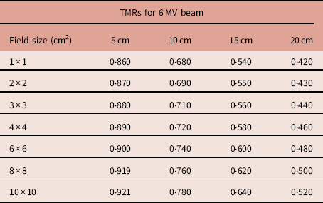
Radiotherapy plans are entirely dependent upon the exact determination of the dosimetric data obtained by the measurement of the dosimetric quantities like PDDs, TARs, TPRs and TMRs as these are essential in the quantification of exposure to the photon beams and calculations of MUs.Reference Rahman, Alam and Akhtaruzzaman 10 – Reference Ding and Krauss 12 In an SAD (source to axis distance) treatment set-ups using energy beams in MV range, the quantities like TPRs and TMRs are used for dosimetric calculations. The measured values of tissue maximum ratios by CC01 at various depths and fields shaped by MLCs ranging from 1 to 10 cm2 are enlisted in the tables given below for both 6 and 18 MV beam energies.
Table 1 shows the TMR values for 6 MV beam, which were measured by CC01 at the depth of 5, 10, 15 and 20 cm by varying the field sizes from 1 to 10 cm2. TMR at the depth of 5 cm increased with the increasing field sizes from 1 to 10 cm2. Similarly, at the other depths of 10, 15 and 20 cm, TMRs increased with the increasing field sizes owing to the production of more scattered photons.Reference Sharma, Kumar and Dagaonkar 13 – Reference Mesbahi 15 TMR decreased with the increasing depth due to attenuation of the photon beam in a water phantom.Reference Rahman, Alam and Akhtaruzzaman 10 , Reference Pichandi, Kadirampatti, Jerin, Balaji and Kilara 16 , Reference Khan and Gibbons 17
Tissue maximum ratios (TMRs) measured by CC01 for 6 MV beam through multi-leaf collimator fields at various depths

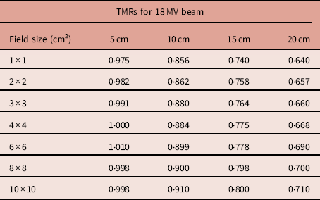
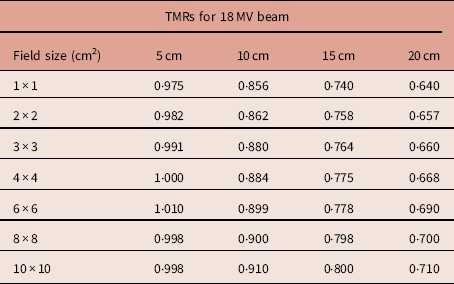
Table 2 shows the TMR values for 18 MV beam, at the depths of 5, 10, 15 and 20 cm by varying the field sizes from 1 to 10 cm2. For this beam energy, TMRs increased with the increasing field sizes but decreased with increasing depths. But 18 MV beam showed higher TMR values as compared with 6 MV beam for a given set of field sizes and depths. This is because attenuation is less pronounced for higher energy beam than that for lower energy beam.Reference Khan and Gibbons 17 At 5 cm depth, mean of TMR was 0·89 for 6 MV beam whereas for 18 MV beam, TMR mean was 0·99 for a given set of field sizes. Similarly, at the depth of 20 cm, 6 MV beam showed a mean of 0·46 and 18 MV beam gave TMR mean of 0·68. 18 MV beam showed 13·95% more TMR value than 6 MV beam at 5 cm depth for 1 cm2 field size. At 20 cm depth for 1 cm2 field, TMR due to 18 MV beam was 52·4% higher than the TMR due to 6 MV beam. For 10 cm2 field size, at the depth of 5 cm, TMR due to 18 MV beam was 8·69% higher than the TMR due to 6 MV beam. At 20 cm depth, for 10 cm2 field size, 36·5% higher TMR was noticed for 18 MV beam as compared with 6 MV beam.
Tissue maximum ratios (TMRs) measured by CC01 for 18 MV beam through multi-leaf collimator fields at various depths
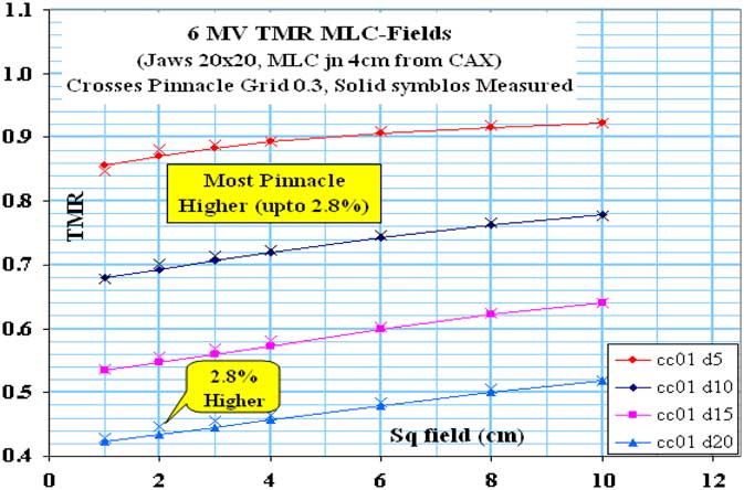
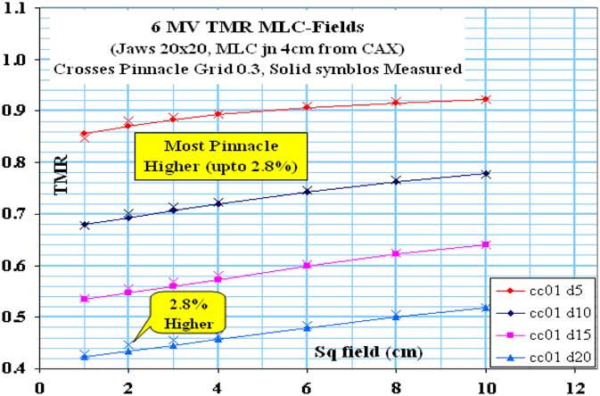
In Figure 1, percentage difference between CC01 measured and pinnacle calculated TMR varied from 0·11 to 2·8% for 6 MV beam for a given set of field sizes at the depths of 5, 10, 15 and 20 cm. There observed no difference between CC01 measured and pinnacle calculated TMR for 10 cm2 field size at all the given depths. The highest observed difference of 2·8% between measured and calculated TMRs occurred at 20 cm depth for 2 cm2 field size.
Representation of CC01 measured and Pinnacle calculated tissue maximum ratios (TMRs) for 6 MV beam. Abbreviation: MLC, multi-leaf collimator.
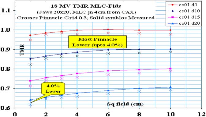
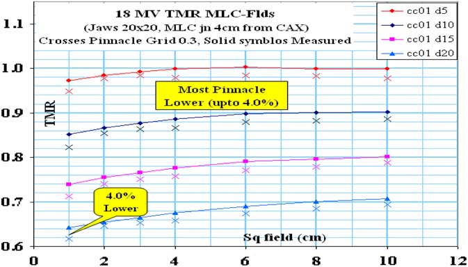
In Figure 2, for 18 MV beam, percentage difference between CC01 measured and Pinnacle calculated TMR were found in a range from 0·2 to 4·4%. For all the given set of field sizes, CC01 measured and pinnacle calculated TMR were not found same at all the depths of 5, 10, 15 and 20 cm. It can be seen that among all the given field sizes, measured and calculated TMRs deviated largely for 1 cm2 field size at the given set of depths and also the highest observed difference of 4% was noticed at the depth of 20 cm for this field size.
Representation of CC01 measured and Pinnacle calculated tissue maximum ratios (TMRs) for 18 MV beam. Abbreviation: MLC, multi-leaf collimator.
Discussion
The notion of TMR is originated in the case of higher energy photon beams and it varies only with varying field sizes and depths.Reference Sharma, Kumar and Dagaonkar 13 , Reference Pichandi, Kadirampatti, Jerin, Balaji and Kilara 16 , Reference Jayaraman and Lanzl 18 It can be observed from the above presented results that TMR due to 18 MV beam at the depth of 5 cm decreased with the increasing field sizes from 6 to 10 cm2. This unanticipated decrease in TMR with increasing field sizes is due to multiple factors like incident scattered radiation, varying balance with depth of phantom scatter and the existence of secondary electrons from the beam. This behavior is significant at depths below the build-up region. At depths above the build-up region, TMR increases with increasing field sizes. It has also been documented in British Journal of Radiology supplement 17. 19 – Reference Osei 21 Whereas for 6 MV beam, TMR increased with increasing field sizes at all the given depths. This is because all the depths of 5, 10, 15 and 20 cm lie above the build-up region for 6 MV beam.
TMR due to 6 and 18 MV beam showed a considerable difference from each other at larger depth than at smaller depth. This happened due to the fact that 18 MV beam produced secondary electrons which penetrate deep in a medium whereas secondary electrons produced by 6 MV travel a shorter distance inside the medium to shed off all their energies.
It was seen from Figure 1 that for 6 MV beam, measured and calculated TMR data were in good agreement for 6, 8 and 10 cm2 field sizes at all the given depths but showed deviations for field sizes smaller than 6 cm2. Furthermore, for 18 MV beam, measured and calculated TMRs deviated from each other at all the given depths and field sizes with the maximum deviation of 4%. But all the percentage differences remained within tolerance limits (±5%) of dose delivery. 22 , Reference Johansson, Hariot, Van Dam, Lepinoy, Setenac and Sernbo 23
Conclusion
For both 6 and 18 MV beam, there was good agreement between the measured and Pinnacle calculated TMRs for the field sizes ranging from 1 to 10 cm2. This exploration can be extended to the determination of other dosimetric parameters like TARs, TPRs in small fields.
Conflicts of Interest
None.
Acknowledgements
The authors would like to acknowledge the Department of Radiation Physics M D Anderson Cancer Centre, University of Texas, Houston USA and the Department of Clinical of Clinical and Radiation Oncology, Shaukat Khanum Memorial Cancer Hospital and Research Centre, Lahore, Punjab, Pakistan.