Case report
An 18-year-old woman was referred to the pediatric cardiology department of a tertiary care hospital due to apparent dilatation of the right atrium found on obstetric morphological ultrasound. She was previously healthy and had no family history of cardiovascular disease or chromosomal abnormalities. Her partner was a non-consanguineous healthy man. The colour doppler fetal echocardiography performed at 32 weeks of gestation confirmed cardiomegaly as a result of a significant enlargement of the right atrium. The tricuspid valve presented a normal morphology with physiological regurgitation. A sinus rhythm was detected without arrhythmias during the study. No other cardiac or extracardiac malformations were found. Pregnancy follow-up was uneventful, with regular appointments until birth with stable parameters and normal intra-uterine fetal growth.
A male infant was delivered at term via spontaneous vaginal delivery with a birth weight of 3335 g and Apgar Score of 8/10. The physical examination after birth was normal. He was admitted to the neonatal ICU for clinical monitoring. Postnatal transthoracic echocardiogram confirmed an aneurismatic dilatation of the right atrium without signs of intracardiac thrombus formation or pleural effusion, a normal anatomy of the tricuspid valve, a patent foramen ovale with a left-to-right shunt and a normal biventricular systolic function. He presented an electrocardiogram with sinus rhythm and overload signs in the inferior leads. Thrombosis prophylaxis with acetylsalicylic acid (5 mg/kg/day) was started on day 5. Twenty-four hour Holter monitoring registered 13 premature atrial contractions without other abnormalities. He was discharged after 10 days.
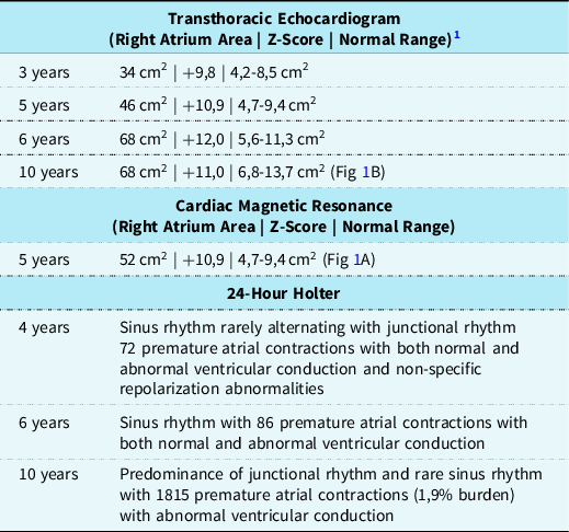
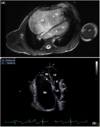
He was regularly evaluated by the Pediatric Cardiology team. Serial evaluations including transthoracic echocardiogram, 24-hour holter and cardiac MRI are summarised in Table 1. Echocardiograms presented progression of the right atrium enlargement with a stable right atrium area in the last two exams. The patient never presented signs of high right ventricle diastolic pressure. He presented a normal estimated pulmonary arterial systolic pressure of 24 mmHg. The inferior vena cava was normal. Cardiac stress test performed at 10 years of age was unremarkable. It was decided to continue medical management due to clinical stability and absence of intracardiac thrombi or compression of adjacent anatomical structures, adjusting the acetylsalicylic acid dose to body weight. He is currently on 100 mg/day.
Serial evaluations on Pediatric Cardiology outpatient visits.

Discussion
Idiopathic dilatation of the right atrium is a rare congenital anomaly first described by Bailey in 1955. Reference Cantinotti, Scalese and Murzi1,Reference Bailey2 It is defined as an isolated diffuse enlargement of the right atrium in the absence of other predisposing conditions such as tricuspid valve disease, structural CHD, pulmonary arterial hypertension, or acquired inflammatory changes in the myocardium. Prenatal diagnosis is extremely rare and very few cases have been reported. Reference Sulu, Baspinar and Kervancıoglu3,Reference Walter, Bartrons and Gómez4 So far, aetiology is unknown and the real pathogenesis has not been clarified. Reference Walter, Bartrons and Gómez4 Patients with this condition are usually asymptomatic, yet they can present with atrial arrhythmias, palpitations, fatigue, shortness of breath, chest pain, severe tricuspid regurgitation, congestive heart failure, thrombus formation leading to pulmonary embolism, paradoxical embolism or stroke, and even sudden death. Reference Srinivasa, Kharge and Patil5,Reference Divekar, Soni and Ross6 The progression varies from spontaneous regression, stabilisation of atrial size, and progressive enlargement. Reference Walter, Bartrons and Gómez4 Diagnosis is based on echocardiography and can be confirmed with other techniques like CT angiography or cardiac MRI. Periodic follow-up with echocardiogram and Holter monitoring should be focussed on detecting atrial enlargement, new symptoms, and uncontrolled arrhythmias needing intervention. In asymptomatic children, treatment is controversial. Antiplatelet drugs have been suggested as prophylactic treatment to prevent the risk of thrombus formation. Reference Walter, Bartrons and Gómez4 In patients with an added risk of thrombophilia or atrial thrombosis, anticoagulation therapy should be associated. Antiarrhythmic medication can be used to control symptoms. Surgical reduction might be necessary for some symptomatic patients or asymptomatic patients at high risk of progression, such as those presenting with initial severe dilatation, significant enlargement over a short period, compression of adjacent structures, or uncontrolled arrhythmias. Reference Walter, Bartrons and Gómez4 Since it is a simple intervention with few post-operative complications, some authors defend surgical reduction as the treatment of choice in order to avoid the occurrence of thrombosis and arrhythmias. Reference Walter, Bartrons and Gómez4,Reference Srinivasa, Kharge and Patil5 Nonetheless other authors report significant post-operative atrial arrhythmias and recommend that this should be taken into consideration when considering surgery in asymptomatic patients. Reference Divekar, Soni and Ross6 Taking into account the wide range of clinical presentations, it is reasonable to individualise management based on each case. Reference Hofmann, Heilmann and Häusler7
Despite being an uncommon event, the authors report an idiopathic dilatation of the right atrium prenatally diagnosed by fetal echocardiogram and confirmed after birth by transthoracic echocardiogram. Our patient was mainly asymptomatic during the 10-year follow-up, as many paediatric cases are described in the literature. He started antiplatelet prophylaxis as a newborn and this approach was maintained since there are no indications on the recommended duration of therapy available in the reviewed literature. The patient was not referred for surgical treatment, given his asymptomatic clinical course at the time. The authors wish to highlight the importance of the prenatal diagnosis that in this case allowed an anticipated strategy to prevent complications and improve outcomes. The medical plan included follow-up in a third-level hospital with paediatric cardiology and obstetrical evaluation and delivery. The later allowed admission in the neonatal ICU after birth for clinical monitoring, evaluation by paediatric cardiology, and early start of antiplatelet prophylaxis.
A: Cardiac magnetic resonance imaging (4-chamber view) at the age of 5. RA – right atrium, * - tricuspid valve, RV – right ventricle, LA – left atrium, # - mitral valve, LV – left ventricle. B: Transthoracic echocardiogram (4-chamber view) at the age of 10. RA – right atrium, * - tricuspid valve, RV – right ventricle, LA – left atrium, # - mitral valve, LV – left ventricle.

Data Availability Statement
The authors confirm that the data supporting the findings of this study are available within the article and its supplementary materials.
Acknowledgement
The authors would like to thank the family for consenting the publication of this case report.
Financial support
None.
Conflict of Interest
The authors have no conflict of interest to declare.