Introduction
Due to the fact that it is not necessary to be exposed to the outside of the incubator for microscopic observation, the time-lapse monitoring system with a built-in microscope and real-time video recording in the incubator has a more stable embryo culture environment and also provides more traceable morphokinetic parameters (Ciray et al., Reference Ciray, Campbell, Agerholm, Aguilar, Chamayou, Esbert and Sayed2014; Jiang et al., Reference Jiang, Wang, Wang, Shen, Wang, Zheng, Yang, Ma and Zhang2023). This means that embryos can develop in a more controlled and consistent environment, leading to improved development potential and better selection of euploid embryos (Zaninovic et al., Reference Zaninovic, Irani and Meseguer2017; Zou et al., Reference Zou, Pan, Ge, Xu, Gu, Li, Fu, Gao, Sun and Sun2022). The advantages of time-lapse monitoring in improving embryo development potential and euploid embryo selection had been widely recognized in the field of IVF-ET. Many centres had adopted morphokinetic embryo evaluation schemes and even integrated artificial intelligence to predict embryo development potential (Kragh and Karstoft, Reference Kragh and Karstoft2021; Yuan et al., Reference Yuan, Yuan, Song, Huang and Yan2023). Some studies had also shown that specific morphokinetic parameters of embryos are correlated with embryo aneuploidy, and when combined with preimplantation genetic testing for aneuploidy (PGT-A), the accuracy of embryo selection was further improved (Kimelman et al., Reference Kimelman, Confino, Okeigwe, Lambe-Steinmiller, Confino, Shulman, Zhang and Pavone2019; Yuan et al., Reference Yuan, Yuan, Song, Huang and Yan2023).
However, there were still centres that remain sceptical about the benefits of time-lapse monitoring (Armstrong et al., Reference Armstrong, Bhide, Jordan, Pacey, Marjoribanks and Farquhar2019; Zhang et al., Reference Zhang, Zhang, Han, Liu, Shen, Yao, Shi, Hu, Wang, Wang, Zhou, Kang, Zhang, Luo, Yu, Liu, Sun, Sun, Wang, Quan and Huang2022). This could be due to variations in culture systems and the lack of standardized operating procedures across different centres. The outcomes observed in these centres may not fully reflect the potential advantages of time-lapse monitoring.
One challenge faced during the use of time-lapse monitoring is the occurrence of microbubbles in the culture dish. This issue is likely caused by the warming of the culture media and the release of gas. The presence of microbubbles between the oil and liquid interface in the culture micropores can interfere with embryo imaging. To address this, the manufacturer recommends prewarming the culture media and dish beforehand to release any dissolved gas, thereby preventing the occurrence of air microbubbles during the culture process. However, prewarming the media can lead to accelerated evaporation of water, which in turn affects the osmotic pressure of the culture droplets (Mestres et al., Reference Mestres, García-Jiménez, Casals, Cohen, Acacio, Villamar, Matia-Algué, Calderón and Costa-Borges2021).
In embryo culture, maintaining the appropriate osmotic pressure is crucial for the development and health of embryos (Taïeb et al., Reference Taïeb, Garske, Contzen, Gossen, Bertinetti, Robinson and Cipitria2021). The recommended osmotic pressures for cleavage and blastocyst media were 275–285 mOsm/kg. The pH should be maintained between 7.2 and 7.4, and the osmotic pressure should not exceed 300 mOsm/kg (Mestres et al., Reference Mestres, García-Jiménez, Casals, Cohen, Acacio, Villamar, Matia-Algué, Calderón and Costa-Borges2021). When using the time-lapse monitoring system, non-humidified culture was often employed to minimize the risk of contamination (Chi et al., Reference Chi, Park, Yoo, Kwak, Son, Kim, Sim, Lee and Koo2020). However, this could result in increased evaporation of water from the culture media, leading to changes in osmolarity (Yumoto et al., Reference Yumoto, Iwata, Sugishima, Yamauchi, Nakaoka, Tsuneto, Shimura, Flaherty and Mio2019). In order to compare the results of IVF embryo culture using different dish-making methods, this study examined the osmotic pressure, embryo quality after fertilization and the outcomes of transferred embryos when using pre-warmed and refrigerated culture media dishes.
Material and methods
Study population
The study population consisted of patients undergoing their first IVF or intracytoplasmic sperm injection (ICSI) cycle at the Center for Reproductive Medicine in Xingtai Meihe Reproductive and Genetic Hospital, located in Xingtai, Hebei Province, China. The criteria for inclusion in the study were women aged 40 years or younger who were experiencing infertility due to factors such as issues with the fallopian tubes, ovarian hyperresponsiveness or male factors. Patients with a history of endometriosis, uterine fibroids, uterine adhesions, endometritis and other endometrial diseases were excluded from the study. Additionally, patients with remedial ICSI, pelvic inflammation, uterine and pelvic lesions, uterine malformations and chromosomal abnormalities were also excluded. Patients were randomized into two groups, according to the oocyte retrieval date. Pre-warmed culture medium was used on even-numbered dates, while refrigerated culture media was used on odd-numbered dates.
All procedures used in this study were conducted in accordance with the principles of conducting experiments with human participants as outlined in the Declaration of Helsinki. This study adopted the consent of the Ethics Committee of Xingtai Meihe Reproductive and Genetic Hospital. All processes in IVF-ET were obtained by written informed consent.
Preparation of time-lapse culture dishes
In the pre-warmed group, both the culture media (G-1 PLUS, Vitrolife, Sterilized) and the culture dish (CultureCoin, AT MEDICAL Ltd., E, Lithuania) used in the study were placed in a humidified incubator (MCO-5M, SANYO Electric Co., Ltd., Japan) set at a temperature of 37 degrees Celsius (°C) with a CO2 concentration of 6%. In the refrigerated group, the culture media was maintained at a set temperature of 2–8, and the culture dish was opened at room temperature of 23–25°C. The preparation of time-lapse culture dishes was done in the same manner in the two groups. Each dish was prepared with 14 drops of 40 ul culture media using a pipette gun (Eppendorf Research plus, Germany). Subsequently, 4 ml of culture oil (Vitrolife, Sterilized) was carefully poured onto each dish using a pipette (Accu-jet pro, Brand, Germany), ensuring that it completely covers the culture drops. Only one petri dish was prepared for each patient. Since only 14 culture microdroplets could be observed in real time, any surplus embryos were occasionally cultured together in additional microdroplets. The dishes were placed in the incubator for at least 12 h for standby and any microbubbles present were removed before use.
Stimulation protocols for IVF
All patients used the standard gonadotropin-releasing hormone (GnRH) agonist protocol. Patients who used the GnRH agonist protocol were injected intramuscularly with GnRH analogue (H20030578, Ipsen Pharma Biotech, Signes, France) from the mid-luteal phase. Once downregulation was achieved, patients commenced treatment with recombinant follicle-stimulating hormone (FSH) (JS20160044, Merck Serono S.p.A., Modugno, Apulia, Italy) to promote ovulation. At the clinician’s discretion, the administration of 5000–10,000 IU human chorionic gonadotropin (hCG, H44020672, Lizhu Pharmaceutical Trading Co., Zhuhai, China) was used to trigger the ovary as soon as one-third of all follicles reached a diameter of ≥ 18 mm. The retrieval of the oocyte was done using needle aspiration guided by transvaginal ultrasound, 36–37 h after hCG injection.
In vitro fertilization and embryo transfer
IVF/ICSI was carried out at 3∼4 h after oocyte retrieval, and the fertilization method was performed depending on sperm parameters. IVF: It was performed by 30–50 ul microdroplets (IVF PLUS, Vitrolife, Sterilized) with 1–2 oocytes per drop and 10 000 sperm. The cumulus and coronal cells surrounding the oocytes were removed from 4 to 6 h after fertilization. Then the zygotes or embryos were placed into time-lapse culture dishes and incubated in a time-lapse imaging incubator (Miri TL6, ESCO, Singapore). Fertilization was assessed approximately 16–18 h after insemination, and zygotes with two pronuclei indicated normal fertilization. Cleavage-stage embryo morphology was observed on the third day after oocyte retrieval as follows: good, 7∼10 blastomeres of uniform size, < 10% fragmentation and no evidence of multinucleation; fair, 6 or > 10 blastomeres of uniform size, 10∼25% fragmentation and no multinucleation; poor, 2-5blastomeres, > 25% fragmentation, blastomeres size not stage specific or multinucleation (Alpha Scientists in Reproductive Medicine and ESHRE Special Interest Group of Embryology, 2011). On the fifth or sixth day, the blastocysts assessed as 3BB or better were considered good, and 3BC or better were considered fair using the blastocyst grading system described by Gardner (Gardner et al., Reference Gardner, Lane, Stevens, Schlenker and Schoolcraft2019). If there was a minimum of four good embryos on the third day, blastocysts were chosen for transfer on the fifth day; otherwise, cleavage-stage embryos were selected for transfer on the third day. Either one good embryo or two fair good embryos were chosen for transfer. If patients displayed symptoms or were at risk of ovarian hyperstimulation syndrome (with an oocyte count exceeding 20 or estradiol levels surpassing 7000 ng/ml on HCG day), the embryo transfer procedure was halted, and all viable embryos were cryopreserved. In fresh embryo transfer cycles, Flavestin (H20041902, Zhejiang Xianju Pharmaceutical Co., LTD, Xianju, Zhejiang, China) was administered via intramuscular injection to provide luteal support starting from the day of oocyte retrieval. Based on the estradiol levels on the day of HCG injection, the suitable estradiol supplement (H20160679, DELPHARM Lille S.A.S., Cedex, France) was provided on the day of transfer. Luteal support was maintained from the day of ovulation for a minimum of 8 weeks or until a negative hCG test was confirmed.
Measurements
A pregnancy test was performed on the 14th day after ET, with a serum b-HCG level > 5 IU/L considered to be positive. Subsequently, clinical pregnancy was defined as a gestational sac detected on the 28th day after ET. The implantation rate was calculated as number of gestational sacs/number of transferred embryos. The clinical pregnancy rate was calculated as number of clinical pregnancy cycles/total number of transfer cycles. The live birth rate was calculated as number of cycles with live births/total number of transfer cycles.
The preterm birth rate was calculated as number of cycles with gestational age < 28 weeks/number of clinical pregnancy cycles. Microbubbles occurrence rate was calculated as number of cases where microbubbles appear during the equilibrium process of the culture media or during embryo culture/total number of cases.
Culture media osmotic pressure test
In order to detect osmotic pressure, the culture dishes were prepared separately using the same method and conditions as described below. The dishes were divided into four groups based on whether the culture media was pre-warmed and the volume of oil overlay. Groups R-3.5 and r-4.0 were covered with 4 ml and 3.5 ml of culture oil, respectively, on the refrigerated media. On the other hand, P-3.5 and P-4.0 were treated with 4 ml and 3.5 ml of oil, respectively, on the pre-warmed media. Each group was repeated in triplicate. Once prepared, the dishes were balanced for 6 h in a 37°C, 6% CO2 incubator. Afterwards, they were transferred to a time-lapse imaging incubator (Miri TL6, ESCO, Singapore).
Before the dishes were prepared and at 1, 3 and 5 days of incubation, the culture media in the droplets of each dish was extracted. The osmotic pressure of the samples was then tested using a freezing point depression osmometer (GONOTEC, Germany). All samples were measured under the same calibration conditions. Initially, 50 ul of the sample was meticulously transferred into pristine and dry test tubes, ensuring the absence of any air bubbles in the liquid. Subsequently, the test tubes containing the sample liquid were delicately positioned on a test tube rack (equipped with a thermistor probe) with the lid oriented upwards. The thermistor probe was then fully immersed in the sample liquid for precise temperature monitoring.
Upon reaching the supercooling phase, the sample liquid commenced automatic crystallization. The temperature fluctuation was continuously monitored on the display screen until the crystallization temperature reached –7°C. The device seamlessly recorded the freezing point temperature, which was subsequently converted into an osmotic pressure value using the predetermined calibration factor. Finally, the calculated osmolality was read on a digital display.
Statistical analysis
Statistical analyses were performed by SPSS version 26 (IBM corporation, Armonk, NY, USA). The normally distributed continuous data were reported as mean ± standard. T test was used to compare categorical variables. The non-normally distributed continuous variables were expressed as median (25th quartile, 75th quartile), and comparisons were performed using the Wilcoxon rank sum test. Count data were indicated as a percentage (%), and comparisons were performed using the χ2 test. Statistical significance was accepted at P < 0.05.
Results
Baseline characteristics
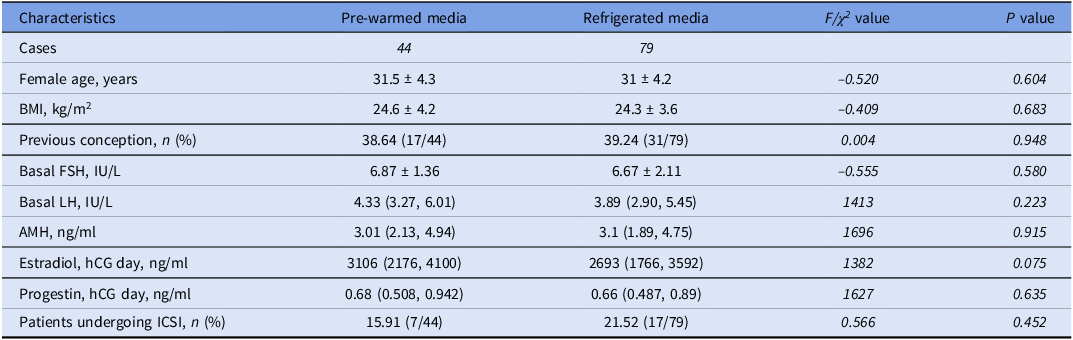
The pre-warmed group included 44 cases with 27 cases of fresh embryo transfer, while the refrigerated group included 79 cases with 51 cases of fresh embryo transfer. We examined various baseline characteristics in both groups, including female age, body mass index (BMI), proportion of primary infertility, basal follicle-stimulating hormone (FSH) and luteinizing hormone (LH) levels, anti-Müllerian hormone (AMH) levels, estradiol (E2) and progesterone (P) levels on trigger day and proportion of ICSI (15.91% vs. 21.92%, P = 0.452). The results, as presented in Table 1, showed no significant differences between the two groups in these baseline characteristics. This suggests that the two groups of patients were comparable in terms of their demographic and clinical profiles.
Baseline characteristics

Abbreviations: BMI, body mass index; FSH, follicle-stimulating hormone; LH, luteinizing hormone; AMH, anti- Müllerian hormone; ICSI, intracytoplasmic sperm injection.
Statistics presented as mean ± standard, median (P25, P75) or percentage (number/total number).
Comparison of ovarian stimulation and embryo outcomes
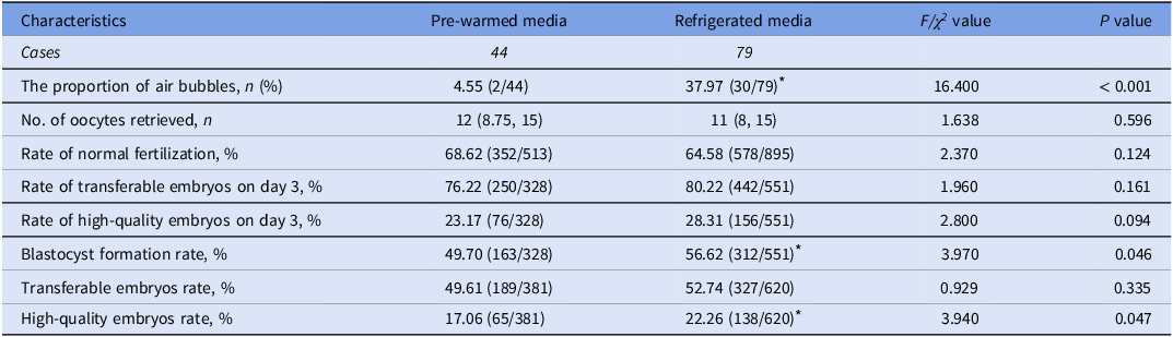
The proportion of air microbubbles affecting imaging significantly decreased (4.55% vs. 37.97%, P < 0.001) after incubating with pre-warmed culture media. There were no significant differences between the two groups in the number of oocytes retrieved and the rate of normal fertilization. With the longer incubation time of zygotes, the rate of transferable (80.22% vs. 76.22%) and high-quality embryos (28.31% vs. 23.17%) in the refrigerated group were observed slightly higher on the third day, although the difference was not statistically significant. The blastocyst formation rate (56.62% vs. 49.70%, P = 0.046) and total high-quality embryo rate (22.26% vs. 17.06%, P = 0.047) in the refrigerated group were significantly higher than those in the pre-warmed group, as shown in Table 2. The refrigerated group showed a gradual superiority in better embryonic outcomes with a longer in vitro incubation time.
The embryo outcomes between the two groups

Statistics presented as median (P25, P75) or percentage (number/total number).
* Statistically significant compared with the pre-warmed media group.
Pregnancy outcomes in two groups
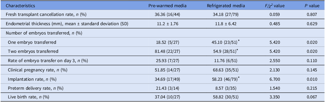
There was no significant difference between the two groups in the cancellation rate of fresh cycle transfers, endometrial thickness on the day of transfer and the proportion of cleavage embryo transfers. The proportion of single embryo transfer in the refrigerated culture media group was significantly higher than that in the pre-warmed culture media dish method (45.10% vs. 18.52%, P = 0.020), which also indicates that the high-quality embryo rate in the refrigerated culture media dish group was higher because our hospital’s transfer strategy recommends single embryo transfer for high-quality embryos and double embryo transfer for non-high-quality embryos. The implantation rate in the refrigerated culture media dish group was significantly higher than that in the pre-warmed culture media dish group (58.23% vs. 34.69%, P = 0.010) (Table 3). Although there was no statistically significant difference in the clinical pregnancy rate, live birth rate and preterm delivery rate in the refrigerated group (P > 0.05), an increasing trend was observed in the clinical pregnancy rate (68.63% vs. 51.85%) and live birth rate (58.82% vs. 37.04%), along with a decreasing trend in preterm delivery rate (8.57% vs. 21.43%) (Table 3).
Fresh transplant outcomes between the two groups

Statistics presented as median (P25, P75) or percentage (number/total number).
* Statistically significant compared with the pre-warmed media group.
Culture media osmotic in the different groups
The culture media in the droplets was extracted before the dishes were prepared and at 1, 3 and 5 days of incubation to investigate whether the use of refrigerated media and increasing the volume of oil overlay could protect against osmolarity destabilization. The dishes were categorized into four groups based on whether the culture media was refrigerated and the volume of oil overlay. Groups R-3.5 and R-4.0 were covered with 4 ml and 3.5 ml of culture oil, respectively, on refrigerated media, while groups P-3.5 and P-4.0 received 4 ml and 3.5 ml of oil, respectively, on pre-warmed media. Each group was replicated in triplicate. The results, depicted in Fig. 1, revealed significant differences in osmolality among the four groups at each time point, with the most pronounced change observed in group P-3.5 and the smallest change observed in group R-4.0. After 5 days of incubation, the osmotic pressure of group R-4.0 (287 ± 1 mOsm/kg) was significantly lower than that of groups P-3.5 (294 ± 1 mOsm/kg), P-4.0 (297.67 ± 0.58 mOsm/kg) and P-3.5 (301.00 ± 2.00 mOsm/kg) (Figure 2A). From day –1 to day 1, osmolality increased, with the P-3.5 group showing a significant elevation compared to the other three groups, which did not exhibit any significant differences among themselves. From day 1 to day 3, there were no significant differences in osmolality changes among the four groups. However, from day 3 to day 5, the osmolality in the P-3.5 group was significantly higher than that in the R-4.0 group, while no significant differences were observed among the other three groups (Figure 2B). These findings suggest that the refrigerated media covered with 4 ml seems to be advantageous for maintaining osmotic pressure stability.
Comparison of osmolality among the four groups at different incubation durations. (A) The osmolality of the culture droplets after 1 day of incubation. (B) The osmolality of the culture droplets after 3 days of incubation. (C) The osmolality of the culture droplets after 5 days of incubation. *P < 0.05.

The change in osmolality among the four groups at different incubation durations. (A)The osmolality of the culture droplets was tested before the preparation of the dish, after 1, 3 and 5 days of incubation, respectively. (B) The change in osmolality from day –1 to day 1, from day 1 to day 2 and from day 3 to day 5. *P < 0.05.

Discussion
In recent years, the time-lapse imaging culture system has gradually been widely used in IVF and embryo transfer technology (Kimelman et al., Reference Kimelman, Confino, Okeigwe, Lambe-Steinmiller, Confino, Shulman, Zhang and Pavone2019; Jiang et al., Reference Jiang, Wang, Wang, Shen, Wang, Zheng, Yang, Ma and Zhang2023). Many embryo evaluation schemes combined with embryo dynamics have been proposed and effectively used to predict embryo ploidy and developmental potential (Ottolini et al., Reference Ottolini, Rienzi and Capalbo2014; Yuan et al., Reference Yuan, Yuan, Song, Huang and Yan2023). However, there were few reports on the operation process of the time-lapse imaging culture system, which might be the main reason why the time-lapse imaging culture system was suspected and limited in application. This study compared the changes in the osmotic pressure of culture droplets and the outcome of embryo culture using two methods of dish preparation: pre-warmed culture media and refrigerated culture media. It was found that using refrigerated culture media to prepare dishes was beneficial to protecting the osmotic pressure of culture droplets, and the rate of high-quality embryos and implantation rate were significantly higher than using pre-warmed culture media to prepare dishes.
This study included a total of 123 patients, with 44 receiving a warm culture medium and 79 receiving a cold culture medium. For the sake of convenience, the random grouping was determined by the oocytes retrieved date: patients with even-numbered dates were assigned to the pre-warmed culture medium, while those with odd-numbered dates were assigned to the refrigerated culture medium. The difference in group sizes may arise from a higher number of odd-numbered days compared to even-numbered days, or it could be due to variations in the daily number of patients undergoing oocyte retrieval.
Embryo culture media are routinely stored at 2–8°C, while embryo culture is conducted at 37°C (Gardner and Kelley, Reference Gardner and Kelley2017). Due to the different solubility of gas at different temperatures, gas may overflow during the equilibrium process of the culture media, forming microbubbles, affecting embryo observation and imaging. Therefore, the manufacturer recommends using a culture media that has been balanced at 37°C to reduce the generation of air microbubbles during the culture process. The study compared the proportion of air microbubbles appearing in the culture droplets after pre-warmed and refrigerated culture media were prepared, and the proportion of air microbubbles appearing in the culture droplets after pre-warmed culture media were prepared significantly decreased (4.55% vs. 37.97%, P < 0.001).
Maintaining stable osmotic pressure is an effective measure to improve the developmental potential of embryos in in vitro culture (Yumoto et al., Reference Yumoto, Iwata, Sugishima, Yamauchi, Nakaoka, Tsuneto, Shimura, Flaherty and Mio2019). Pre-warmed culture media can cause water loss during dish making, resulting in changes in osmotic pressure. This study has shown that the osmotic pressure of pre-warmed culture media dish-making group was significantly higher than that of the refrigerated culture media dish-making group on the first, third and fifth days after fertilization, regardless of a low or high volume of oil overlay. From day –1 to day 1, osmolality increased, with the P-3.5 group exhibiting a significant elevation compared to the other three groups. From day 1 to day 3, there were no significant differences in osmolality changes among the four groups. Therefore, the change in osmotic pressure primarily occurs during dish preparation, and it is possible that different volumes of oil overlay offer varying degrees of protection against osmotic pressure. This study also showed that increasing the volume of oil overlay from 3.5 ml to 4 ml effectively reduced the increase in osmotic pressure, regardless of whether the culture medium was pre-warmed or refrigerated.
The evaporation of water not only affects osmotic pressure but also affects pH levels, and both of these factors play crucial roles in embryo development (Yumoto et al., Reference Yumoto, Iwata, Sugishima, Yamauchi, Nakaoka, Tsuneto, Shimura, Flaherty and Mio2019). Water evaporation can lead to changes in the concentration of solutes inside the embryo’s cells, altering the osmotic pressure and potentially disrupting the delicate balance required for proper development (Gatimel et al., Reference Gatimel, Moreau, Parinaud and Léandri2020; Aklima et al., Reference Aklima, Onojima, Kimura, Umiuchi, Shibata, Kuraoka, Oie, Suganuma and Ohta2021). Additionally, the change in pH levels resulting from water evaporation can impact enzyme activity and cellular functions necessary for embryonic growth, further influencing the development process (Gatimel et al., Reference Gatimel, Moreau, Parinaud and Léandri2020; Aklima et al., Reference Aklima, Onojima, Kimura, Umiuchi, Shibata, Kuraoka, Oie, Suganuma and Ohta2021). The osmotic pressure of refrigerated culture media with a high volume of oil overlay was best maintained, while that of pre-warmed culture media with a low volume of oil overlay was poorly maintained. The osmotic pressure of pre-warmed culture media with a low volume of oil overlay exceeds 300 mOsm/kg, on the fifth day of cultivation. The oil can protect the osmotic pressure stability in a non-humidified cultivation environment (Boumerdassi et al., Reference Boumerdassi, Huet, Millin, Sarandi, Bennani Smires and Sifer2021). Currently, the time-lapse imaging culture systems mostly use non-humidified culture to facilitate cleaning and reduce microbial growth (Boumerdassi et al., Reference Boumerdassi, Huet, Millin, Sarandi, Bennani Smires and Sifer2021). However, some studies have compared the outcomes of non-humidified culture and humidified culture, and the clinical outcomes of humidified culture were better (Chi et al., Reference Chi, Park, Yoo, Kwak, Son, Kim, Sim, Lee and Koo2020). It may be that humidified culture is closer to the physiological state, effectively avoiding water loss and stabilizing the osmotic pressure of the culture environment.
High osmotic pressure can cause developmental arrest in embryos (Baltz, Reference Baltz2012). This study compared the embryo outcomes of two dish-making methods. The results showed that the blastocyst formation rate (56.62% vs. 49.70%, P = 0.046) and the total high-quality embryo rate (22.26% vs. 17.06%, P = 0.047) in the refrigerated culture media dish group were slightly higher than those in the pre-warmed culture media dish group. This might be due to the higher osmotic pressure in the pre-warmed culture medium group. Additionally, the proportion of single embryo transfer in the refrigerated culture media dish group was significantly higher than that in the pre-warmed culture media dish group (45.10% vs. 18.52%, P = 0.020). It further indicates that the refrigerated culture media dish group has a higher rate of high-quality embryos, as our hospital’s transfer strategy recommends single embryo transfers for high-quality embryos and double embryo transfers for non-high-quality embryos.
The two dish-making methods have both advantages and disadvantages. During the process of making dishes with refrigerated culture media, air microbubbles easily appear and affect the camera. During the process of removing air microbubbles, the culture media is exposed outside the incubator. Making dishes with refrigerated culture media was more conducive to stabilizing the osmotic pressure of culture droplets. There was a significant statistical difference between the two groups (P > 0.05), but the clinical pregnancy rate (68.63% vs. 51.85%) and live birth rate (58.82% vs. 37.04%) in the refrigerated culture media group showed an increasing trend. The implantation rate in the refrigerated culture media dish group was significantly higher than that in the pre-warmed culture media dish group (58.23% vs. 34.69%, P = 0.010).
However, the range of osmolality within which human embryos can develop normally is relatively broad. Some commercial media were formulated with osmolalities as low as 265 mOsm, while others fall within the 275–280 range. A value of 290 was considered physiological, and embryos will typically develop to the blastocyst stage at an osmolality of 300. It was important to note that lower osmolality ranges may be associated with more robust blastocyst cavity expansion, which may not always be a positive indicator of outcome. Therefore, it is worth examining studies on the fallopian tube and uterine milieu to determine the normal osmolality that embryos experience in vivo. Moreover, it is worthwhile to investigate whether this osmolality is location-specific within the reproductive tract. Furthermore, future exploration will delve into potential factors beyond osmotic pressure that could influence differences in embryo and pregnancy outcomes in pre-warmed culture media, as well as possible alterations at the molecular level of the embryos.
In conclusion, refrigeration of culture medium in dish preparation is beneficial for maintaining the osmotic pressure of culture droplets, and the rate of high-quality embryos is significantly higher than in pre-warmed culture media dishes. A high volume of oil overlay in refrigerated culture media is beneficial to the osmotic stability of embryo culture media.
Data availability
The datasets generated and analyzed during the current study are not publicly available since the dataset will be used for other retrospective analyses. The data are available from the corresponding author upon reasonable request.
Acknowledgements
We would like to express our gratitude to all those who helped us during the writing of this manuscript. Thanks to all the peer reviewers for their opinions and suggestions.
Author contributions
YG, FD and SW contributed to the conception and design of the study. FD and YG organized the database. GL and ZY performed the statistical analysis. YG wrote the first draft of the manuscript. YG, FD, GL, ZY, LT and BZ wrote sections of the manuscript. All authors contributed to the manuscript revision and read and approved the submitted version.
Funding
The work was supported by the Health Commission of Hebei Province (Grant No. 20221865).
Competing interests
All authors declare no conflict of interest.
Ethical standards
All procedures used in this study were conducted in accordance with the principles of conducting experiments with human participants as outlined in the Declaration of Helsinki. This study adopted the consent of the Ethics Committee of Xingtai Infertility Specialized Hospital (approval number: 2021-05). All processes in IVF-ET were obtained by written informed consent.