Introduction
Although Bolivia is one of the countries with the greatest biodiversity in the world, its lichenicolous fungi have remained almost completely unexplored for a long time. Because of this, our current interests have focused on that group of fungi (see Flakus Reference Flakus2010). So far only 20 species and one variety have been reported from this country; most of these have been recorded recently and in scattered publications (Nylander Reference Nylander1859; Hawksworth & Santesson Reference Hawksworth and Santesson1990; Hafellner & Obermayer Reference Hafellner and Obermayer1995; Triebel et al. Reference Triebel, Rambold and Elix1995; Diederich Reference Diederich1996, Reference Diederich2003; Diederich & Etayo Reference Diederich and Etayo2000; Feuerer & Sipman Reference Feuerer and Sipman2005; Flakus et al. Reference Flakus, Kukwa and Czarnota2006, Reference Flakus, Ahti, Kukwa and Wilk2008; Etayo Reference Etayo2010; Farkas Reference Farkas2010). In this paper four species are described as new to science. Additionally, the host lichen Trypethelium ochroleucum (Eschw.) Nyl. is reported for the first time from Bolivia.
Material and Methods
Most of the specimens examined were collected by the authors in Bolivia and are deposited in KRAM, LPB, UGDA, hb. Flakus and hb. Etayo. Morphology and anatomy of the fungi were examined using NIKON SMZ800 and NIKON ECLIPSE 80i (DIC) microscopes. Squash preparations were studied in water, KOH solution (K) or Congo Red, and ascus structure in Lugol's iodine solution without (I) or with pre-treatment with KOH (K/I). All measurements were made on material mounted in water.
The New Species
Capronia etayoi Flakus & Kukwa sp. nov.
MycoBank No: MB564361
Capronia leptogii similis sed ascosporis latioribus, hyalinis ad pallide cinereis et hospite differt.
Typus: Bolivia, Dept. La Paz, Prov. Bautista Saavedra, ANMIN Apolobamba, near Taypi Cañuma village, 15°03′20″S, 69°09′07″W, 4506 m, high Andean open vegetation, on thallus of terricolous Dictyonema minus, 5 July 2010, A. Flakus 17508 & P. Rodriguez (KRAM—holotypus; LPB—isotypus).
(Fig. 1)
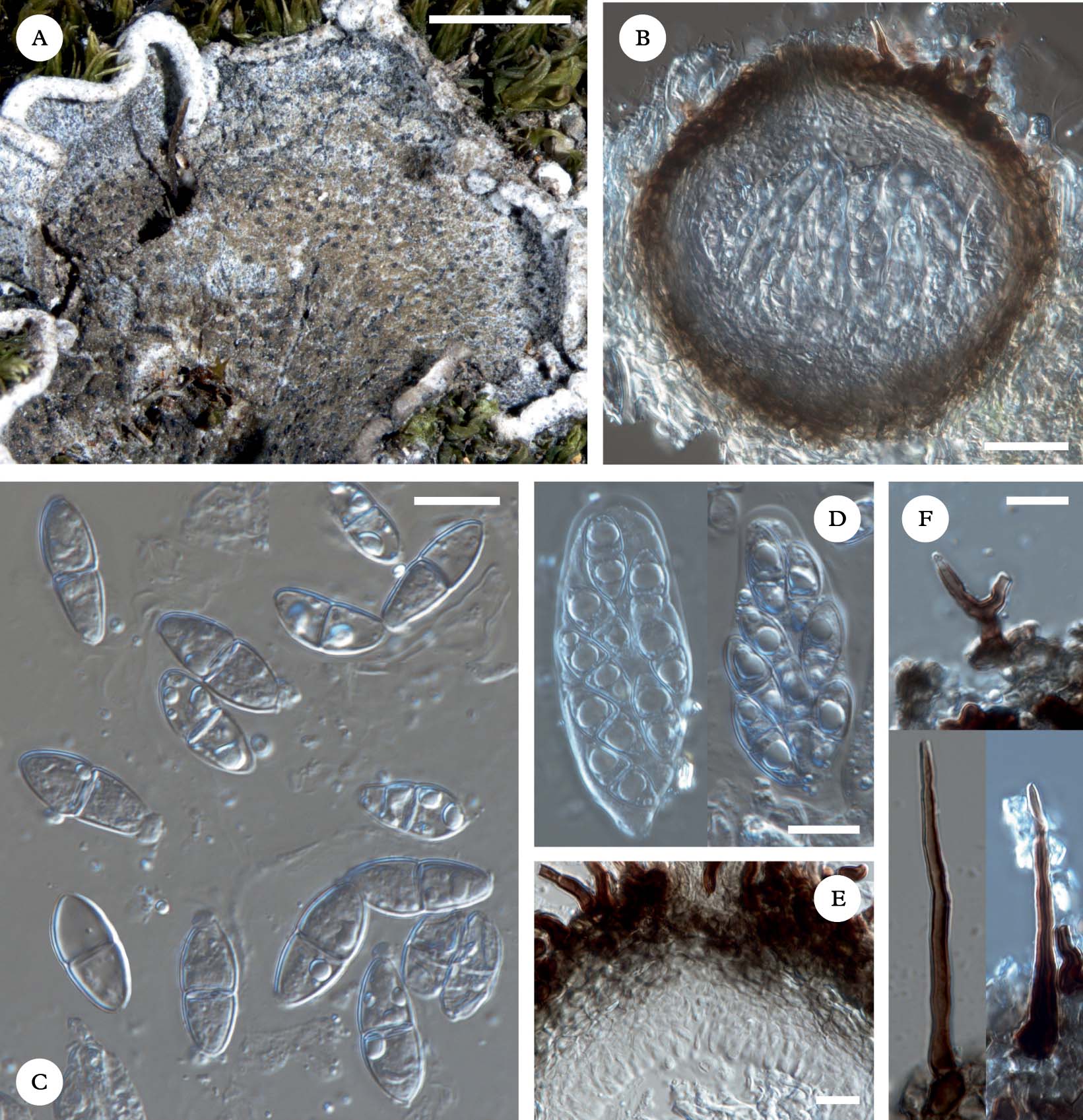
Capronia etayoi (holotype). A, habit of the fungus causing necrosis on the host thallus; B, section of an ascoma; C, mature ascospores; D, asci with ascospores; E, paraphysoids; F, simple and forked setae. Scales: A=2 mm; B=25 µm; C–F=10 µm. In colour online.
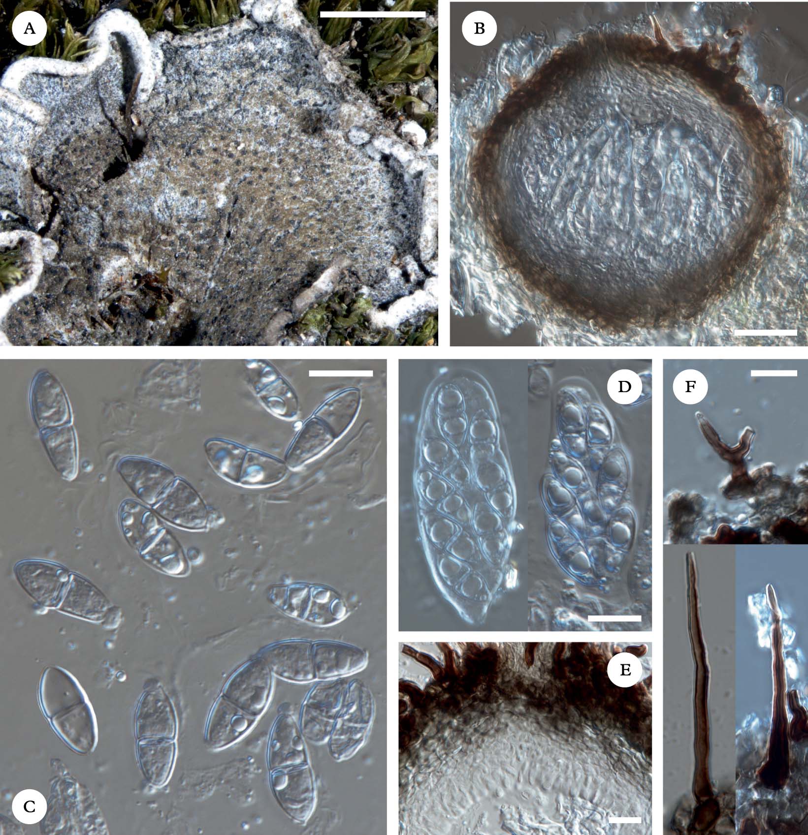
Ascomata perithecioid, ostiolate, arising singly, dispersed on the host thallus, half immersed to superficial, subglobose to globose, black, almost glabrous to densely covered by black setae arising mainly from the upper part, 110–130 μm diam. Setae simple or very rarely forked, non-septate or rarely 1–2-septate, smooth-walled, dark brown, rounded to slightly pointed at the apex and with thickened base, 25–55×2–4 μm. Exciple wall 8–17 μm thick, pseudoparenchymatous, composed of c. 4–7 layers of thin-walled cells, the outermost layers brown to dark brown, of short, angular cells, the inner layers pale brown to colourless, of more elongated cells. Hamathecium gelatinized, I−, pseudoparaphyses absent. Periphysoids clavate, hyaline, 0–1-septate, 6–9(–11)×3–4 μm. Asci 8-spored, obpyriform to broadly clavate, bitunicate, wall apically thickened, I− (only epiplasm I+ orange), 35–60×10–18 μm. Ascospores hyaline to pale grey (when older), constantly 1-septate, sometimes constricted at the septum, smooth-walled, usually with 1–2(–3) guttules in each cell, 12–19×5·0–7·5 μm.
Host. The species inhabits only the upper surface of terricolous thalli of Dictyonema minus, causing necrotic olive-brown areas on the parasitized host thallus.
Etymology. This new species is named after Dr Javier Etayo (Pamplona), a great Spanish lichenologist, for his magnificent contributions to the knowledge of lichenicolous fungi in South America.
Distribution and habitat. The species is known from three Bolivian localities, always in the high Andean open vegetation.
Notes. The 1-septate ascospores of Capronia etayoi are reminiscent of those produced by C. leptogii Etayo & Diederich and C. baeomycetis Diederich; however, in the former they are narrower (15–18×3·5–5·0 µm) and permanently grey (Etayo Reference Etayo2002), and in the latter considerably smaller (8–10×3·5–4·0 µm) (Aptroot et al. Reference Aptroot, Diederich, Sérusiaux and Sipman1997). Both species also inhabit different hosts, Leptogium spp. and Baeomyces spp., respectively. Also, C. peltigerae (Fuckel) D. Hawksw. is similar as it has hyaline ascospores, but they are longer (19–24×6–8 µm) and 1–3-septate (Hawksworth Reference Hawksworth1980; Halıcı et al. Reference Halıcı, Hawksworth, Candan and Türk2010).
Two other members of the genus are known as lichenicolous on Dictyonema. The first, Capronia santessoniana Etayo, which has recently been described from Peru, can be easily distinguished by its grey, fusiform, (6–)7–8(–12)-septate, much longer, but narrower [(28–)35–40(–45)×3·0–4·5 µm] ascospores (Etayo Reference Etayo2010). A second, still unnamed taxon mentioned by Etayo (Reference Etayo2010) from the Venezuelan Paramo, differs by having 3-septate ascospores.
Additional specimens examined (paratypes). Bolivia: Dept. La Paz: Prov. Murillo, near Cumbre pass, 16°19′18″S, 68°04′42″W, 4550 m, Andean Puna vegetation, A. Flakus 8531 (LPB, hb. Etayo, hb. Flakus); Prov. Bautista Saavedra, ANMIN Apolobamba, near Villa Amarca village, 15°16′47″S, 69°01′47″W, 4643 m, Puna Húmeda vegetation, A. Flakus 17357 & P. Rodriguez (KRAM, LPB).
Lichenosticta jurgae Kukwa & Flakus sp. nov.
MycoBank No: MB564362
Lichenosticta alcicornaria similis sed conidiis minoribus et hospite differt.
Typus: Bolivia, Dept. Santa Cruz, Prov. Cordillera, Charrata village near campamento de guardaparques, PN y ANMI Kaa-Iya del Gran Chaco, 18°28′05″S, 62°05′43″W, 307 m, Chaqueño forest, on corticolous Lecanora sp., 2 December 2010, A. Flakus 20088 & J. Quisbert (KRAM—holotypus; LPB—isotypus).
(Fig. 2)
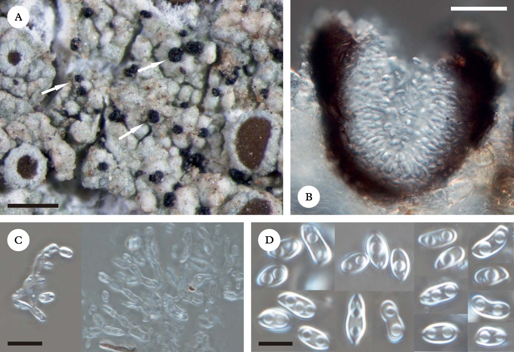
Lichenosticta jurgae (holotype). A, habit of pycnidia; B, section of a pycnidium; C, conidiophores with conidiogenous cells and young conidia; D, mature guttulate conidia. Scales: A=300 µm; B=25 µm; C=10; D=5 µm. In colour online.
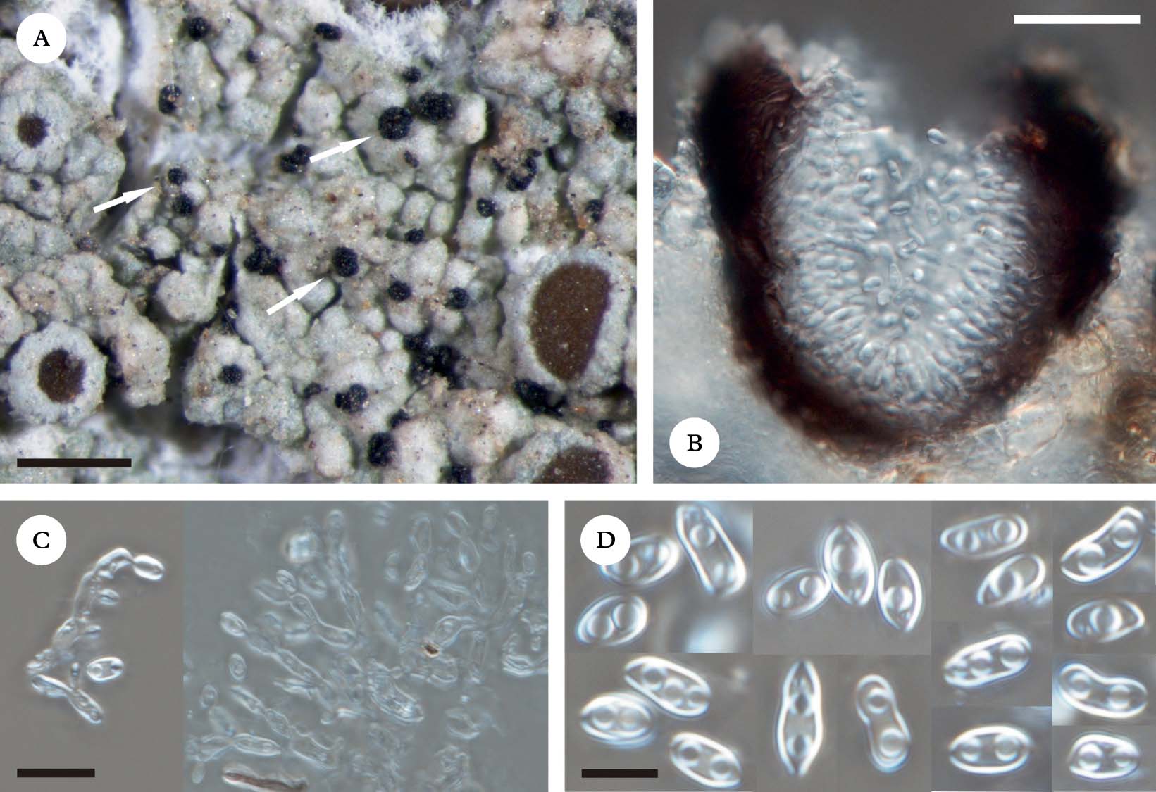
Conidiomata pycnidial, sessile or partially immersed, dispersed to aggregated, black, usually glossy, subglobose to broadly pyriform, ostiolate, 60–100(–110) μm diam. Pycnidial wall pseudoparenchymatous, irregular in outline, 5–10 μm thick, up to 20 μm thick in upper part, brown to dark brown (with slightly pinkish tinge), darker in exposed parts, composed of several layers of cells variable in diameter and shape; individual cells c. 2–5 μm wide, in inner part subhyaline to pale brown and with slightly elongated lumina, in outer part strongly pigmented, thick-walled (especially near ostiolum) and with shorter lumina. Conidiophores arising from the inner walls of pycnidia, hyaline, irregularly branched. Conidiogenous cells enteroblastic, phialidic, integrated into chains, cylindrical, hyaline, 5–7×2·0–2·5 μm. Conidia acro-pleurogenous, simple, hyaline, broadly ellipsoid to oblong or rarely slightly constricted in the middle, often slightly attenuated at the base (then lacriform or pyriform), rounded at the apex, smooth-walled, usually 1–4 guttulate, (4–)5–6(–8)×(2·0–)2·5–3·0(–3·5) μm (n=100); conidial wall thin, c. 0·3 μm wide.
Host. The species grows on the thallus of an epiphytic member of the Lecanora subfusca group. The fungus causes slight bleaching of the host thallus.
Etymology. The species is named to honour our friend, Dr Jurga Motiejūnaitė (Vilnius), an eminent Lithuanian lichenologist, who stimulated the second author to work on lichenicolous fungi, and whose help has always been appreciated.
Distribution and habitat. So far the species is known only from the type locality in the Chaqueño forest at low altitude (Bolivia).
Notes. The genus Lichenosticta Zopf previously comprised two species growing on fruticose lichens (Hawksworth Reference Hawksworth1981; Zhurbenko Reference Zhurbenko2010). The new species differs from the widespread L. alcicornaria (Linds.) D. Hawksw. and the recently described L. dombrovskae Zhurb. by having smaller conidia [(6·0–)6·5–10·0(–11·0)×(2·0–)3·0–4·5(–6·0) μm in L. alcicornaria; (4·5–)6·5–8·5(–11·0)×(3·0–)4·0–4·5(–5·5) μm in L. dombrovskae] and different host selection (Cladonia spp. in L. alcicornaria; Stereocaulon spp. in L. dombrovskae). It can also be distinguished from L. dombrovskae by a thinner conidial wall (0·5–0·8 μm in L. dombrovskae) (Hawksworth Reference Hawksworth1981; Zhurbenko Reference Zhurbenko2010).
One could assume that the pycnidia of L. jurgae actually may belong to its host; in Lecanora Ach. a wide range of hyaline conidium types are known, but conidiogenous cells are short, elongate-ampulliform, sessile or produced on short, branched conidiophores (e.g., Edwards et al. Reference Edwards, Aptroot, Hawksworth, James, Smith, Aptroot, Coppins, Fletcher, Gilbert, James and Wolseley2009), whereas in Lichenosticta they are cylindrical and integrated into chains (Hawksworth Reference Hawksworth1981; Zhurbenko Reference Zhurbenko2010). In the Lecanora subfusca group, to which the host of Lichenosticta jurgae belongs, conidia are filiform and often curved, thus very different from those produced by L. jurgae (e.g. Brodo Reference Brodo1984).
Phaeosporobolus trypethelii Flakus & Kukwa sp. nov.
MycoBank No: MB564363
Phaeosporobolus lichenicola supra thallos specierum generis Trypethelio vigens. Similis species P. alpinus, sed stromatibus maioribus et irregulariter prolixaribus, et conidiis regulariter orbicularibus cum cellulis pluralibus et valdae verruculosae differt.
Typus: Bolivia, Dept. Beni, Prov. Ballivian, near Reyes village, 14°18′10″S, 67°18′49″ W, 192 m, savanna vegetation, on thallus of Trypethelium ochroleucum, 29 November 2004, A. Flakus 3724 (KRAM—holotypus; LPB, hb. Flakus—isotypi).
(Fig. 3)
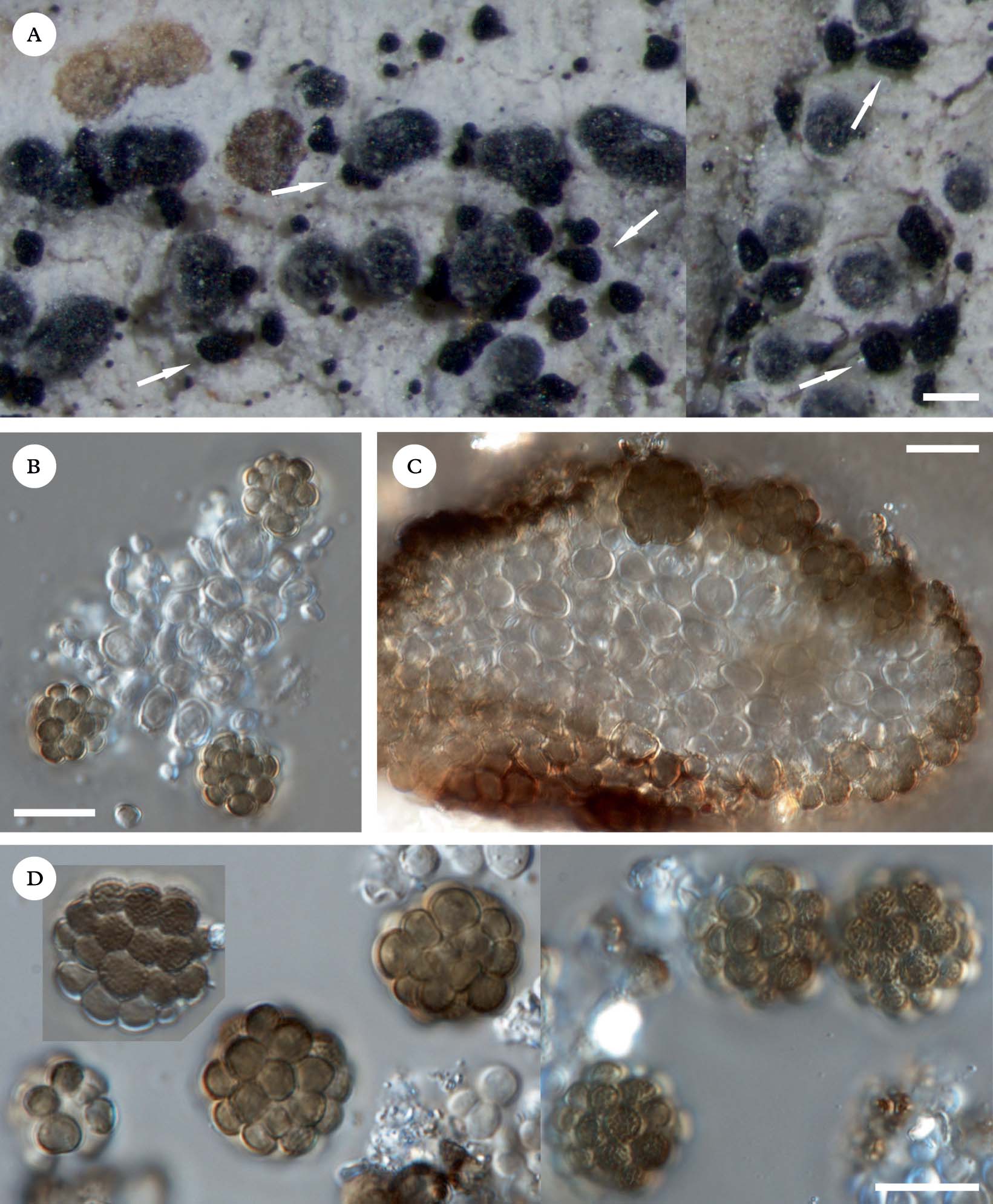
Phaeosporobolus trypethelii (holotype). A, habit of stromatic conidiomata; B, conidiogenous cells with young conidia; C, section of stroma; D, muriform conidia. Scales: A=100 µm; B–D=10 µm. In colour online.
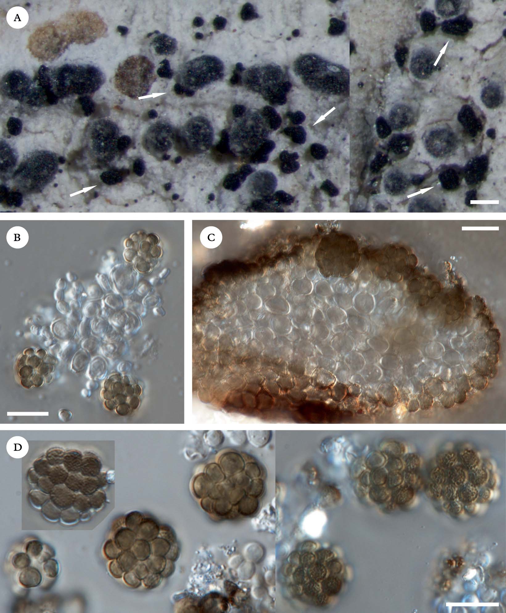
Conidiomata stromatic, superficial, dispersed to aggregated, black, irregularly convex and subglobose when young, horizontally elongated and strongly flattened (cushion-like) when older, usually constricted below, (30–)45–110 μm diam., 30–80 μm high, not covered by a pellicle-like layer of hyphae, composed of compacted subglobose to ellipsoid, brown cells; individual cells 3·5–7·0×3–5 μm, in outermost layer somewhat darker and strongly irregularly verruculose. Conidiophores micronematous, not clearly distinguished from the hyaline, subspherical cells of the central part of the stroma. Conidiogenous cells hyaline, subglobose to subcylindrical, 5·0–7·5×4·5–7·0 μm, at first producing apically a few hyaline, small, subspherical cells, which become more numerous and browner, and form conidia. Conidia muriform, regularly globose, 12–16 μm diam., composed of 12–20(–25) densely compacted subglobose, slightly thick-walled cells, outer layer composed of more trapezoidal cells with extended wall faced outward, individual cells 2·5–5·0 μm diam., strongly warted-rugose, partly or fully hyaline or pale brown when young, dark brown when mature.
Host. The new species has been found on the thallus of corticolous Trypethelium ochroleucum; no damage to the host thallus has been observed. The host species is reported in this paper for the first time from Bolivia.
Distribution and habitat. So far the species has been found only once in Bolivia in savanna vegetation.
Notes. The new species is most similar to P. alpinus R. Sant et al., which can be distinguished by numerous characters, such as smaller regular stroma [(20–)30–75 μm diam.], irregularly subglobose conidia, with a smaller number of cells [10–15(–20)], and different hosts, usually Ochrolechia and Pertusaria species (see Alstrup & Hawksworth Reference Alstrup and Hawksworth1990), although it has been reported in a large variety of lichen genera (e.g. Kocourková Reference Kocourková2000). In some specimens of P. alpinus conidial cells have been found to be ornamented (von Brackel Reference von Brackel2011), being echinulate and not verruculose as in P. trypethelii. According to Alstrup & Hawksworth (Reference Alstrup and Hawksworth1990), the species should produce conidia with±smooth cells; specimens with ornamented conidial cells need more study. The type of conidial ornamentation requires more studies using SEM to determine their taxonomic value.
Phaeosporobolus usneae D. Hawksw. & Hafellner, known from epiphytic macrolichens, differs by a stroma surrounded by a brown pellicle-like layer and bigger ellipsoid conidia (19–27×13–17 μm) (Hawksworth & Hafellner Reference Hawksworth and Hafellner1986). Two other recently described species, P. minutus Etayo from Chile and P. fellhanerae R. C. Harris & Lendemer from the USA, are easy to distinguish from P. trypethelii. Phaeosporobolus minutus has small conidiomata (up to 60 μm diam.) and very small conidia (6·5–10·5×5–8), composed of (3–)4–5(–8) cells and P. fellhanerae has ellipsoid, smaller (11–12×7·5–8·5 μm) conidia with smooth cell walls. Additionally, they grow on different hosts: P. minutus on corticolous Coccotrema and Pertusaria species, and P. fellhanerae on saxicolous Fellhanera granulosa (Etayo & Sancho Reference Etayo and Sancho2008; Harris & Lendemer Reference Harris and Lendemer2009).
Von Brackel (Reference von Brackel2011) recognized an unnamed Phaeosporobolus species on Fuscidea stiriaca, but his material has echinulate conidia, whereas the conidial cells of P. trypethelii are strongly warted-rugose.
Spirographa usneae Flakus, Kukwa & Etayo sp. nov.
MycoBank No: MB564364
Spirographa fusisporella similis sed ascosporis latioribus et hospite differt.
Typus: Bolivia, Dept. Cochabamba, Prov. Chapare, near Incachaca village, 17°14′13″S, 65°49′02″W, 2294 m, Yungas montane cloud forest, on thallus of Usnea sp., 10 June 2006, A. Flakus 8176.2 (LPB— holotypus).
(Fig. 4)
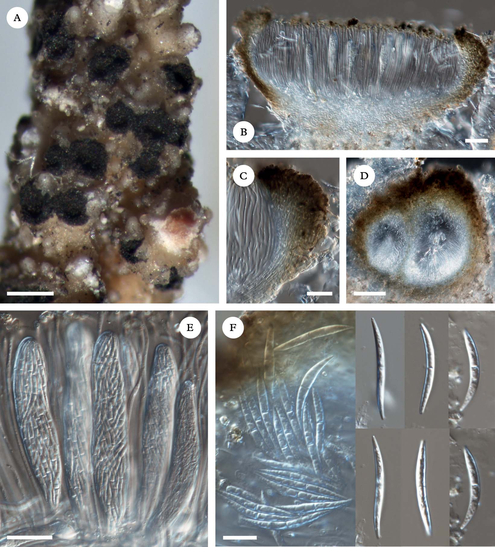
Spirographa usneae (holotype). A, habit of apothecial ascomata; B, section of a mature ascoma; C, section of exciple; D, section of young, closed ascoma; E, multispored asci with spirally arranged ascospores; F, mature ascospores. Scales: A =200 µm; B=50 µm; C=25 µm; D=10 µm; E=25 µm; F=10 µm. In colour online.
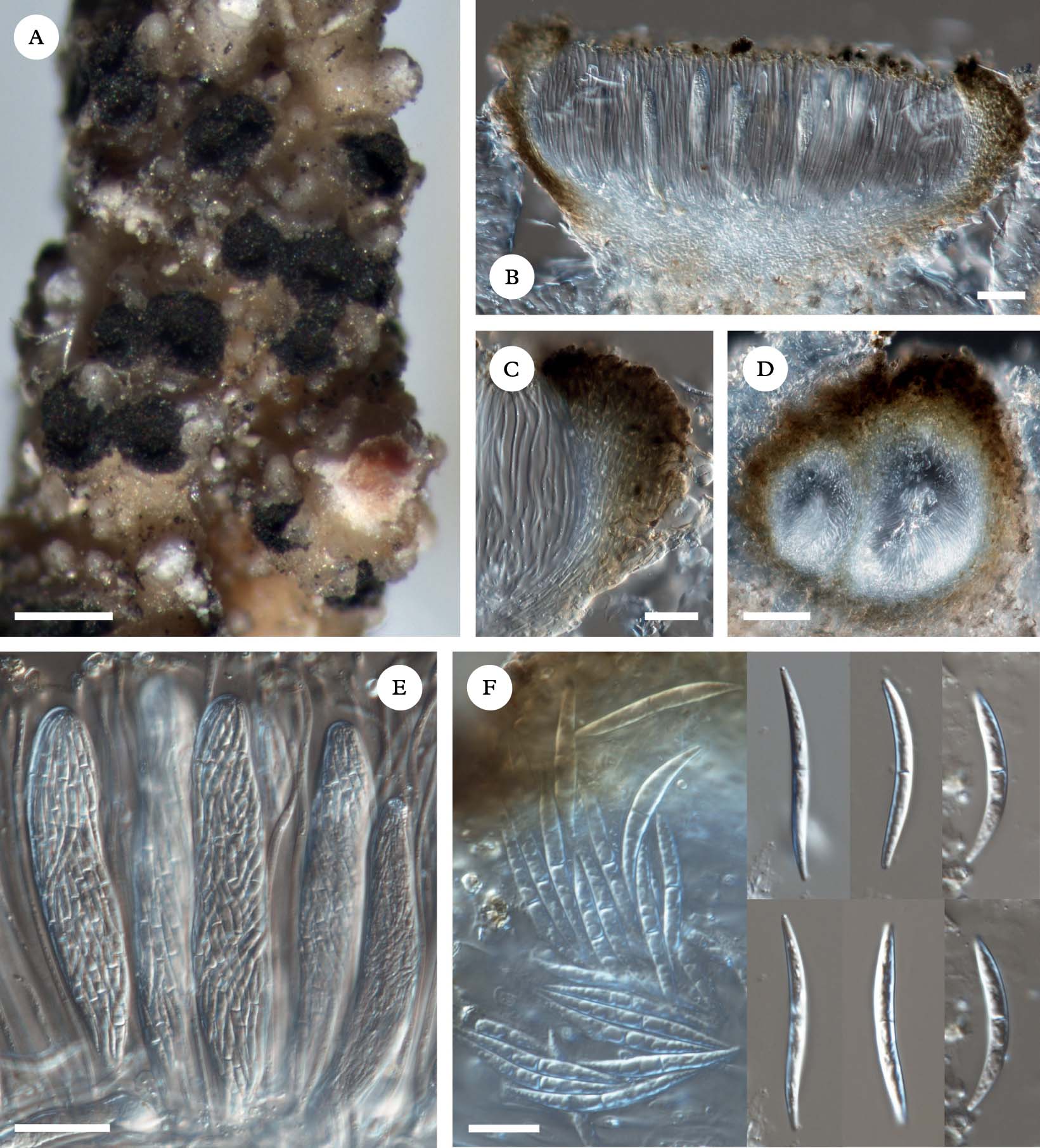
Ascomata 100–250 µm diam., cleistohymenial, often aggregated on the host thallus, initially immersed, then breaking through the thallus cortex of the host and almost sessile when mature. Disc exposed, concave, concolorous with the margin, black, matt, with a rough surface. Margin black, prominent, thick, with a rough to fissured surface, without hairs. Exciple paraplectenchymatous (composed of thin hyphae, c. 1·5–2·0 µm wide), laterally 35–50 µm wide, basally c. 5–15 µm wide, with a honey-brown to olive-brown pigment, K−, N−, darker in outer layer and hyaline in inner layer and at the base. Hymenium hyaline, 90–110 µm tall, K/I−, I−. Subhymenium hyaline. Epihymenium with honey-brown pigmented granules, K− (partly soluble in K), N−. Paraphyses hyaline, simple, septate, sometimes slightly branched in the upper part, 1·5–2·0 µm thick, apically slightly thicker (up to 3 µm) and covered by a granular pigment. Asci clavate to cylindrical, functionally unitunicate, wall apically not thickened, K/I−, I− (only endoascus slightly orange), c. 32-spored, 65–100×10–20 µm. Ascospores hyaline, spirally arranged in ascus, 1(–3)-septate with pointed ends, not constricted at the septum, fusiform, straight to curved or slightly sigmoid, multiguttulate, smooth, without perispore, 15–32×2–3 µm.
Host. The new species has been collected on thalli of epiphytic Usnea species, the parasitized host thalli having an orange tinge.
Distribution and habitat. The new species has been found in Bolivia and Ecuador, in both cases in the Yungas forests.
Notes. The new species is characterized by predominantly curved, rarely straight or sigmoid ascospores, which are 2–3 µm wide, a tall hymenium, relatively long asci and the host selection (Usnea spp.). Initially we considered placing the taxon in the genus Pleoscutula Vouaux, because of the shape of the ascospores. However, a closer examination of the exciple structure and the presence of at least some sigmoid ascospores led us to describe it in Spirographa Zahlbr. According to Holien & Triebel (Reference Holien and Triebel1996), both genera are very similar, and more detailed studies might lead to the conclusion that they are synonymous.
So far only one other species has been recognized in the genus Spirographa, S. fusisporella (Nyl.) Zahlbr. It can easily be distinguished from S. usneae by its distinctly helicoid and narrower (22–31×1·0–2·5 µm) ascospores, a lower hymenium (50–65 µm) and smaller asci (47–55×5·5–12·0 µm) (Holien & Triebel Reference Holien and Triebel1996; Kocourková Reference Kocourková2000; Diederich Reference Diederich, Nash, Ryan, Diederich, Gries and Bungartz2004). Spirographa fusisporella has been reported from unrelated lichen genera (mostly crustose and few foliose), and since all other parasitic members of the order Ostropales, where Spirographa is placed at present, are host specific, it is possible that it represents a group of several still unrecognized species (see Diederich Reference Diederich, Nash, Ryan, Diederich, Gries and Bungartz2004).
Due to the predominantly curved ascospores, the new species can also be confused with Pleoscutula arsenii Vouaux and P. hypotrachynae Etayo & Diederich; however, the former species has lower hymenium (up to 60 µm), shorter and wider ascospores (9–13×2·5–4·0 µm) and grows on Heterodermia species, whereas the latter has a slightly lower hymenium (80–90 µm), smaller ascospores (13–19×2·0–2·5 µm) and parasitizes Hypotrachyna species (Hafellner Reference Hafellner1982; Etayo Reference Etayo2002).
Additional specimen examined. Ecuador: Prov. Tungurahua: camino Baños a Puyo, confluencia de ríos Verde y Pastaza, Pailón del Diablo, en Usnea de color rojizo, 1600 m, J. Etayo 19937 & Z. Palice (hb. Etayo).
We are greatly indebted to J. Etayo (Pamplona), P. Diederich (Luxembourg) and two anonymous reviewers for valuable comments on the manuscript, and to R. I. Meneses Q., the Director of Herbario Nacional de Bolivia, Instituto de Ecología, Universidad Mayor de San Andrés, La Paz, for generous cooperation. We also thank P. Rodriguez (Frankfurt am Main), J. Quisbert (LPB) and F. Saavedra (La Paz) for help during fieldwork. This research received support from the National Centre for Research and Development (NCBiR) in Poland under the LIDER Programme for the years 2010–2013 (no. 92/L–1/09).