Introduction
Spinal stereotactic ablative body radiotherapy (SABR) is increasingly used to treat primary and secondary spinal tumours. Reference Sahgal, Bilsky and Chang1–Reference Osborn, Lee and Yamada3 In the UK, since 2015, SABR for spinal oligometastatic disease has become more widely available. 4 The high, ablative dose of radiation delivered with SABR is delivered precisely to the tumour while minimising dose to the spinal cord, maintaining delivery precision within 1·5 mm and 1o within a 95% confidence interval. Reference Foote, Letourneau and Hyde5,Reference Hyde, Lochray and Korol6 Conventional radiotherapy is unable to achieve this level of precision and thus is limited to delivery of palliative doses due to the potential risk of myelopathy. The Canadian Association of Radiation Oncology defines SABR as ‘the precise delivery of highly conformal and image-guided hypofractionated external beam radiotherapy, delivered in a single or few fraction(s), to an extracranial body target with doses at least biologically equivalent to a radical course when given over a protracted conventionally fractionated schedule’. Reference Sahgal, Roberge and Schellenberg7 To date, the rationale behind delivering ablative doses to oligometastases includes to achieve a prolonged progression-free survival, to delay the time to systemic therapy and to improve both symptomatic control and quality of life. This is being evaluated by various trials including the UK CORE trial 8 which is currently in follow-up and the Canadian COMET trial (which has shown a significant improvement in median overall survival, 28 versus 41 months). Reference Palma, Olson and Harrow9
Delivering an ablative dose to spinal oligometastases is associated with an increased risk of adverse effects compared to palliative doses. The most common acute side effect reported is a risk of temporary pain flare, although a course of dexamethasone at the time of treatment has been shown to significantly reduce this. Reference Chiang, Zeng and Zhang10 The most commonly reported late side effect is the risk of vertebral compression fracture (VCF). Reference Sahgal, Whyne and Ma11
Bone is a complex structure composed primarily of two components, minerals and collagen. Reference Sahgal, Whyne and Ma11 While minerals give strength and stiffness to bone, the collagen is responsible for its toughness. Radiotherapy primarily affects the collagen, potentially reducing the toughness of bone. Reference Currey, Brear and Zioupos12,Reference Guo and Kim13 High doses of radiation, such as those delivered through SABR, can also result in osteoradionecrosis of bone and tumour tissue. Reference Sahgal, Whyne and Ma11,Reference Currey, Foreman, Laketic, Mitchell, Pegg and Reilly14 Within the literature, several factors predicting an increased risk of VCF have been described. These include lytic bone lesions, extent of vertebral body involvement, age more than 55 years, presence of a pre-treatment fracture or spinal deformity, histological type and dose per fraction of more than 20 Gy. Reference Sahgal, Whyne and Ma11,Reference Rose, Laufer and Boland15–Reference Boehling, Grosshans and Allen17 With increasing knowledge of these factors, it may be possible to predict which patients are at a high risk of VCF to allow appropriate intervention, most commonly surgical, prior to SABR.
Survival rates, among a wide range of tumour types, are ever improving with the development of novel therapies, which are increasingly being approved for regular use. As a result of this, late side effects from treatments, such as SABR, are becoming more common, and it is increasingly important to recognise and prevent these. Internationally outcomes from a number of retrospective and some prospective spine SABR cohorts have been published, with data including the observed VCF rates. The rate of VCF in international publications broadly ranges from 11 to 39%. Reference Rose, Laufer and Boland15,Reference Boehling, Grosshans and Allen17,Reference Sahgal, Atenafu and Chao18 The Spinal Instability Neoplastic Score (SINS) Reference Fisher, DiPaola and Ryken19 is calculated based on degree of pain, location, type of bone metastases, collapse of vertebral body and spinal alignment. SINS Reference Fisher, DiPaola and Ryken19 is used widely to screen patients who are at high risk of developing fractures, and a spinal surgical opinion is recommended for all cases with a score of 7 or above prior to SABR. Reference Fisher, DiPaola and Ryken19 The aim of this study was to determine VCF rates at one UK institution.
Materials and Methods
All patients undergoing SABR for spinal oligometastases between July 2014 and July 2019 at one UK institution were included in a retrospective analysis. Each patient was discussed and their appropriateness for SABR agreed at both the local tumour site-specific multidisciplinary team (MDT) meeting at a hospital in the surrounding region and the institution’s SABR MDT meeting. All patients had a known histologically confirmed primary malignancy with up to two spinal oligometastases. The spinal oligometastases were diagnosed with conventional imaging techniques including computerised tomography (CT), nuclear medicine bone scanning or positron emission tomography scans and then subsequently confirmed on thin 1-mm slice magnetic resonance imaging scans with specialist radiologist review. The SINS was collated for all patients. Patients with SINS of 7 or more were referred for spinal surgical opinion. Patients with SINS of 6 or less were accepted for SABR.
All patients were treated with SABR delivered by a CyberKnifeTM (Accuray, Inc Sunnyvale, California) using a treatment technique described in the UK SABR Consortium guidelines. 20 These guidelines recommend using dose of 24 Gy in one fraction or 24–27 Gy in three fractions or 0–35 Gy in five fractions. Inclusion of vertebral body, pedicles, lamina and transverse processes in the clinical target volume (CTV) depended on the extent of vertebral involvement. The cohort included both previously unirradiated patients and those receiving re-irradiation. SABR was delivered in three alternate day fractions in previously unirradiated patients and five alternate day fractions in those who had previously received a conventional palliative dose.
Treatment planning was calculated using the CyberKnife (Accuray, Inc Sunnyvale, California) Precision treatment planning system. Targets and organs at risk (OARs) volumes were contoured on 1-mm axial CT dataset, with a fused 1-mm axial MRI scan used to aid delineation. Gross tumour volume (GTV) as demonstrated on imaging was expanded to a CTV based on the SABR guidelines. 20 A planned target volume (PTV) was defined as the CTV + 2 mm (but excluding thecal sac). A 2-mm margin was added to the spinal cord to create a cord PRV (planning risk volume). The plans were optimised to ensure 95% of the PTV, and 98% of the GTV received the prescribed dose with the dose prescription reduced in order to achieve OAR tolerances as specified in the SABR Consortium guidance. 20 The maximum point dose in the plan was constrained to 143% of the prescribed dose, meaning the periphery of the PTV was encompassed by the 70–80% isodose line.
The CyberKnife VSI (Accuray, Inc Sunnyvale, California) treatment delivery system was used for treatments. This utilises digital reconstructed radiographs taken from the planning CT and on-treatment planar X-rays to perform intrafraction motion correction with a spine tracking algorithm. The geometric uncertainty of the treatment delivery has been verified to be less than 1 mm based on routine end-to-end quality assurance measurements.
The primary end point of this study was the VCF rate, while secondary end points included baseline SINS prior to SABR and time to VCF. Site of metastases, dose fractionation schedules, site of primary disease and how many VCFs were managed were also reported.
Patients were followed up after SABR according to the local follow-up schedule, comprising clinical review 6 weeks post-SABR and baseline imaging (ideally, MRI spine) at 3 months post-SABR and then 3 monthly thereafter until 12 months post-SABR. Imaging beyond 12 months follow- up was guided by the treating oncological site specialist team.
Results
Forty-eight metastatic lesions between C1 and L5 were treated in 41 patients, with a median follow-up of 20·5 (range 1–58) months. Median age at the time of treatment was 71 (range 46–88) years. Basic demographics are detailed in Table 1.
Basic demographics

A maximum of two vertebral bodies were treated synchronously. SINS was calculated as detailed by Fisher et al. Reference Currey, Foreman, Laketic, Mitchell, Pegg and Reilly14 for all but three patients, in whom data were missing. The median baseline SINS was 3 (range 1–7). Of the two patients with SINS of 7, one was referred for surgical review and was advised to undergo surgery, but he declined and opted for SABR. Another patient with SINS of 7 was treated with SABR with no documented discussion of surgical review prior to treatment.
Fisher et al. Reference Fisher, DiPaola and Ryken19 have divided the spine into sections as an aid to predict stability. The most common site of oligometastatic disease was the semi-rigid spine. No sacral oligometastases were included. Both patients with SINS of 7 were at junctional sites. SABR to the spine was delivered using a CyberKnifeTM. Different dose regimens as recommended by the UK SABR Consortium 20 were delivered. Of the 41 patients treated, only 3 had received previous spinal radiotherapy. The median dose in this cohort was 26 Gy in three fractions.
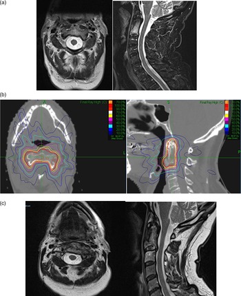
Two VCFs were observed in this cohort during the period of follow-up. One, at C2, was identified at a follow-up of 3 months (pre-SABR SINS:4), as an incidental finding on the baseline MRI scan post-SABR (Figure 1). This was defined as a Grade 1 spinal fracture according to the Common Terminology Criteria for Adverse Event Criteria (CTCAE) Version 5.0. 21 The patient had been suffering with mild pains prior to SABR which had persisted but were no worse following treatment. His case was discussed with and reviewed by the local spinal surgical team who advised no surgical intervention for the un-displaced fracture. His pain slowly improved with appropriate analgesia. Subsequent imaging confirmed that the fracture was healed, and that the metastasis had responded to SABR.
Images of an example patient who received 27 Gy in three fractions at C2 secondary to prostate cancer. Pre-SABR SINS = 4. (a) Pre-operative T2-weighted MRI. (b) Dose distribution image (orange 70%, pink 50%, light blue 30% and dark blue 10% of max dose 38·57 Gy). (c) Post-treatment MRI revealing VCF.

The second patient suffering a VCF developed a fracture of L3 at a follow-up of 14 months (pre-SABR SINS:3). This was a Grade 2 CTCAE event. He had been suffering with back pains for a few months prior to undergoing a CT scan which revealed the fracture. He was treated with analgesia and also had a surgical review with a recommendation for conservative management. Subsequent imaging revealed stable disease at L3, and the pain resolved.
No patients in this cohort had a fracture prior to SABR. The remaining 39 patients remained fracture-free at the time of last follow-up or death. At the time of death or last follow-up, where imaging available, 42 out of 48 sites (88%) had local control.
Discussion
Following conventional palliative radiotherapy to spine, the risk of fracture has been found to be low at 3·2% (with 8 Gy in one fraction) and 2·8% (with 20 Gy in five fractions). Reference Chow, Harris and Fan22 SABR for selected patients is now favoured over conventional radiotherapy, offering the potential for reduced symptoms, improved local control rates and survival. Reference Palma, Olson and Harrow9 Patients at high risk of spinal instability as defined by SINS should be considered for surgical stabilisation first regardless of the expected dose of radiotherapy, although this is of particular importance in the context of ablative radiation doses. With the increasing use of spinal SABR, VCF has become a major concern given the increased risk of VCF compared to conventional doses of radiation.
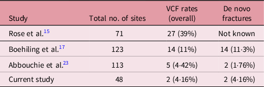
The data presented here reveal a low rate of VCF, in line with previous publications as shown in Table 2. The median time to VCF reported by Rose et al., Reference Rose, Laufer and Boland15 Boehling et al. Reference Boehling, Grosshans and Allen17 and Abbouchie et al. Reference Abbouchie, Chow and Tacy23 was 25 months, 3 months and 9·2 months, respectively, compared to these data, where the fractures happened at 3 months and 14 months. In the series published by Boehling et al., Reference Boehling, Grosshans and Allen17 median time to fracture progression was 3 months while the median time from SABR to new fracture was 14 months as measured by Kaplan–Meier curves. In the same series, only 14 (11%) patients were found to have new fractures. Similarly, in a series published by Rose et al., Reference Rose, Laufer and Boland15 fracture progression was seen in 27 (39%) patients; however, it is unclear whether these fractures were present prior to SABR and progressed or whether they occurred after SABR. The cohort presented here did not include any patients who had pre-existing fractures. Therefore, a direct comparison with time to fracture progression with both Rose et al. Reference Rose, Laufer and Boland15 and Boehling et al. Reference Sahgal, Bilsky and Chang1 is not possible. Abbouchie et al. Reference Abbouchie, Chow and Tacy23 recently published data and reported two de novo fractures and progression of three already existing fractures.
Comparison of VCF rates in studies reported in literature

The radiation doses delivered in this series ranged from 18 Gy to 30 Gy and all were delivered in multiple fractions. In Rose et al., Reference Rose, Laufer and Boland15 only single fractions were delivered with a median dose of 24 Gy. Boehling et al. Reference Boehling, Grosshans and Allen17 reported doses of 18, 27 and 30 Gy, delivered in 1, 3 and 5 fractions. Abbouchie et al., Reference Abbouchie, Chow and Tacy23 despite having a higher biologically effective dose (BED) than Boehling et al., Reference Boehling, Grosshans and Allen17 showed a low VCF rate. A limitation of the data presented here was the heterogeneity of the dose delivered. Patients treated during the early part of the cohort presented here were treated prior to publication of the UK SABR Consortium guidelines 20 , and so some patients were treated in two fractions. Following commencement of the NHS commissioning through evaluation SABR programme, 4 the three-fraction schedule as per the Consortium guidelines became the standard of care. The cohort presented here received a relatively lower BED compared to the majority of those published in the literature which may account for a lower number of subsequent VCF (see Table 3). Similarly, in the data presented by Rose et al., Reference Rose, Laufer and Boland15 all doses were delivered in a single fraction with a higher BED possibly explaining the higher rates of VCF. Table 3 shows BEDs calculated using the median dose fractionation reported by these respective papers and their VCF rates. The α/β ratio used for the purpose of calculation is 2 Gy.
BED based on median doses offered in various studies and VCF

Fisher et al. Reference Fisher, DiPaola and Ryken19 have recommended that SINS score of 7 or more is potentially unstable and therefore it is accepted that this would warrant a surgical review prior to SABR. SINS data were missing in two patients in this cohort and of the two patients with SINS of 7 or more, only one was referred for a surgical review prior to SABR. Neither of these patients has, to date, developed a VCF following SABR. The data presented here included three patients who had received prior conventional radiotherapy to their spinal oligometastasis. None of these developed a VCF. While Rose et al. Reference Rose, Laufer and Boland15 did not include such patients, Boehling et al. Reference Boehling, Grosshans and Allen17 did include patients who had received conventional radiotherapy to same sites prior to SABR; however, despite the inclusion of these re-irradiation patients, the VCF rate is lower in Boehling et al. Reference Boehling, Grosshans and Allen17 as compared to Rose et al. Reference Rose, Laufer and Boland15 Again this may be as a result of the lower BED delivered.
Contrary to Boehling et al. Reference Boehling, Grosshans and Allen17 and Rose et al., Reference Rose, Laufer and Boland15 where the most common primary cancer was renal cell carcinoma, prostate cancer was the most common primary in this cohort. Only 1% patients had prostate cancer in Boehling et al. Reference Boehling, Grosshans and Allen17 , while Rose et al. Reference Rose, Laufer and Boland15 had 13% prostate cancer patients (See Table 4). The most common histological primary tumour type in Abbouchie et al. Reference Abbouchie, Chow and Tacy23 was prostate cancer constituting 49·6% of all patients which closely resembles this study. At present, there is no enough data available in the published literature to assess how different tumour types respond to SABR based on their radio sensitivity. Further trials are needed to consider this. Both patients who developed VCF had prostate cancer and were treated with 26 and 27 Gy in three alternate day fractions. More than 50% of vertebral body was involved in the L3 vertebra without any collapse, while the C2 lesion had less than 50% of its body involved.
Comparison of primary tumour histological subtypes

The distribution of the location of the oligometastatic lesions was similar to those in the literature with the thoracic spine being the most commonly treated in Rose et al. Reference Rose, Laufer and Boland15 (54%), Boehling et al. Reference Boehling, Grosshans and Allen17 (66%), and Abbouchie H. et al. Reference Abbouchie, Chow and Tacy23 (56·6%) and 54% in this study.
Saghal et al. Reference Sahgal, Atenafu and Chao18 defined predictors for VCF following SABR. These include dose per fraction of more than 19 Gy, lytic lesions, baseline spinal misalignment and pre-existing compression fracture. None of our patients had a baseline spinal fracture or a misaligned spine or were given a dose of more than 19 Gy in one fraction. Four patients (8%) had lytic lesions at baseline but none of those developed VCF afterwards.
This study is limited by its retrospective nature. Only two patients had VCF, which limits the ability to make any firm conclusions with regard to causative factors, but it could be hypothesised that this low rate is a result of the lower BED delivered compared to many of the other published series (see Table 3). Furthermore, no formal evaluation of the use of bisphosphonates or systemic therapy was undertaken which might impact on the rate of VCF.
Conclusion
SABR for spine oligometastases, within the UK, is being performed safely with low VCF rates compared to international data. This may be as a result of strict adherence to criteria for delivery of SABR in spinal oligometastases with low pre-treatment SINS and inclusion of patients without pre-existing fractures of metastatic sites.
Acknowledgements
None.