Malignant mesothelioma (MM) is a rare, highly aggressive cancer arising from the mesothelial cells that form the lining of the pleural, and less frequently the peritoneal cavities. Mesothelioma is an emergent neoplasm, because it was rarely diagnosed before the middle of the 20th century. The incidence has risen steadily since 1970, and there are currently an estimated 3000 new cases per year in the USA. The peak incidence of mesothelioma might have occurred in the USA during the last decade, and the peak incidence in much of the developed world is expected to occur in the next 10–20 years. These data are thought to reflect the widespread occupational asbestos exposure in the Western world from the 1940s to the 1970s, as well as the inherent latency period of approximately 30 years between asbestos exposure and disease manifestation which is typical of MM. Approximately 80% of mesothelioma cases can be directly attributed to asbestos fibre exposure. Additional suspected causes or co-carcinogens include other mineral fibres such as erionite, simian virus 40 (SV40) and radiation (Ref. Reference Robinson and Lake1). Moreover, a mesothelioma epidemic in Turkey has demonstrated a likely genetic predisposition to mineral fibre carcinogenesis (Ref. Reference Carbone2).
Mesothelioma arises from multipotential mesothelial cells that are capable of differentiating into epithelial, sarcomatoid or biphasic (mixed) neoplasms. There is lack of consensus on a staging system for MM; however, most patients present with advanced stage of disease. Advanced age, poor performance status, male sex and sarcomatoid histological subtype are all poor prognostic factors. Despite modest advances in clinical treatment, the mean overall survival for patients with MM is approximately 12 months (Ref. Reference Ducko, Sugarbaker, Third, Alexander Patterson, Deslauriers, Lerut, Luketich and Rice3). There is also lack of consensus regarding treatment of MM. Suitable surgical candidates with disease limited to one hemithorax can undergo surgical resection by extrapleural pneumonectomy (EPP) or pleurectomy/decortication as part of a multimodality treatment approach. Radiation therapy alone is generally ineffective because of the large volume of primary tumour and its proximity to vital mediastinal structures. However, radiation therapy, particularly intensity-modulated radiation therapy (IMRT), has been demonstrated to reduce local recurrence following resection by EPP (Ref. Reference Rice4). Historically, chemotherapy response rates have been less than 20%. However, improved response rates of 41% have been demonstrated with the addition of the folate antimetabolite pemetrexed (Ref. Reference Vogelzang5). Highly selected patients appear to benefit from trimodality therapy consisting of aggressive surgical debulking followed by adjuvant radiation and chemotherapy (Ref. Reference Sugarbaker6). Failure of conventional therapies has led to interest in novel treatment approaches including intrapleural administration of immunotherapy and gene therapy, as well as intraoperative adjuncts such as intrapleural chemotherapy and photodynamic therapy (Refs Reference Vachani, Moon and Albelda7, Reference Friedberg8).
Studies on human cell lines and tissues as well as animal models of MM have demonstrated genetic and epigenetic events that contribute to the multistep process of mineral fibre carcinogenesis. These events include inactivation of tumour suppressor genes, modulation of signal transduction pathways including receptor tyrosine kinases (RTKs), avoidance of apoptosis and inhibition of the ubiquitin–proteasome degradation pathway. This review will focus on the molecular pathogenesis of MM. Preclinical and clinical trials of targeted therapies such as tyrosine kinase, histone deacetylase (HDAC) and proteasome inhibitors will also be discussed.
Aetiology of mesothelioma
Asbestos
The vast majority of cases of mesothelioma can in some manner be linked to asbestos exposure. Materials consisting of asbestos fibres have been present since ancient times. In fact, the word asbestos is derived from a Greek term meaning inextinguishable or unquenchable, a reference to its fire-resistant properties. It was these heat-resistant and insulating properties that made asbestos a valuable commodity, particularly as the industrial revolution began. In the USA, mining and subsequent use of asbestos increased steadily during the first half of the 20th century, escalated rapidly following World War II, and peaked in 1973, after which it precipitously declined (Fig. 1). Asbestos refers to a group of crystalline-hydrated silicate minerals that occur in one of two forms: serpentine or amphibole. Chrysotile is the only serpentine asbestos, and exists as a long, curly and pliable fibre most suitable for making fabrics. Amphibole fibres are short, straight and stiff, and have been used to make pipes and tiles. The major commercial amphiboles are amosite, crocidolite and anthophyllite. Mixtures of chrysotile and amphiboles were used to produce an array of roofing, insulation and fire-proofing materials. Evidence exists that all asbestos fibre types can demonstrate pulmonary toxicity in a dose-dependent manner. Moreover, all fibre types possess carcinogenic potential; however, exposure to amphibole fibres is more likely to cause mesothelioma than is exposure to chrysotile fibres (Ref. Reference Cugell and Kamp9).
Asbestos production and mesothelioma incidence. Asbestos production in the USA in the last century and mesothelioma incidence from 1980 projected to 2055. Redrawn from Ref. Reference Cugell and Kamp9 with permission.

The pulmonary hazards of asbestos exposure, including asbestosis and bronchogenic carcinoma, were recognised and published by physicians in the early 20th century. However, the link between asbestos exposure and mesothelioma was not established until 1960, when Wagner reported 33 cases of pleural mesothelioma occurring in a relatively short time period in an area of South Africa where crocidolite was mined (Ref. Reference Wagner, Sleggs and Marchand10). In 1964, Selikoff and colleagues reported on the link between asbestos exposure and thoracic neoplasia (bronchogenic carcinoma and mesothelioma) in insulation workers in the New York area in a variety of industries, including shipbuilding (Ref. Reference Selikoff, Churg and Hammond11). Subsequently, this group reported mesothelioma as the cause of 10 of 307 consecutive deaths among these same workers, concluding that mesothelioma was indeed a complication of relatively light and intermittent (occupational) exposure to asbestos, including chrysotile, which was the dominant fibre in American industry at the time (Ref. Reference Selikoff, Churg and Hammond12).
Although smoking cigarettes has been proved to increase the likelihood of developing bronchogenic carcinoma, particularly in individuals exposed to asbestos, mesothelioma is not associated with smoking. It is also interesting that although most cases of mesothelioma are associated with asbestos exposure, only a small minority (approximately 5%) of exposed individuals develop mesothelioma (Ref. Reference Gazdar and Carbone13). Asbestos exposure induces benign manifestations such as pleural effusion or plaques in some individuals, whereas causing MM in others. Obviously, other aetiologic factors, including genetics, have a role in mesothelioma pathogenesis.
Simian virus 40
SV40 is a polyoma virus of monkey origin that has been identified in a number of human tumours. SV40 contributes to the transformation of human cells by perturbing several intracellular pathways, including disabling the p53 and retinoblastoma (Rb) tumour suppressor pathways. In the 1960s, SV40 was found to be a contaminant in poliovirus vaccines that were prepared in primary cultures of rhesus monkey kidney cells. Contaminated vaccines were administered to children and adults in many countries including the USA. In fact, in the USA before 1963, approximately 90% of children and 60% of adults received at least one contaminated vaccination. The prevalence of SV40 infections in humans is not known. However, indirect evidence of widespread distribution of SV40 throughout the human population exists in that SV40-positive tumours have been detected throughout the world, except in countries that reportedly did not use SV40-contaminated vaccine (Ref. Reference Gazdar, Butel and Carbone14).
The human tumours most frequently found to have SV40 sequences are brain and bone tumours, lymphoma and MM. SV40 is also a potent oncogenic virus in rodents, and a similar spectrum of tumours is induced in hamsters following viral inoculation. In fact, the incidence of mesothelioma is 100% in hamsters following intrapleural inoculation. Human mesothelial cells contain high endogenous levels of p53 and are unusually susceptible to SV40-mediated transformation, with asbestos acting as a co-carcinogen. Despite powerful evidence regarding the biological effects of SV40 in mesothelial cells, considerable scepticism exists within the scientific community regarding a causal relationship between the presence of SV40 viral sequences and development of mesothelioma (Ref. Reference Gazdar and Carbone13).
Genetic predisposition: the Cappadocia epidemic
In 1978, an unprecedented epidemic of mesothelioma was discovered in three villages located in Cappadocia, Turkey. Mesothelioma accounts for >50% of all deaths in these villages. Mineralogical studies of the volcanic rock in these villages demonstrated the presence of a fibrous mineral called erionite which shares some physical properties with crocodolite. Curiously, large erionite deposits are present in other parts of the world, including the western USA, but had never been associated with development of mesothelioma in these regions. The mesothelioma epidemic in Cappadocia was initially linked solely to exposure to erionite contained in the stones used to build houses in the region. However, construction and examination of careful pedigrees demonstrated that mesothelioma occurred in certain families, but not in others. Studies have confirmed that the cause of the mesothelioma epidemic in Cappadocia is genetic predisposition to erionite-induced carcinogenesis, which is transmitted in an autosomal-dominant manner (Ref. Reference Carbone2).
Molecular pathogenesis of mesothelioma
The mechanisms whereby inhaled asbestos fibres induce pleural disease, including mesothelioma, are diverse and are likely to be multifactorial. The traditional explanation includes migration of fibres from the airway, through the visceral pleura and eventual uptake from the parietal pleura. Alternative routes of fibre translocation to the parietal pleura include lymphatic and haematogenous dissemination (Ref. Reference Cugell and Kamp9). There are several features of asbestos fibres that contribute to their carcinogenicity, including chemical composition, fibre length and form, and their biopersistence. Local responses to these characteristics include frustrated phagocytosis of fibres, generation of reactive oxygen and nitrogen species, which may be genotoxic, initiation of inflammatory mechanisms, stimulation of growth factors and their receptors, and initiation of signal transduction pathways, which stimulate proliferation and avoidance of apoptosis (Ref. Reference Godleski15).
Chromosomal alterations
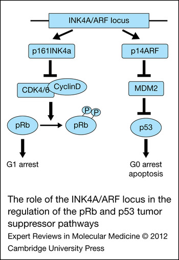
Allele loss, with subsequent loss of heterozygosity (LOH) at tumour suppressor loci, is a common occurrence in oncogenesis. Mutations and deletions of the p53 and pRb tumour suppressor pathways are prominent features in many human malignancies; however, p53 and pRb remain genetically intact in most mesotheliomas (Ref. Reference Lee16). Gene copy number alterations are present in mesothelioma. Common chromosomal regions of allele loss include 1p, 3p21, 6q, 9p21, 15q11-15 and 22q (Ref. Reference Zucali17). Homozygous deletion of the 9p21 region is frequently present in mesothelioma cell lines and tumour specimens. Loss of 9p21 results in loss of the INK4a/ARF locus, which encodes two distinct proteins, p16INK4a and p14ARF, translated from alternatively spliced mRNA. p16INK4a inhibits the cyclin-dependent kinase- mediated inactivation of pRb. p14ARF stabilises p53 through its actions on Mdm2. As the INK4a/ARF locus plays an important role in the activity of both the p53 and pRb tumour suppressor pathways (Fig. 2), a single mutational event could lead to the functional loss of both of these two key regulatory pathways (Ref. Reference Lee16).
The role of the INK4A/ARF locus in the regulation of the pRb and p53 tumour suppressor pathways. Reproduced from Ref. Reference Lee16 with permission from Lippincott Williams and Wilkins © 2007.

Recent publications have demonstrated that allele loss at 3p21 is the result of mutation of the BAP1 (BRCA 1-associated protein 1) gene. Somatic inactivating mutations in BAP1 were found in 23% of mesotheliomas (Ref. Reference Bott18). Furthermore, germline BAP1 mutations were identified in two American families with a high incidence of mesothelioma (Ref. Reference Testa19).
Bcl-XL and resistance to apoptosis
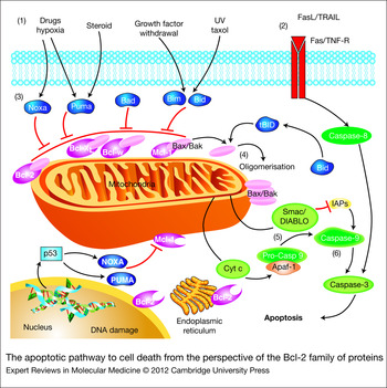
Conventional chemotherapeutic agents and radiation therapy have been shown to exert their cytotoxic effects by inducing apoptosis via the mitochondrial (intrinsic) pathway (Fig. 3) (Ref. Reference Kang and Reynolds20). Alterations in expression levels of genes and proteins that regulate this pathway of programmed cell death occur frequently in tumour cells. These alterations favour inappropriate cell survival through increased expression of antiapoptotic proteins. This, in turn, might lead to resistance to chemotherapeutics and radiation, because these therapies use apoptosis as a final common death pathway (Ref. Reference Mow21). Apoptotic resistance is therefore not only a hallmark of cancer, but also a key mechanism of treatment failure (Ref. Reference Hanahan and Weinberg22).
The apoptotic pathway to cell death from the perspective of the Bcl-2 family of proteins. (a) The intrinsic pathway is initiated by various signals, principally extracellular stimuli. (b) The extrinsic pathway is activated by Fas ligand or TRAIL, subsequently activating caspase-8. Caspase-8 transforms BID into truncated BID. In addition, caspase-8 initiates a cascade of caspase activation. (c) BH3-only proteins (BIM, BID, BAD, NOXA, PUMA) engage with antiapoptotic Bcl-2 family proteins to relieve their inhibition of BAX and BAK to activate them. (d) Next, BAX and BAK are oligomerised and activated, leading to mitochondrial outer membrane permeabilisation. (e) Once mitochondrial membranes are permeabilised, cytochrome c and/or Smac (DIABLO) is released into the cytoplasm, wherein they combine with an adaptor molecule, apoptosis protease-activating factor 1 and an inactive initiator caspase, procaspase-9, within a multiprotein complex called the apoptosome. Smac (DIABLO) inhibits inhibitors of apoptosis proteins to activate caspase-9. (f) Caspase-9 activates caspase-3, which is the initiation step for the cascade of caspase activation. Intrinsic and extrinsic pathways converge on caspase-3. Bcl-2 family proteins are also found on the endoplasmic reticulum and the perinuclear membrane in haematopoietic cells, but they are predominantly localised to mitochondria. Reprinted from Ref. Reference Kang and Reynolds20 with permission from American Association for Cancer Research © 2009.

The Bcl-2 family consists of 23–25 genes coding for proteins that, in conjunction with other constituents of programmed cell death pathways, regulate apoptotic homeostasis. The longer splice product of the bcl-x gene (BCL2L1, located on the short arm of chromosome 20, 20pter-p12.1), Bcl-XL, is an important antiapoptotic member of the family and is overexpressed in several solid tumours including MM (Ref. Reference Cao23). The physiological role of antiapoptotic proteins is to prevent apoptosis by inhibiting release of soluble mitochondrial intermembrane proteins such as cytochrome c and DIABLO into the cytoplasm. The release of these proteins leads to caspase activation. Interactions of the various members of the Bcl-2 family have been revealed to be more complex than originally thought, but the role of BAX and BAK as effectors of mitochondrial membrane permeability remains central. The exact mechanism by which BAX and BAK affect membrane permeability is not completely understood, but it is known to involve membrane incorporation of these proteins, as well as autodimerisation and interaction with voltage-dependent anion channel (VDAC) proteins (Refs Reference Kim24, Reference Youle and Strasser25). Antiapoptotic proteins such as Bcl-XL act by sequestration of BAX and BAK ‘activator’ proteins tBID, BIM and PUMA. This activity is antagonised by the interaction of ‘inactivator’ proteins such as BIK, NOXA and BAD with Bcl-XL. When challenged with proapoptotic stimuli in a wide variety of human tumour cell lines, Bcl-XL is at least as potent as Bcl-2 in prevention of apoptosis. In systems where Bcl-2 and Bcl-XL have been alternatively and co-overexpressed, Bcl-XL is more important for the prevention of apoptosis (Ref. Reference Huang, Cory and Strasser26). Although there is significant homology between Bcl-2 and Bcl-XL, Bcl-XL has proved to be uniquely important in human disease, and has been the focus of our studies in mesothelioma, in which Bcl-2 is not typically overexpressed. Our laboratory has demonstrated the therapeutic potential of Bcl-XL downregulation and functional inhibition both in vitro and in preclinical models of mesothelioma. In combination therapies, these models have proven successful in helping to overcome resistance to conventional chemotherapy.
Increased expression of proapoptotic members of the Bcl-2 family should favour programmed cell death in the presence of an appropriate stimulus such as cytotoxic chemotherapy. In fact, transduction of mesothelioma cell lines with an adenoviral vector containing the proapoptotic protein BAK induces decreased cellular viability and increased apoptosis in vitro (Ref. Reference Pataer27). Antisense oligonucleotide (ASO) therapy directed at mRNA encoding Bcl-XL has been shown to chemosensitise a number of tumour cell types, including mesothelioma. ASOs were used in vitro to downregulate Bcl-XL protein expression, decrease viability and engender apoptosis in human mesothelioma cell lines (Ref. Reference Smythe28). Furthermore, exposure of human mesothelioma cells to Bcl-XL ASOs in vitro was demonstrated to sensitise them to the conventional chemotherapeutic agent cisplatin in a synergistic manner (Ref. Reference Ozvaran29). Finally, the combination of Bcl-XL ASOs and cisplatin was demonstrated to reduce the growth of established flank tumour xenografts in mice as well as extend survival in an orthotopic xenograft mouse model of mesothelioma (Ref. Reference Littlejohn30). Similar results have been obtained with small interfering RNA (siRNA)-induced inhibition of Bcl-XL.
Pharmacological agents that neutralise the functions of antiapoptotic Bcl-2 family proteins have emerged as a promising new class of anticancer agents. These direct inhibitors of Bcl-XL function have a number of theoretical advantages over ASO- and siRNA-based approaches, including facilitation of systemic delivery and crossreactivity with other antiapoptotic members. 2-Methoxy antimycin A3 is a small molecular ligand that inhibits binding of proapoptotic family members such as BAK by occupying the binding cleft of Bcl-XL and Bcl-2. Treatment of mesothelioma cell lines with this compound results in apoptotic cell death without altering expression of Bcl-2 family proteins. Furthermore, co-administration of 2-methoxy antimycin A3 and cisplatin results in synergistic inhibition of tumour growth in an in vivo mesothelioma tumour model (Ref. Reference Cao31). Pharmacological inhibitors of antiapoptotic Bcl-2 family members continue to undergo further refinement and have shown promise in a number of tumour types, including mesothelioma.
Histone deacetylase inhibitors
Histones are a family of proteins that serve as structural and regulatory components of chromatin. The fundamental complex of chromatin is the nucleosome, which consists of 146 base pairs of DNA wrapped around an octamer of histone subunits (Ref. Reference Marks, Miller and Richon32). Histone acetylation, regulated by histone acetyltransferases and HDACs, affects the relative condensation of chromatin. In short, when histones are acetylated, chromatin is decondensed and DNA is available for transcription. HDACs facilitate chromatin condensation, preventing transcription of genes including tumour suppressors (Ref. Reference Zucali17). In addition to effects on histone proteins and the structure of chromatin, HDAC inhibitors (HDACis) also modulate the acetylation of nonhistone proteins such as transcription factors. This ultimately leads to a number of biological effects such as promotion of apoptosis, cell cycle inhibition and inhibition of angiogenesis (Ref. Reference Paik and Krug33).
Sodium butyrate is an HDAC inhibitor known to alter Bcl-2 family gene expression in a variety of tumour types. Exposure of mesothelioma cell lines to sodium butyrate leads to decreased mRNA transcription and protein expression of Bcl-XL as well as induction of apoptosis (Ref. Reference Cao34). In a subsequent study, cellular death and apoptosis of mesothelioma cell lines were augmented by the combination of sodium butyrate with proapoptotic gene therapy, namely adenoviral transfer of the proapoptotic Bcl-2 family members BAX and BAK (Ref. Reference Mohiuddin35). Similar in vitro effects have been demonstrated in mesothelioma using the HDACi suberoylanilide hydroxamic acid (SAHA, Vorinostat). Others have demonstrated a synergistic response between HDACis and combination chemotherapy in mesothelioma. Treatment with valproic acid, another known HDACi, in combination with pemetrexed and cisplatin led to complete suppression of epithelioid mesothelioma growth in a mouse xenograft model (Ref. Reference Vandermeers36).
An increasing amount of preclinical data demonstrating the utility of HDAC inhibition in vitro and in mouse xenograft models has led to early-phase clinical trials in patients with mesothelioma. On the basis of compelling evidence from two Phase I trials involving mesothelioma patients who received Vorinostat, a multicentre, randomised, placebo-controlled Phase III trial of Vorinostat in patients with advanced mesothelioma was performed and its results have recently been made available. Patients who progressed or relapsed following treatment with pemetrexed and platinum therapy were randomised 1:1 to receive Vorinostat or placebo. Unfortunately, overall survival was not significantly different in the Vorinostat and placebo groups (Ref. Reference Paik and Krug33).
Receptor tyrosine kinases
Peptide growth factors are important in maintaining tumour cell viability, particularly in the face of apoptotic stimuli. These growth factors are well known to induce intracellular signal transduction pathways such as the phosphoinositide 3-kinase (PI3 K) and mitogen-activated protein kinase (MAPK) pathways through their interaction with specific cell surface transmembrane RTKs. Several growth factors and their receptors have been shown to play a significant role in the oncogenesis, progression and resistance to therapy of MM. Among them, epidermal growth factor (EGF), hepatocyte growth factor (HGF), vascular endothelial growth factor (VEGF) and insulin-like growth factor (IGF) have been shown to be targets for therapy on the basis of promising preclinical data (Ref. Reference Villanova, Procopio and Rippo37).
Epidermal growth factor
One of the most thoroughly studied targets in cancer therapeutics is the EGF receptor (EGFR) and its ligand, EGF. EGFR is well known to be overexpressed in many human cancers, among them colon, breast, lung and upper aerodigestive tract malignancies. In 1990, Dazzi and colleagues found that 68% of mesothelioma specimens stained positively for EGFR by means of immunohistochemistry and that EGFR positivity was more common in the epithelial subtype (Ref. Reference Dazzi38). In studying the immunohistochemical expression of EGFR and its ligand, transforming growth factor-alpha (TGF-α), Cai and associates found that 76% of mesotheliomas expressed TGF-α, whereas 45% expressed EGFR, indicating the possibility of an EGFR autocrine loop (Ref. Reference Cai39). EGFR expression has also been linked to asbestos exposure in tissue culture. SV40-transformed human mesothelial cells exposed to asbestos fibres in vitro overexpress EGFR compared with control cells, and EGFR expression is related to increasing fibre length of crocidolite asbestos (Ref. Reference Pache40). Similar results have been obtained in vivo in rat pleural mesothelial cells (Ref. Reference Faux41). A preclinical study using gefitinib (Iressa), an orally bioavailable EGFR kinase inhibitor, demonstrated growth inhibition and G1 cell-cycle arrest in four mesothelioma cell lines (Ref. Reference Janne42). EGFR kinase inhibition led to apoptotic cell death by downregulation of PI3K–Akt signalling in mesothelioma in vitro (Ref. Reference Rascoe43). Finally, gefitinib was noted to potentiate the radiation response of mesothelioma xenografts in nude mice, with many animals demonstrating complete regression with no tumour regrowth (Ref. Reference She44).
On the basis of the aforementioned preclinical data, pharmacological inhibition of EGFR was thought to be a promising strategy in mesothelioma therapy. Moreover, the identification of activating mutations in the kinase domain of EGFR as a biomarker of response to tyrosine kinase inhibitor therapy in non-small-cell lung cancer (NSCLC) patients was equally promising (Refs Reference Lynch45, Reference Paez46). Although activating mutations of EGFR have been reported in patients with malignant peritoneal mesothelioma (Refs Reference Foster47, Reference Foster48), no such activating mutation of EGFR has been discovered in patients with pleural mesothelioma, and the results of EGFR inhibitors in Phase II clinical trials have been disappointing (Ref. Reference Velcheti49). The cancer and leukaemia group B (CALGB) 30101 Phase II trial enrolled 43 chemotherapy-naive patients to receive 500 mg gefitinib (Iressa) daily. The 3-month progression-free survival was 40%, which was not different than historical controls, and the authors concluded that single-agent gefitinib was not active in MM. A total of 97% of the patients in CALGB 30101 were found to have high expression of EGFR when scored by immunohistochemistry (Ref. Reference Govindan50). A Southwest Oncology Group Phase II trial enrolled 63 chemotherapy-naive patients to receive erlotinib (Tarceva). Despite high EGFR expression in 75% of participants, only 42% of patients had stable measurable disease, and the median progression-free survival of 2 months was significantly lower than that observed with standard first-line chemotherapy (Ref. Reference Garland51).
Phosphatase and tensin analogue (PTEN) is a tumour suppressor gene that has been localised to chromosome 10q23. LOH at 10q23 has been demonstrated in a number of tumour types, including mesothelioma. In a large tissue array study of clinical mesothelioma samples, 62% demonstrated absent PTEN expression whereas 14% demonstrated weak expression. Examination of clinical data from this cohort revealed that loss of PTEN expression was an independent predictor of poor survival in mesothelioma patients (Ref. Reference Opitz52). Our laboratory has previously demonstrated that adenoviral gene transfer and forced overexpression of PTEN engenders apoptosis in mesothelioma by Akt hypophosphorylation and decreased Akt kinase activity (Ref. Reference Mohiuddin53). It has been hypothesised that loss of PTEN and resultant constitutive Akt activation might explain the resistance seen with EGFR tyrosine kinase inhibitors, because they act upstream of PTEN (Ref. Reference Agarwal, Lind and Cawkwell54).
Hepatocyte growth factor
HGF is a multifunctional growth factor known to induce cellular growth and proliferation, motility and morphogenesis. HGF induces these biological functions through binding to its transmembrane tyrosine kinase receptor, c-Met (Ref. Reference Zucali17). c-Met is overexpressed and activated in a majority of cases of mesothelioma when compared with normal tissues. In addition, the circulating serum levels of HGF in mesothelioma patients are two-fold that of healthy control patients. On HGF stimulation and c-Met phosphorylation, the PI3 K and MAPK signal transduction pathways are activated in mesothelioma cell lines. Moreover, c-Met siRNA and a pharmacological c-Met inhibitor (SU11274) are effective in inhibiting cell growth and migration of these same cell lines (Ref. Reference Jagadeeswaran55).
An association between c-Met and Bcl-XL levels in malignant tissues has been established. In mesothelioma, the HGF–cMet axis appears to upregulate Bcl-XL expression at the transcriptional level. Specifically, through the activation of MAPKs, members of the ETS family of transcription factors are phosphorylated. This leads to nuclear importation of the factors ETS-2 and PU.1, both of which increase Bcl-XL promoter activity in mesothelioma. Conversely, the transcriptional repressor Tel is phosphorylated and exported from the nucleus to the cytoplasm (Ref. Reference Cao23)
Insulin-like growth factors
IGFs represent a family of peptides produced by various tissues throughout the body. IGFs possess growth stimulatory activities similar to insulin and can work in an autocrine, paracrine or endocrine fashion. As such, IGF has been reported to be an important growth factor in many tumour types. Both normal mesothelial and mesothelioma cell lines express mRNA encoding IGF-1 and IGF-1R, indicating the possibility of an autocrine loop (Ref. Reference Lee16). An IGF-1 receptor antisense expression vector led to a decrease in proliferation and tumourigenicity in a hamster mesothelioma cell line (Ref. Reference Pass56). We observed increased IGF-1R expression in mesothelioma cell lines relative to a transformed mesothelial line, as well as decreased cellular viability and apoptosis in a sarcomatous-type mesothelioma line following IGF-1R inhibition (Ref. Reference Rascoe43). Others have demonstrated dose-dependent growth repression, inhibition of IGF-1R phosphorylation and decreased activity of downstream PI3 K–Akt and MAPK signal transduction pathways following treatment with an orally bioavailable IGFR inhibitor, NVP-AEW541 (Ref. Reference Whitson57).
Vascular endothelial growth factor
VEGF is an essential regulatory component of physiological angiogenesis. Furthermore, its role in tumour pathogenesis, growth and metastasis are well documented, and VEGF is overexpressed in most human malignancies. The effects of VEGF are mediated through binding to two tyrosine kinase receptors: VEGFR-1, or Flt-1 (fms-like tyrosine kinase-1), and VEGFR-2, or KDR/Flk-1 (kinase-insert domain receptor/fetal liver kinase-1) (Ref. Reference Villanova, Procopio and Rippo37).
Mesothelioma patients have higher serum levels of VEGF than normal controls and, in fact, have higher VEGF levels than other solid tumour patients (Ref. Reference Linder58). VEGF and its two receptors are expressed in mesothelioma cell lines, biopsy specimens and pleural effusions. VEGF levels in the pleural effusions of mesothelioma patients were seven-fold higher than levels in effusions in patients with nonmalignant disease. Moreover, linear regression analysis has demonstrated an inverse correlation between serum VEGF levels and survival in mesothelioma patients (Ref. Reference Strizzi59).
A number of angiogenesis inhibitors directed at VEGF and its receptors have been developed. The anti-VEGF monoclonal antibody bevacizumab (Avastin) has demonstrated modest survival benefit and is approved for use in metastatic colorectal carcinoma and NSCLC. Unfortunately, despite the aforementioned promising preclinical data, a Phase II trial combining cisplatin and gemcitabine with and without bevacizumab in unresectable, chemotherapy-naive mesothelioma patients yielded no differences in progression-free or overall survival (Ref. Reference Karrison60). A similar trial comparing bevacizumab and placebo in patients receiving current first-line chemotherapy (cisplatin or pemetrexed) is ongoing. Studies investigating several small-molecule pharmacological inhibitors of VEGF RTKs have demonstrated only modest activity to date (Ref. Reference Kelly, Sharon and Hassan61).
Proteasome inhibitors
Investigation has revealed that the ubiquitin–proteasome pathway has a key role in regulating homeostasis of cellular proteins that involve cell cycle, survival and apoptosis. Therapeutically, targeting the proteasome with a specific inhibitor, bortezomib (Velcade), has been successful in selectively inducing apoptosis in a variety of human cancer cells including mesothelioma. Bortezomib is a selective inhibitor of the 20S proteasome. Its actions are pleiotropic and include inhibition of nuclear factor (NF)-κB activation by preventing degradation of its inhibitor IκB (Ref. Reference Zucali17). Inhibition of constitutively activated NF-κB by bortezomib resulted in cytotoxicity and apoptosis in vitro and regression of mesothelioma xenografts in mice (Ref. Reference Sartore-Bianchi62). Furthermore, bortezomib potentiated the activity of pemetrexed and cisplatin in mesothelioma cell lines (Ref. Reference Gordon63). Bortezomib is currently under investigation in a number of mesothelioma trials, both as a single agent and in combination with chemotherapy. Although bortezomib is approved for the treatment of multiple myeloma, cellular resistance to boretezomib-induced apoptosis might limit its successful application as a therapeutic agent in this context (Ref. Reference Richardson64). In vitro development of a bortezomib-resistant mesothelioma cell line has demonstrated that evasion of the unfolded protein response and concomitant reduction in proapoptotic gene induction accounts for resistance in bortezomib-adapted mesothelioma cells (Ref. Reference Zhang65).
Conclusions and future directions
Despite modest advances in clinical treatment over the last decade, malignant pleural mesothelioma remains a vexing clinical problem. The mean overall survival for patients with malignant pleural mesothelioma (MPM) is approximately 12 months. Highly selected patients appear to benefit from aggressive surgical debulking followed by IMRT to achieve local control of disease followed by systemic chemotherapy. Modest improvements in the treatment of unresectable mesothelioma have been made by using combination chemotherapy with cisplatin and pemetrexed. This combination is the current standard of care in the adjuvant setting as well.
Studies of human cell lines and tissues as well as animal models of MPM have demonstrated genetic and epigenetic events that contribute to the multistep process of mineral fibre carcinogenesis. These events include inactivation of tumour suppressor genes, modulation of signal transduction pathways including RTKs, avoidance of apoptosis and inhibition of the ubiquitin–proteasome degradation pathway. Preclinical investigations of targeted therapies such as tyrosine kinase, HDAC and proteasome inhibitors have been promising. However, randomised clinical trials using many of these same agents have been disappointing to date. Pharmacological inhibitors of antiapoptotic Bcl-2 family members continue to undergo refinement, and there is a hope that they will emerge as a promising new class of anticancer agents. Preclinical data suggest that they could demonstrate therapeutic effect in a number of tumour types, including mesothelioma.
Autophagy, a concerted process of intracellular breakdown within specialised double-membrane vesicles, occurs in response to events such as metabolic stress. It is an evolutionarily conserved prosurvival mechanism, regulated downstream of mammalian target of rapamycin (MTOR) in the PI3 K–Akt cell-survival pathway. While generally cytoprotective, excessive autophagy results in a type of programmed cell death that is morphologically distinct from apoptosis (Ref. Reference Sinha and Levine66). It has been noted that, depending on context, autophagy can augment either cellular demise or protection. Apoptosis and autophagy are not mutually exclusive programmed cell death pathways, as evidenced by a specific physical interaction between regulators of the two pathways: Beclin-1 (autophagy) and Bcl-XL (apoptosis). Bcl-XL has recently been determined to inhibit autophagy through a direct functional and physical interaction with Beclin-1, a protein essential for the initiation of autophagy (Ref. Reference Maiuri67). Beclin-1, which was characterised as a haploinsufficient tumour suppressor, is a known cytosolic mediator of autophagy, and a recent addition to the BH3-only members of the Bcl family of proteins. These proteins govern intrinsic apoptosis by selective interaction with the BH3 binding pocket of Bcl-XL (Ref. Reference Oberstein, Jeffrey and Shi68). Autophagy is probably important in response to toxic insults such as chemotherapy or irradiation, but its exact role in the context of a growing solid tumour remains unclear (Ref. Reference Degenhardt69). Beclin-1 is known to promote cell survival in solid tumours by facilitation of autophagy, yet has also been shown to suppress tumourogenicity (Refs Reference Oberstein, Jeffrey and Shi68, Reference Degenhardt69). In malignant glioma cells, it has been shown that the antitumour effect of temozolomide can be suppressed by inhibiting early autophagy, but that inhibition of late autophagy enhances cytotoxicity (Ref. Reference Kanzawa70). Inhibition of autophagy in radiation-resistant cell lines of breast, lung, pharyngeal and cervical cancers resensitised them to radiation treatment (Ref. Reference Apel71). Thus, it is becoming clear that a greater understanding of autophagy in the context of chemotherapy-induced apoptosis within a growing solid tumour could add to our understanding of chemotherapeutic response and development of resistance.
Ongoing studies in our laboratory have demonstrated that both apoptosis and autophagy occur in MM following HDAC inhibition through mutually exclusive processes. Autophagy appears to occur much earlier than apoptosis, suggesting that autophagy has a cytoprotective role in mesothelioma cells following cytotoxic therapy, thus subverting their entry into the apoptotic pathway.
Acknowledgements and funding
We thank the peer reviewers for their comments and suggestions.