Introduction
The ductus venosus is an embryonic vascular structure that connects the umbilical cord to the inferior vena cava during fetal life. In a normal fetus, about one-third of the placental blood returning from the umbilical cord bypasses the portal venous circulation through the ductus venosus. Reference Kiserud and Acharya1 In most healthy babies, the closure of the ductus venosus begins shortly after birth, and complete closure is achieved within the first 3 weeks of life. Reference Gillespie, Golden, Sivarajan and Rome2 If the ductus venosus remains open and the shunt is large, liver failure, hypoxaemia, and encephalopathy may develop. Reference Uchino, Matsuda and Endo3 It may be necessary to close the shunt when the patient is symptomatic. Surgical treatment and transcatheter embolisation have been reported for ductus venosus closure. Reference Araki, Kamada, Okamoto, Arai and Oba4–Reference Yoshimoto, Shimizu and Saeki6
Case report
Clinical presentation
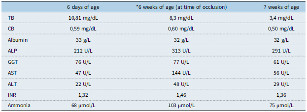
A patient weighing 3500 g at 36 weeks of birth, with an APGAR score of 5/7 at the first and fifth minutes after delivery and persistent respiratory distress, was admitted to our centre. Echocardiographic evaluation of the patient showed dextrocardia, mild isthmus hypoplasia, persistent left superior vena cava, and patent foramen ovale. Abdominal ultrasound showed that the right portal vein was extremely hypoplastic at the intrahepatic level; the left portal vein, which was well developed and dilated, extended toward the apex of the heart, continuing with the umbilical vein at the level of the falciform ligament, and poured directly into the right atrium. A 5-millimetre vascular structure that shows filling in the left portal vein branch and umbilical vein trace on dynamic abdominal computed tomography opens to the right atrium in the vicinity of the inferior vena cava opening point at the diaphragmatic level. With these findings, it was accepted that the patient had a patent ductus venosus. Due to the increase in ammonia and transaminase levels during the patient’s follow-up (Table 1), it was decided to close the patent ductus venosus by transcatheter method, considering that interventional therapy is safer.
Improvement in hepatic biomarkers after vascular plug closure of patent ductus venosus (*)

TB = Total bilirubin; CB = Conjugated bilirubin; ALP = Alkaline phosphatase; GGT = Gamma-glutamyl transferase; AST = Aspartate aminotransferase; ALT = Alanine aminotransferase; INR = International normalised ratio; µmol/L = Micromole per litre.
Procedure
The procedure was performed in the paediatric cardiac catheterisation hall under general anaesthesia using biplane fluoroscopy. The procedure was performed by paediatric cardiologists. Access was performed from the right femoral vein and the right jugular vein. It was thought that the right internal jugular access provided alignment with the patent ductus venosus. A 0.018-inch guide wire was used. First, the right Judkins catheter was advanced from the right femoral vein via a guide wire from the inferior vena cava to the portal vein through the right atrium and ductus venosus. Selective angiography performed here showed that the ductus venosus was open (Figure 1). Its narrowest place was measured at 5.2 millimetres. A 0.014-inch guide wire from the right jugular vein was advanced through the ductus venosus to the portal vein, and the 5 × 20 mm Tyshak balloon angioplasty catheter was inflated so that the tip of the right Judkins catheter in the ductus venosus was more distal. A balloon occlusion test was performed for 10 minutes, and portal pressure was measured at the end of the test, and it was found to be 8 mmHg on average (Figure 2). Later, while the balloon was being inflated, a contrast agent injection was made into the portal vein, and it was seen that the intrahepatic portal veins were in sufficient development. It was decided that it would be appropriate to close the ductus venosus. The guide wire was advanced from the right jugular vein and placed in the portal system. The 5F sheath was tried to be replaced with the 5F long sheath, but the long sheath was replaced with the 6F sheath because it could not be advanced to the portal system. A 0.018-inch guide wire was advanced from the right jugular vein to the portal system, and a 6F guiding catheter was advanced to the portal system via the wire. An 8 mm Lifetech Cera Vascular® Plug System was inserted through the catheter into the ductus venosus. After the angiograms performed after the procedure showed that the device was in the appropriate position. The device was released, and the process was terminated (Figure 3).
(a–b): Angiographic identification of a patent ductus venosus and its systemic drainage. Frontal ( a ) and lateral ( b ) angiographic views obtained during vascular catheterisation, demonstrating the anatomy of the patent ductus venosus. The white arrow indicates contrast flow from the portal venous system into the ductus venosus. The black arrow shows drainage of the ductus venosus into the right atrium via the inferior vena cava.

Balloon occlusion test for assessment prior to ductus venosus closure. Fluoroscopic image demonstrating balloon inflation within the patent ductus venosus. The balloon catheter was advanced from the right internal jugular vein into the portal venous system, while the diagnostic catheter placed via the femoral vein remained in position for contrast injection and pressure monitoring.

(a-b): Angiographic confirmation of successful ductus venosus closure. ( a ), Angiographic image obtained prior to device release, demonstrating complete occlusion of the ductus venosus with no contrast passage, indicating effective sealing by the deployed plug; ( b ), post-deployment lateral view confirming the final position of the lifetech cera vascular® plug within the ductus venosus.

The patient was taken to the neonatal intensive care unit again after the procedure. Improvement in the patient’s liver function tests and a decrease in ammonia levels were observed. On the control abdominal ultrasound, it was confirmed that the ductus venosus was permanently closed. The patient was monitored for 1 week and discharged without any complications. The patient’s follow-up is continuing.
Discussion
The ductus venosus bypasses the liver in fetal life and closes after birth. Within minutes after birth, the blood pressure in the umbilical cord decreases, and functional closure of the canal begins. However, anatomically, obliteration may not be completed for 15 to 20 days. Reference Yoshimoto, Shimizu and Saeki6,Reference Gitzelmann, Arbenz and Willi7 Closure may occur as late as 37 days after birth in premature babies. Reference Vade, Lim-Dunham and Iqbal8 Patent ductus venosus is a rare condition that removes portal venous blood from the liver and directs it directly to the inferior vena cava. Reference Yoshimoto, Shimizu and Saeki6 Complications due to portosystemic shunts can be grouped as neonatal cholestasis, liver tumours, hepatopulmonary syndrome, pulmonary hypertension, and portosystemic encephalopathy. Reference Yoshimoto, Shimizu and Saeki6,Reference Franchi-Abella, Branchereau and Lambert9 Patent ductus venosus is an intrahepatic portocaval shunt and may cause hyperammonemia and encephalopathy. This occurs due to the partial or complete redirection of portal blood into the systemic circulation. Reference Gitzelmann, Arbenz and Willi7
The management of portosystemic shunts in paediatric patients consists of various treatment options such as surgical ligation and banding, liver transplantation, and transcatheter embolisation. Reference Franchi-Abella, Branchereau and Lambert9 The type of surgical treatment is related to the degree of intrahepatic portal vein hypoplasia. If the intra- and extrahepatic portal systems are severely hypoplastic, the preferred treatment method is liver transplantation. Reference Yoshimoto, Shimizu and Saeki6 The transcatheter closure method, which is much less invasive than surgery, may be preferred. In the literature, it has been shown that in some patients with a shunt less than 5 mm, embolisation can be performed successfully by interventional means. Reference Ikeda, Yamaguchi and Sera5
Transcatheter occlusion is a much less invasive method than surgery. Transcatheter closure allows the verification of the adequacy of the intrahepatic portal venous system both by direct pressure measurement during balloon occlusion and by angiography. Reference Gillespie, Golden, Sivarajan and Rome10 A patent ductus venosus occlusion associated with an inadequate intrahepatic portal venous system can be rapidly fatal due to mesenteric venous congestion and intestinal ischaemia. Reference Yoshimoto, Shimizu and Saeki6 The transjugular approach can be used for transcatheter occlusion and eliminates the need for complex surgery. This reduces post-procedural complications, particularly in younger patients. Lifetech Cera Vascular® is advantageous in terms of easy deployment and good occlusion thanks to the nitinol mesh with PTFE membranes.
Financial support
This research received no specific grant from any funding agency, commercial or not-for-profit sectors. There is no conflict of interest in this article.