1. Introduction
Nucleases catalyzing DNA and RNA cleavage are indispensable for life (Table 1). Nuclease activities are integral parts of DNA replication; the 5′ to 3′ exo- and endonucleases are needed to remove RNA primers (Kao & Bambara, Reference Kao and Bambara2003; Shen et al. Reference Shen, Singh, Liu, Qiu, Zheng, Finger and Alas2005), and the 3′ to 5′ exonuclease for proofreading (Reha-Krantz, Reference Reha-Krantz2010). Two other major DNA metabolic processes, recombination and repair, are initiated by nucleases (Marti & Fleck, Reference Marti and Fleck2004; Mimitou & Symington, Reference Mimitou and Symington2009). Nuclease activity is also required for structural alterations of nucleic acids, for example, topoisomerization (Champoux, Reference Champoux2001; Schoeffler & Berger, Reference Schoeffler and Berger2008; Wang, Reference Wang2002), site-specific recombination (Grindley et al. Reference Grindley, Whiteson and Rice2006) and RNA splicing (Patel & Steitz, Reference Patel and Steitz2003), during which a phosphodiester bond is temporarily broken and reformed after strand passing or transfer to a new target. In addition, nuclease activities are essential in RNA processing, maturation and RNA interference (RNAi) (Abelson et al. Reference Abelson, Trotta and Li1998; Chu & Rana, Reference Chu and Rana2007; Moore & Proudfoot, Reference Moore and Proudfoot2009; Nowotny & Yang, Reference Nowotny and Yang2009). RNA and DNA degradation are essential components of microbial defense mechanisms (James et al. Reference James, Kleanthous and Moore1996; Sorek et al. Reference Sorek, Kunin and Hugenholtz2008; Tock & Dryden, Reference Tock and Dryden2005). Nucleases are even essential for programmed cell death (Parrish & Xue, Reference Parrish and Xue2006). Defective DNase and RNase activities have been associated with various autoimmune diseases due to an incomplete removal of endogenously produced nucleic acids (Crow & Rehwinkel, Reference Crow and Rehwinkel2009; Stephenson, Reference Stephenson2008).
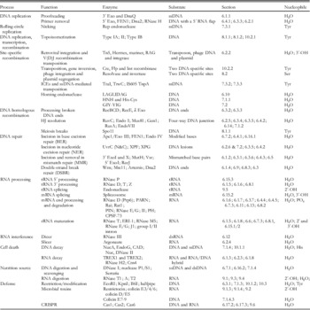
Classification of nucleases by function. DNase and RNase are classified according to biological processes they are involved from DNA replication to cell defense. Each process is further divided into functional pathways. Enzymes of different catalytic mechanisms or different tertiary structures are separated by semi-colons for each pathway or process. Detailed descriptions of each category are referred to specific sections and under subsections.

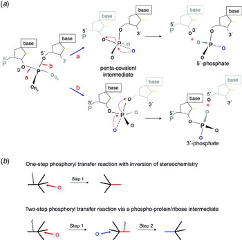
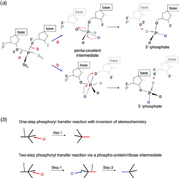
RNA and DNA present only two types of phosphodiester bonds for cleavage, 5′ or 3′ of a scissile phosphate (Fig. 1a), and the fundamental chemistry is bimolecular nucleophilic substitution or SN2 in short. Nonetheless, structures and catalytic mechanisms of RNA and DNA nucleases are greatly varied and complex. Nucleases can be protein or RNA and use water, (deoxy)ribose, inorganic phosphate, or the side chains of Ser, Tyr or His as a nucleophile. Catalysis may or may not require metal ions. In addition, nuclease activities are strictly regulated by stringent substrate specificity (Pingoud et al. Reference Pingoud, Fuxreiter, Pingoud and Wende2005; Stoddard, Reference Stoddard2005), confined localization, or by potent inhibitors (Chowdhury et al. Reference Chowdhury, Beresford, Zhu, Zhang, Sung, Demple, Perrino and Lieberman2006; Kolade et al. Reference Kolade, Carr, Kuhlmann, Pommer, Kleanthous, Bouchcinsky and Hemmings2002; Widlak & Garrard, Reference Widlak and Garrard2005) to avoid unwanted or uncontrolled degradation of cellular DNA and RNA.
Adjacent nucleotides in RNA and DNA are linked by 3′ and 5′ O-P phosphodiester bonds. (a) Nucleases degrade nucleic acid by breaking either one of these two bonds labeled as ‘a' and 'b’. A nucleophile attacking in-line from the 5′ side breaks the 3′ O–P bond and produces 5′-phosphate and 3′-OH. Alternatively, a nucleophile attacking the scissile phosphate from the 3′ side breaks the 5′ O–P bond and produces 3′-phosphate and 5′-OH. Rp stands for pro-Rp and Sp for pro-Sp. (b) The SN2 type associative reaction inverts the stereo configuration of phosphate. A two-step trans-esterification reaction can set the stereochemistry back to the original.
Individual nuclease families or superfamilies, for example, topoisomerases (Schoeffler & Berger, Reference Schoeffler and Berger2008), sequence-specific recombinases (Grindley et al. Reference Grindley, Whiteson and Rice2006), metal-ion-independent ribozymes (Cochrane & Strobel, Reference Cochrane and Strobel2008), Holliday junction (HJ) resolvases (Declais & Lilley, Reference Declais and Lilley2008) and DNA nucleases (Horton, Reference Horton, Rice and Correll2008) have been recently reviewed. This particular review is intended to summarize DNases and RNases with known atomic structures and focus on catalytic mechanisms. The aim is to compare and contrast structural and mechanistic diversity in the context of shared functionality.
2. Nucleolytic reactions
2.1 Basic chemistry
A nuclease is a phosphodiesterase that cleaves one of the two bridging P–O bonds, 3′ or 5′, in a nucleic acid polymer (Fig. 1a). Cleavage of phosphodiester bonds is thought to be done by a general acid–base catalysis, where the general base activates the nucleophile by deprotonation and the general acid facilitates product formation by protonating the leaving group. Perhaps because of the stability of nucleic acid phosphodiester bonds, the cleavage reaction is usually of the associative SN2 type (Gerlt et al. Reference Gerlt, Coderre and Mehdi1983). The reaction can be divided into three stages: nucleophilic attack, the formation of a highly negatively charged penta-covalent intermediate and the breakage of the scissile bond. Cleavage of the P–O3′ bond, which generates 5′-phosphate and 3′-OH products, requires a nucleophile to be on the 5′ side poised for the in-line attack (Fig. 1a). In the penta-covalent bipyramid intermediate, the O5′ moves to the center plane, which consists of the phosphorus and two non-bridging oxygens, and the attacking nucleophile and O3′ leaving group are opposite each other at the apices of the bipyramid. In the cleaved product, the stereo configuration of the phosphorus is inverted in this one-step reaction. Although phosphate in nucleic acid is achiral, due to two chemically indistinguishable non-bridging oxygen atoms, the stereo inversion is evident when one of the non-bridging oxygen is replaced by a heavy isotope or sulfur (Eckstein, Reference Eckstein1985; Gerlt et al. Reference Gerlt, Coderre and Mehdi1983). Hence, the two non-bridging oxygens are referred to as pro-Sp and pro-Rp. A double inversion, which returns the phosphorus to its original configuration, requires a two-step reaction and occurs if an enzyme–nucleic acid covalent intermediate forms (Fig. 1b). To generate 3′-PO4 and 5′-OH products, the reaction configuration has to be reversed, and the nucleophile has to attack on the 3′ side of the scissile phosphate (Fig. 1a).
2.2 Nucleophiles
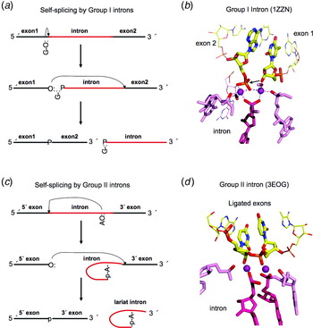
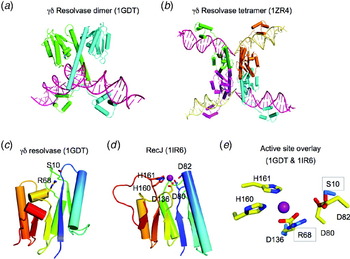
Nucleases utilize a variety of nucleophiles to cleave a scissile phosphate bond (Fig. 2a). The most common nucleophiles are water molecules deprotonated by a general base for direct hydrolysis. A hydroxyl group of the 3′ end of DNA or RNA can also be the nucleophile as during the RNA splicing, DNA strand transfer or hairpin formation (Doudna & Cech, Reference Doudna and Cech2002; van Gent et al. Reference Van Gent, Mizuuchi and Gellert1996) (Fig. 2b, c). For DNA cleavage, the side chains of Ser, Tyr and His have been observed to serve as nucleophiles to form a covalent DNA phosphoryl-protein intermediate, which is subsequently resolved either by phosphoryl transfer reaction back to DNA during recombination and topoisomerization (Fig. 2d) or by hydrolysis in two-step cleavage reactions (Grindley et al. Reference Grindley, Whiteson and Rice2006; Stuckey & Dixon, Reference Stuckey and Dixon1999).
Phosphodiester bond cleavage. (a) Nucleases can attack a scissile phosphate in the middle of nucleic acids (endo) or at the 5′ or 3′ ends (exo). The nucleophiles can be the hydroxyl groups of water molecules, Ser or Tyr side chains, hydroxyls of ribose (2′ or 3′) or deoxyribose (3′) in RNA and DNA, or free nucleophiles. PLD family nucleases use the imidazole of His as the nucleophile. When a protein side chain serves as a nucleophile, the phosphodiester bond to be cleaved is transferred to the protein. Similarly, when the hydroxyl of a nucleotide or nucleic acid serves as the nucleophile, it is transferred to the nucleic acid. Both are indicated as R. (b) DNA transposition by the cut-and-paste mechanism. The donor DNA uses its cleaved 3′-OH to attack a phosphate in a target DNA. Thus, the donor DNA is inserted into the target in this strand transfer reaction. RNA splicing is similar. The cleavage of the 5′ exon–intron junction frees the 3′-OH of the first exon. This 3′-OH than attacks the 3′ exon–intron junction and results in a linked two exons and free intron. (c) DNA Hairpin formation. When a 3′-OH of a duplex DNA attacks a phosphate of its complementary strand, the two strands of that duplex are linked and become one. (d) Type-II topoisomerization. Both strands of one DNA segment are cleaved and form covalent bonds with a dimeric (or tetrameric) topoisomerase. The second DNA segment shown in light blue passes through the cleaved segment thus changing the linking number and topology. The cleaved DNA is then religated and the topoisomerase regenerated. (e) The 2′-OH immediate adjacent to a scissile phosphate can serve as a nucleophile and hydrolysis of 5′ O–P bond leads to 2′, 3′ cyclic phosphate and 5′-OH. (f) RNase PH and PNPase are 3′ exonucleases. They use inorganic phosphate as nucleophiles and degrade RNA to nucleotide diphosphates.

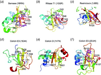
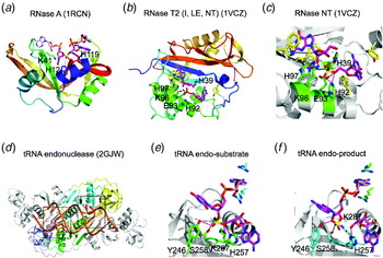
The 2′ hydroxyls of RNA or free ribonucleotides are additional nucleophiles utilized by RNases. The 2′-OH adjacent to a scissile phosphate often serves as the nucleophile and leads to the formation of a labile 2′,3′-cyclic phosphate (Figs 2e, 3b, and c), which is then hydrolyzed to produce a 3′ phosphate as exemplified by RNase A (Raines, Reference Raines1998) or occasionally a 2′ phosphate as catalyzed by the yeast RNA ligase during tRNA splicing (Abelson et al. Reference Abelson, Trotta and Li1998). Finally, RNase PH and polynucleotide phosphorylases (PNPases) use inorganic phosphate as a nucleophile to degrade single-stranded RNA (ssRNA) by phosphorolysis and produce nucleoside 5′-diphosphates (Deutscher et al. Reference Deutscher, Marshall and Cudny1988) (Fig. 2f). The pK a of these nucleophiles are 6·0 (His), 10·0 (Tyr), 12–14 (2′-OH of ribose), 13 (Ser) and 16 (H2O) (Saenger, Reference Saenger1984; Voet & Voet, Reference Voet and Voet2004). If water is the nucleophile, a general base is necessary to deprotonate it for efficient cleavage. Similarly, the 2′-OH of ribose also needs deprotonation to be an efficient nucleophile.
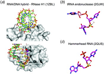
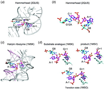
Double- versus single-stranded nucleic acid substrate. (a) To cleave a double helix, the nucleophile can attack from outside of the duplex only. Shown here is an RNA/DNA hybrid in a mixed A and B form, which is approached by RNase H1 from the minor groove side. A water molecule is coordinated by the Mg2+ ion in the active site to attack the phosphate from the 5′ side and produce a 5′ phosphate and 3′-OH. (b) A scissile phosphate in pre-tRNA to be cleaved by tRNA endonuclease using the 2′-OH as the nucleophile. The nucleotides surrounding the scissile phosphate are not base paired and splayed. The phosphate itself is distorted. (c) Self-cleaving hammerhead ribozyme distorts the scissile phosphate in a similar way, because the double helix conformation is incompatible with the reaction coordinates of using a 2′-OH to attack the adjacent phosphate. PDB accession codes are shown in parentheses.

2.3 Double- versus single-stranded substrate
If the scissile phosphate is in a double helix, the options for placement of an external nucleophile (that is not within the nucleic acid itself) are limited. Looking down the helical axis, a nucleophile can approach the nucleic acid backbone only from outside the double helix, because the inside is occupied by sugars and bases (Fig. 3a). If a nucleic acid is single stranded and bases are unstacked, the approach of a nucleophile is not as restricted. For example, RNA cleavage using the 2′-OH adjacent to a scissile phosphate can only occur with ssRNA, because the reaction coordinate is incompatible with the double helix conformation and requires a distorted backbone phosphate with bases surrounding the scissile phosphate unstacked and splayed (Buckle & Fersht, Reference Buckle and Fersht1994; Cochrane & Strobel, Reference Cochrane and Strobel2008; Correll et al. Reference Correll, Yang, Gerczei, Beneken and Plantinga2004) (Fig. 3b and c).
Potential reaction coordinates for nicking double-stranded nucleic acids are far less varied than possibilities for cleaving single-stranded nucleic acids. Most often DNA is in a double helix. Even RNA substrates in pre-mRNA splicing or RNAi-mediated cleavage are often base-paired in a double-helical conformation (Nowotny & Yang, Reference Nowotny and Yang2009; Patel & Steitz, Reference Patel and Steitz2003; Stahley & Strobel, Reference Stahley and Strobel2006). To date, the nucleases that cleave double-stranded substrates all approach RNA and DNA from the minor groove side, where scissile phosphates are easier to reach than from the major groove side (Fig. 3a). Sequence-specific interactions, which usually take place in the major groove, are often accomplished by additional domains attached to catalytic centers (Lee et al. Reference Lee, Chang, Joseph, Ghirlando, Rao and Yang2005).
2.4 Products of 5′ or 3′ phosphate
Nucleolytic cleavage products are most often 5′-phosphates and 3′-OH groups. This type of cleavage is likely to be preferred (over 3′-phosphate and 5′-OH), likely because firstly the 3′-OH can be passed onto other nucleic acid enzymes and be directly used as the nucleophile, e.g. RNA and DNA polymerases, DNA ligases, transposases, spliceosomes, CCA-adding enzymes and aa-tRNA synthetases. Secondly, the 5′-phosphate is a ready substrate for DNA ligation at the end of replication, repair and recombination. The two main exceptions are RNA cleavage via the 2′,3′-cyclic phosphodiester intermediate and DNA cleavage by Tyr-dependent site-specific recombinases and Type IB topoisomerases (TopIBs). For the DNA enzymes, a 3′-phosphotyrosyl bond is a transient intermediate and is reversed back to a phosphodiester bond in the product (Champoux, Reference Champoux2001; Grindley et al. Reference Grindley, Whiteson and Rice2006). In the RNA cases, either the 3′-phosphate is an end product or these RNA molecules require further hydrolysis and dephosphorylation. For example, the 2′,3′-cyclic phosphodiester formed in the tRNA splicing is hydrolyzed to form 3′-OH and 2′-phosphate (Abelson et al. Reference Abelson, Trotta and Li1998), which require several enzymatic activities to alter the phosphorylation states in a process referred to as healing (Nandakumar et al. Reference Nandakumar, Schwer, Schaffrath and Shuman2008) before RNA fragments can be ligated.
3. Diversity and classification of nucleases
Nucleases are a very diverse group and include both proteins and catalytic RNA (ribozyes). There is no simple way to classify and divide them. Based on substrate preference, nucleases may be divided into DNase and RNase, yet quite a number of nucleases are sugar non-specific and can cleave both RNA and DNA (Hsia et al. Reference Hsia, Li and Yuan2005; Laskowski, Reference Laskowski, Linn and Roberts1985; Rangarajan & Shankar, Reference Rangarajan and Shankar2001). Depending on whether a 5′ or 3′ end is required for substrate recognition and whether cleavage products are single or oligo nucleotides, they may be divided into exo- and endonucleases. For instance, all self-cleaving ribozymes found to date only endonucleolytically cleave RNA (Cochrane & Strobel, Reference Cochrane and Strobel2008; Lilley, Reference Lilley2005). Exonucleases, which remove one nucleotide at a time from the end of a strand, can be further divided into two groups by the 5′ to 3′ versus 3′ to 5′ polarity. However, the fundamental chemistry of endo- and exo-cleavage is the same, and it is not unusual that a single active site may contain both exonuclease and endonuclease activities. For example, members of the Flap endonuclease 1 (FEN1) family have the 5′ to 3′ exonuclease activity in addition to the endonuclease activity (Harrington & Lieber, Reference Harrington and Lieber1994; Lyamichev et al. Reference Lyamichev, Brow and Dahlberg1993) and Mre11, which is involved in DNA double-strand break (DSB) repair, has both endo and 3′ to 5′ exonuclease activities (Paull & Gellert, Reference Paull and Gellert1998; Usui et al. Reference Usui, Ohta, Oshiumi, Tomizawa, Ogawa and Ogawa1998).
According to catalytic mechanism, all nucleases may be divided into three major classes based on whether none, one or two metal ions are involved. However, each of these classes includes many different families and superfamilies. For instance, two-metal-ion-dependent nucleases include both protein enzymes and ribozymes and can be vastly different in the tertiary structure, metal-ion-coordination motif and cellular function. The following are just a few examples of diversity among nucleases.
3.1 The lack of correlation in structure, mechanism and biological function
Similar biological outcomes are often achieved by molecules with unrelated tertiary structures and drastically different mechanisms. For example, sequence-specific restriction of endonucleases encompasses at least five structural families and employ at a minimum four different catalytic mechanisms (Orlowski & Bujnicki, Reference Orlowski and Bujnicki2008) (Table 1). For an up-to-date complete list of restriction endonucleases, please consult the REBASE database (rebase.neb.com) (Roberts et al. Reference Roberts, Vincze, Posfai and Macelis2009). In parallel, HJ resolvases are composed of five different structural and mechanistic families (Declais & Lilley, Reference Declais and Lilley2008; Ip et al. Reference Ip, Rass, Blanco, Flynn, Skehel and West2008) (Table 1). Conversely, similar tertiary structures may adopt different catalytic properties. For example, the Cas6 and Cas2 nucleases share the ferredoxin-fold, but their active sites are unrelated (Beloglazova et al. Reference Beloglazova, Brown, Zimmerman, Proudfoot, Makarova, Kudritska, Kochinyan, Wang, Chruszcz, Minor, Koonin, Edwards, Savchenko and Yakunin2008; Carte et al. Reference Carte, Wang, Li, Terns and Terns2008). Cas2 is metal-ion dependent and Cas6 is metal independent. Furthermore, structural and mechanistic conservation are not necessarily correlated with identical functions. For example, molecules with the RNase H-like fold that utilizes the two-metal-ion catalysis are involved in RNA processing, DNA cleavage, transposition and HJ resolution (Nowotny et al. Reference Nowotny, Gaidamakov, Crouch and Yang2005; Yang & Steitz, Reference Yang and Steitz1995b).
3.2 Different catalytic mechanisms of ribozymes
RNA is thought to be the primordial nucleic acid and can function both as a genetic material and a catalytic entity (Chen et al. Reference Chen, Li and Ellington2007b; Yarus, Reference Yarus2002). It is not surprising that RNases and ribozymes display great mechanistic diversity. Naturally occurring ribozymes can be metal-dependent or -independent (Lilley, Reference Lilley2005). The self-cleaving hammerhead, hairpin, glmS, hepatitis delta virus (HDV) and Varkud satellite (VS) ribozymes cleave the extended ssRNA by a metal-ion-independent mechanism (Cochrane & Strobel, Reference Cochrane and Strobel2008), whereas group I and group II introns cleave exon–intron junctions embedded in double-stranded RNA (dsRNA) by a two-metal-ion mechanism (Stahley & Strobel, Reference Stahley and Strobel2005; Toor et al. Reference Toor, Keating and Pyle2009). Different catalytic mechanisms lead to the different types of cleavage products. The two-metal-ion-dependent ribozymes generate 5′-phosphate and 3′-OH products, whereas the metal-independent ribozymes use 2′-OH as the nucleophile and generate 2′,3′-cyclic phosphate and 5′-OH products.
3.3 Different catalytic mechanisms of topoisomerases
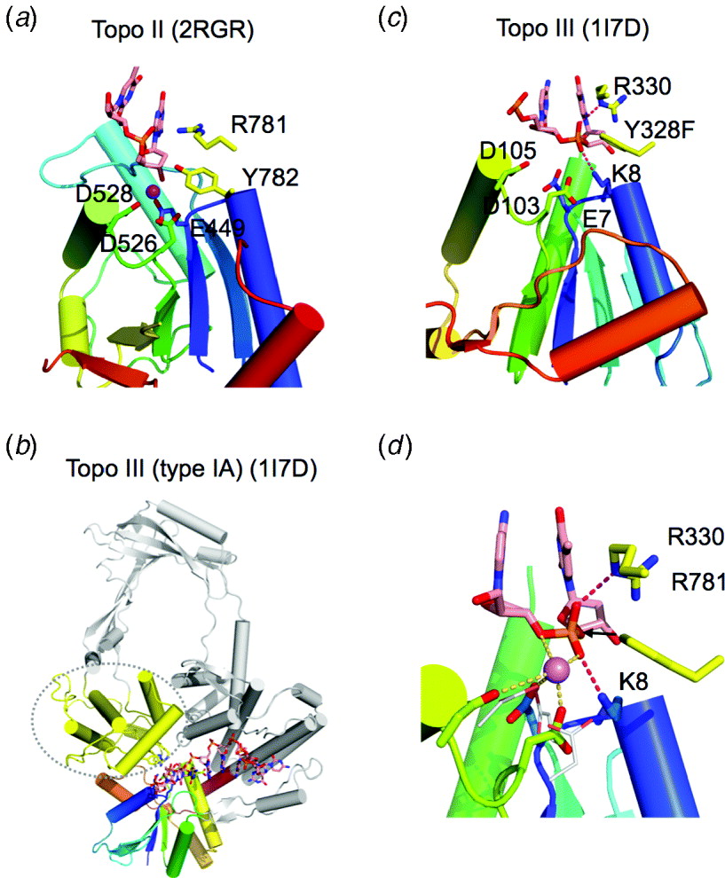
All topoisomerases cleave DNA using a Tyr side chain as the nucleophile to produce a covalent phosphotyrosyl DNA–enzyme intermediate for strand passing (see reviews by Schoeffler & Berger, Reference Schoeffler and Berger2008; Wang, Reference Wang2002). Following cleavage and topological change, DNA is religated using the DNA hydroxyl group generated in the cleavage step as the nucleophile to cleave the phosphotyrosyl bond and religate the DNA, which regenerates a free topoisomerase (Fig. 2d). In this coupled cleavage-religation scheme, the phosphodiester bond energy is preserved and an external energy source is not necessary. The active site of all topoisomerases consists of an essential Tyr nucleophile. Type IA and type II topoisomerases share a conserved catalytic core and require Mg2+ for efficient cleavage and religation, whereas TopIB are different and do not need a divalent cation. Correlated with the metal ion dependence, type II and IA topoisomerases form 5′-phosphotyrosine and 3′-OH cleavage products, whereas type IB form 3′-phosphotyrosine and 5′-OH products (Schoeffler & Berger, Reference Schoeffler and Berger2008). Type IA and type II topoisomerases further differ in ATP and DNA substrate requirements. Type IA usually requires no ATP and cleaves ssDNA only, whereas type II requires ATP and cleaves dsDNA (Schoeffler & Berger, Reference Schoeffler and Berger2008).
3.4 A two-way approach of categorization in this review
Because of the great diversity of nucleases, their classification cannot be explicitly accomplished using any single criterion. For convenience, I separate nucleases into three major classes based on their metal-ion dependence and catalytic mechanism. In each class, nucleases are further divided into families or superfamilies according to sequence and structure conservation and functional diversity. Because the catalytic mechanisms of some nucleases are yet to be determined, uncertainty in their classification is indicated. To complement the classification by catalytic mechanism, nucleases are also divided using their biological function and summarized in Table 1. Diverse catalytic mechanisms involved in each biological pathway are then cross referenced to the relevant sections and subsections.
Before going into the details of each class and superfamily of nucleases according to this metal-centric classification, the properties of Mg2+ and divalent cations and the main characteristics of metal-dependent and -independent catalyses are first summarized.
4. Basic properties of Mg2+ and divalent cations
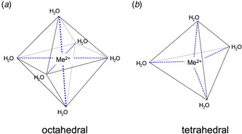
Mg2+ and Ca2+ are the most abundant divalent cations in living organisms (Cowan, Reference Cowan2002; Lyons & Eide, Reference Lyons, Eide, Bertini, Gray, Stiefel and Valentine2006), and Mg2+ is the most abundant divalent cation inside cells (Maguire & Cowan, Reference Maguire and Cowan2002; Romani & Scarpa, Reference Romani and Scarpa1992). Other ions like Fe2+, Zn2+ and Cu2+ are widespread, and Mn2+ and Ni2+ are essential but found at low concentrations (Lyons & Eide, Reference Lyons, Eide, Bertini, Gray, Stiefel and Valentine2006). The principal quality of these divalent cations is the high density of positive charge, which makes them efficient for charge neutralization of phospholipids and nucleic acids. The second common property is hydration and the specific ligand requirement. According to the atomic or covalent radius Ca2+ is the largest (1·9 Å), Mg2+ the second (1·6 Å) and the rest are similarly smaller (1·3 Å). However, when hydrated, Mg2+ is larger than Ca2+, because Mg2+ is associated with multiple shells of ligated water molecules (Maguire & Cowan, Reference Maguire and Cowan2002). In biological systems, these metal ions never exist without water or ligand. Therefore, ionic (Shannon) radii (Pauling, Reference Pauling1961) are inapplicable. The most common ligand coordination geometry is octahedral or tetrahedral (Harding, Reference Harding1999) (Fig. 4). Mg2+ prefers to have six inner-sphere ligands arranged in an octahedral configuration and so does Fe2+ (Harding, Reference Harding1999). Ca2+ and Zn2+ can have octahedral coordination, but Zn2+ is frequently coordinated by four ligands in a tetrahedron (Christianson, Reference Christianson1991), and Ca2+ by seven, eight or even nine ligands (Pidcock & Moore, Reference Pidcock and Moore2001). The number and length of ligand bonds of metal ions are empirically determined and vary within a very narrow range (Harding, Reference Harding1999). Metal-ion coordination is far more sensitive to the ligand type and geometry than hydrogen bonds or salt bridges observed in macromolecules.
Metal-dependent nucleases most often use Mg2+ and Zn2+ to facilitate phosphodiester bond breakage. (a) Diagram of octahedral coordination preferred by Mg2+ and can also be adopted by Zn2+. (b) Tetrahedral coordination favored by Zn2+.

A variety of divalent cations have been found to be involved in enzymatic catalysis, e.g. Fe2+ in non-heme iron enzymes (oxygenases and demethylases) (Lange & Que, Reference Lange and Que1998), Zn2+ in deacetylases and proteases (Hernick & Fierke, Reference Hernick and Fierke2005; Kim & Mobashery, Reference Kim and Mobashery2001), Ni2+ in urease (Jabri et al. Reference Jabri, Carr, Hausinger and Karplus1995), and the Mg2+ ion is most frequently associated with nucleic acid enzymes (Cowan, Reference Cowan2002). This frequent use of Mg2+ is perhaps because of its abundance, solubility, redox stability when compared with Mn2+, Fe2+ and Cu2+, its small size relative to Ca2+, and its rigid coordination geometry when compared with the transition metals Fe2+, Cu2+, Ni2+ and Zn2+ (Maguire & Cowan, Reference Maguire and Cowan2002). The ligand bond of the Mg2+ ion is empirically determined to be 2·07 Å (Harding, Reference Harding1999). The coordination geometry of Mg2+ is far more stringent than hydrogen bonds. The unusual hydration property of Mg2+ ions may also be a factor for its catalytic role. Mg2+ ions exhibit extremely slow exchange rates of inner shell water molecules (Diebler et al. Reference Diebler, Eigen, Ilgenfritz, Maass and Winkler1969; Maguire & Cowan, Reference Maguire and Cowan2002) and often retain a couple of water ligands when coordinated by organic or macro molecules (Harding, Reference Harding1999). It is energetically very costly to replace water ligands of Mg2+ with macromolecular ligands. The pK a value of a water molecule is reduced from 16·0 in free solution to 11·4 when associated with Mg2+ (Pontius et al. Reference Pontius, Lott and Von Hippel1997).
5. Three major classes divided by metal-ion dependence
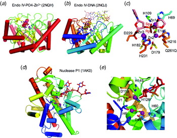
An overwhelming majority of nucleases of diverse function and structure belong to one of the three classes, requiring none, one or two metal ions for catalysis. Three Zn2+ ions have been found in the active site of two nucleases: nuclease P1 and Escherichia coli Endo IV (Garcin et al. Reference Garcin, Hosfield, Desai, Haas, Bjoras, Cunningham and Tainer2008; Ivanov et al. Reference Ivanov, Tainer and Mccammon2007; Romier et al. Reference Romier, Dominguez, Lahm, Dahl and Suck1998). They are treated as a variation of the two-metal-ion mechanism in this review. The metal-independent RNases and DNases are grouped into different sections, because there is nothing in common.
5.1 Two-metal-ion catalysis
Two-metal-ion catalysis was first proposed after observing two metal ions in the active site of 3′ to 5′ exonuclease (DnaQ-like) active site of the Klenow fragment and alkaline phosphatase (Beese & Steitz, Reference Beese and Steitz1991; Freemont et al. Reference Freemont, Friedman, Beese, Sanderson and Steitz1988; Kim & Wyckoff, Reference Kim and Wyckoff1991). It was further suggested to be a general mechanism for ribozymes catalyzing RNA splicing, which have no chemical groups that have pK a's near neutrality to serve as general base and acid for the phosphoryl transfer reaction (Steitz & Steitz, Reference Steitz and Steitz1993). Over the years, the two-metal-ion mechanism has been proven to be utilized by all DNA and RNA polymerases (Steitz, Reference Steitz1998) and many nucleases including self-splicing ribozymes (Stahley & Strobel, Reference Stahley and Strobel2005; Toor et al. Reference Toor, Rajashankar, Keating and Pyle2008).
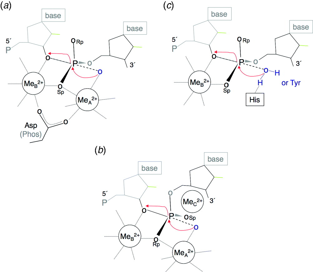
In all cases, the metal-ion-dependent reaction products are 5′-phosphate and 3′-OH groups. The two metal ions (A and B) are ~4 Å apart in the ground state and coordinated between non-bridging oxygen of the scissile phosphate and an absolutely conserved Asp or a phosphate backbone in the case of ribozymes (Fig. 5a). The A metal ion is on the nucleophile side and the B metal ion is on the 3′-O leaving-group side. Additional carboxylates or polar groups help to coordinate the metal ions and exclude water molecules from metal ion B. Two-metal-ion catalysis is fully compatible with the native double-helix structure and requires no phosphosugar distortion, base unstacking or flipping out. The advantages of incorporating two metal ions into catalysis are high substrate specificity and efficient product release (Yang et al. Reference Yang, Lee and Nowotny2006). In addition, two symmetrically placed metal ions in the cut-and-paste recombinases and group I and II introns (ribozymes) allow multiple reactions to be consecutively catalyzed in one active site (Kennedy et al. Reference Kennedy, Haniford and Mizuuchi2000; Steitz & Steitz, Reference Steitz and Steitz1993).
Metal-ion-dependent catalysis. (a) Two-metal-ion mechanism. The pro-Sp oxygen of a scissile phosphate coordinates both metal ions, one on the 5′ side and the other on the 3′ side. The pro-Rp oxygen is exposed to solvent. When labeling, pro-Sp is abbreviated as Sp and pro-Rp as Rp. (b) The three-metal-ion mechanism is a variation of the two-metal-ion mechanism. The scissile phosphate is turned and the pro-Rp oxygen coordinates the two catalytic metal ions and, the pro-Sp oxygen is stabilized by the third divalent cation. (c) The one-metal-ion mechanism is an alternative to the two-metal-ion mechanism. One metal ion (A) is eliminated and may be replaced by a positively charged protein side chain. In the cases where water is the nucleophile, a conserved His often acts as the general base to deprotonate it for nucleophilic attack. Alternatively, Tyr is the nucleophile as in topoisomerases, relaxases and RCR-related recombinases. In all three metal-ion-dependent mechanisms, the nucleophile is always on the 5′ side, and cleavage results in 5′-phosphate and 3′-OH.

In most cases, Mg2+ is the choice for two-metal-ion catalysis, and Ca2+ inhibits phosphoryl transfer (Yang et al. Reference Yang, Lee and Nowotny2006). Some enzymes, e.g. Mre11, require Mn2+ for in vitro nuclease activity (Hopfner et al. Reference Hopfner, Karcher, Craig, Woo, Carney and Tainer2001; Paull & Gellert, Reference Paull and Gellert1998). Because of the stringent coordination geometry and charge requirements of Mg2+, a binding of two such metal ions is most often substrate dependent and highly selective. A number of crystal structures have captured metal ions in non-canonical configurations, which can be correlated with mutated metal-ion coordinated ligands and displacement of the scissile phosphate (Devos et al. Reference Devos, Tomanicek, Jones, Nossal and Mueser2007; Horton & Perona, Reference Horton and Perona2004; Nowotny et al. Reference Nowotny, Gaidamakov, Ghirlando, Cerritelli, Crouch and Yang2007). In comparison, Mn2+ tends to relax substrate specificity and can rescue defective enzymes (Yang et al. Reference Yang, Lee and Nowotny2006 and references therein). This ‘relaxing’ and broadening of substrate specificity by Mn2+ is widely observed in metalloenzymes perhaps due to Mn2+ being a transitional element with less stringent coordination requirements than Mg2+. Many enzymes are active in mixed metal ion experiments as long as a trace of Mg2+ or Mn2+ are present (Brautigam & Steitz, Reference Brautigam and Steitz1998; Pingoud et al. Reference Pingoud, Wende, Friedhoff, Reuter, Alves, Jeltsch, Mones, Fuxreiter and Pingoud2009). It has been shown that the A site can accept various metal ions but the B site is more selective for Mg2+ for catalysis (Brautigam et al. Reference Brautigam, Sun, Piccirilli and Steitz1999).
Although metal ion A was proposed to drive the reaction by deprotonating the nucleophile, a similar pH profile of phosphoryl transfer reaction in the presence of Mg2+ or Mn2+ (Sam & Perona, Reference Sam and Perona1999), which would result in different pK a's of their ligands, is inconsistent with the metal ion being the general base and simply deprotonating the nucleophilic water. Hydrolytic reactions may occur without a general base, and the catalytic rate may be reduced by three to five orders of magnitude (Mildvan et al. Reference Mildvan, Xia, Azurmendi, Saraswat, Legler, Massiah, Gabelli, Bianchet, Kang and Amzel2005). The current interpretation is that metal ion B is at least as important as metal ion A in two-metal-ion catalysis. In addition to the proposed role of stabilizing the penta-covalent transition state, metal ion B appears to destabilize the ground state of a scissile phosphate for cleavage (Yang et al. Reference Yang, Lee and Nowotny2006). Structural studies of an RNase H reaction intermediate and product indicate that the two metal ions are likely to move closer than 3·5 Å and together stabilize the transition state (Nowotny & Yang, Reference Nowotny and Yang2006).
5.2 One-metal-ion catalysis
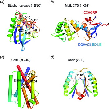
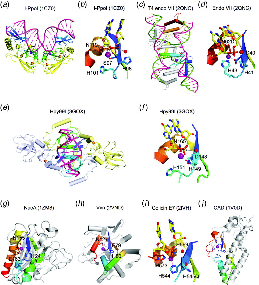
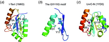
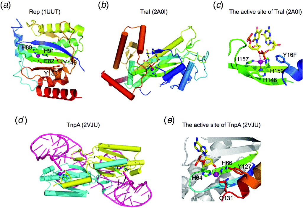
An alternative to the two-metal-ion mechanism is one-metal-ion catalysis, in which metal ion B is retained and metal ion A is absent (Yang, Reference Yang2008) (Fig. 5b). Two major classes of endonucleases are confirmed to use one-metal ion for catalysis: ββα-Me and HUH (Friedhoff et al. Reference Friedhoff, Franke, Meiss, Wende, Krause and Pingoud1999; Koonin & Ilyina, Reference Koonin and Ilyina1993; Kuhlmann et al. Reference Kuhlmann, Moore, James, Kleanthous and Hemmings1999; Monzingo et al. Reference Monzingo, Ozburn, Xia, Meyer and Robertus2007). Although sharing little tertiary structural similarity, both classes of nucleases use at least one His side chain to coordinate the metal ion, and the reaction coordinates (arrangement of the metal ion, scissile phosphate and nucleophile) are nearly superimposable. Like the two-metal-ion mechanism, the active site configuration of ββα-Me and HuH nucleases is compatible with the double-helix structure and produces 5′-phosphate and 3′-OH groups (Fig. 5b). The ββα-Me nucleases most often contain a His as a general base to deprotonate and activate a nucleophilic water, whereas the HUH nucleases invariably use Tyr as the nucleophile to cleave DNA substrates (Yang, Reference Yang2008). Metal ion selection by one-metal-ion-dependent nucleases is often less stringent, and substrate specificity is concomitantly less discriminating than for two-metal-ion enzymes (Yang, Reference Yang2008). A number of ββα-Me nucleases are equally capable of hydrolyzing RNA and DNA (sugar non-specific) with little sequence specificity (Hsia et al. Reference Hsia, Li and Yuan2005). Sequence-specific DNases in the ββα-Me family, for example, homing endonuclease I-PpoI and the type-II restriction endonuclease KpnI, depend on additional DNA-binding domains to achieve their specificity (Sokolowska et al. Reference Sokolowska, Czapinska and Bochtler2009; Stoddard, Reference Stoddard2005).
5.3 Metal-independent catalysis
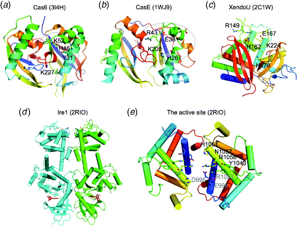
Both RNase and DNase can be metal independent. To date, the RNases that do not require metal ions all use a 2′-OH as the nucleophile to generate 2′,3′ cyclic phosphate intermediates. These enzymes therefore require phosphoribose distortion and base unstacking and unpairing surrounding a scissile phosphate for catalysis to proceed (Fig. 3b and c) (Buckle & Fersht, Reference Buckle and Fersht1994; Calvin & Li, Reference Calvin and Li2008; Cochrane & Strobel, Reference Cochrane and Strobel2008; Correll et al. Reference Correll, Yang, Gerczei, Beneken and Plantinga2004). The DNases that have been shown to cleave DNA without metal ions form phosphoenzyme covalent intermediates via Tyr, Ser or His side chains (Champoux, Reference Champoux2001; Gottlin et al. Reference Gottlin, Rudolph, Zhao, Matthews and Dixon1998; Grindley et al. Reference Grindley, Whiteson and Rice2006; Sasnauskas et al. Reference Sasnauskas, Connolly, Halford and Siksnys2007). The active site and mechanisms for metal-ion-independent cleavage by RNases and ribozymes, in particular, have been extensively characterized by X-ray crystallography (Cochrane & Strobel, Reference Cochrane and Strobel2008; Deshpande & Shankar, Reference Deshpande and Shankar2002; Raines, Reference Raines1998; Xue et al. Reference Xue, Calvin and Li2006; Yoshida, Reference Yoshida2001). The mechanism for DNA cleavage without metal ions has also been revealed by combined biochemical analysis and atomic resolution structures (Grindley et al. Reference Grindley, Whiteson and Rice2006; Schoeffler & Berger, Reference Schoeffler and Berger2008; Stuckey & Dixon, Reference Stuckey and Dixon1999). Despite variations in structures and active site components, some common features emerge from metal-independent RNases and DNases. Water appears to be excluded from the active site, a 2′-OH of RNA or a protein side chain serves as the nucleophile, and the highly negatively charged penta-covalent intermediate is neutralized by positively charged side chains.
6. Two-metal-ion-dependent nuclease superfamilies
This is by far the largest class of nucleases. It includes protein enzymes and ribozymes, sequence-specific and non-specific nucleases, endo- and exonucleases with the 5′ to 3′ or 3′ to 5′ polarity, RNase and DNase. Based on the tertiary fold and catalytic motifs, they are divided into more than a dozen superfamilies and families as listed below.
6.1 DnaQ-like 3′–5′ exonucleases with the DEDD motif
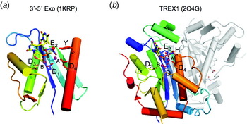
DnaQ-like nucleases share the absolutely conserved sequence motif of DEDD and all carry out 3′–5′ exonucleolytic degradation of DNA or RNA. The topology of the catalytic core, after removing the non-conservative parts, is β1-β2-β3-αA-β4-αB-β5-αC, of which the five strands form a central mixed β-sheet (54123, ↑↑↑↓↑) (Fig. 6a). The first two conserved carboxylates (DE) are one residue apart on the β1 strand, and the remaining two (DD) are located on αB and αC on either side of the β-sheet. The two-metal ions are coordinated between the first Asp of the DEDD motif and the pro-Sp non-bridging oxygen of the scissile phosphate. Each of the remaining three carboxylates coordinates one of the two-metal ions. The nucleophilic water may be oriented and activated by the highly conserved Tyr or His 4 or 5 residues proceeding to the last D of DEDD, which give rise to the name of DEDDy or DEDDh exonucleases. Interestingly, the difference in the sequence motif appears to result in little difference in function. For example, both DEDDy and DEDDh are found in the proofreading exonucleases (see below). Several members of this family form dimers (Fig. 6b), but with vastly different dimeric interfaces. Curiously, most dimeric 3′ exonucleases of the DnaQ superfamily have the DEDDh motif (see below).
The DEDD superfamily consists of DnaQ-like 3′–5′ exonucleases. (a) The core of the 3′–5′ exonuclease domain of E. coli DNA polymerase I (PDB code is shown in parenthesis) represents the DEDDy nucleases. (b) Dimeric TREX1 (2O4G) represents the DEDDh exonucleases. Proteins are shown in ribbon diagrams. One subunit of each enzyme is shown in rainbow colors from the blue N- to red C-terminus. Divalent cations are shown as purple spheres and the nucleophile water as a red sphere. The side chains of the conserved DEDDy/h motifs and nucleic acid/nucleotide are shown in sticks with oxygen in red and nitrogen in blue. All DEDD family members use the two-metal-ion mechanism as represented in the enzyme–nucleic acid or nucleotide complex structures.

6.1.1 DnaQ
DnaQ, also known as MutD or epsilon subunit of E. coli DNA polymerase III holoenzyme, encodes the 3′–5′ exonuclease (DEDDh) that performs proofreading in DNA replication (McHenry, Reference Mchenry1985). The 3′–5′ exonuclease may exist as a domain of DNA polymerase or a separate polypeptide chain in the replisome. As a domain of the Klenow fragment, the 3′–5′ exonuclease (DEDDy) was one of the earliest crystal structures of nucleic acid enzymes determined (Ollis et al. Reference Ollis, Brick, Hamlin, Xuong and Steitz1985) and the structures of Klenow-oligonucleotide complexes led to the proposal of the two-metal-ion mechanism (Beese & Steitz, Reference Beese and Steitz1991; Freemont et al. Reference Freemont, Friedman, Beese, Sanderson and Steitz1988). The structure of epsilon subunit is homologous to the 3′–5′ exonuclease domain of Klenow fragment, and only one metal ion was detected in the absence of ssDNA (Hamdan et al. Reference Hamdan, Carr, Brown, Ollis and Dixon2002). The structure of the Klenow fragment complexed with ssDNA substrate reveals the coordination of two-metal ions (Fig. 6a). Although the substrate DNA is single stranded, its conformation is highly similar to a strand in a double helix (Brautigam & Steitz, Reference Brautigam and Steitz1998; Brautigam et al. Reference Brautigam, Sun, Piccirilli and Steitz1999). These observations indicate that proper binding of two metal ions requires the presence of a cognate DNA substrate. The importance of the scissile phosphate in metal ion coordination is revealed by the sulfur substitution experiments. Replacement of the pro-Sp oxygen with sulfur essentially abolishes the metal-ion binding and the nuclease activity, and replacement of the 3′ bridging oxygen (leaving group) affects the metal-ion binding, particularly in the B site, whereas the replacement of pro-Rp oxygen moderately affects the metal ions, active site configuration and nuclease activity (Brautigam & Steitz, Reference Brautigam and Steitz1998; Brautigam et al. Reference Brautigam, Sun, Piccirilli and Steitz1999).
6.1.2 E. coli ExoI and ExoX
The 3′ ExoI is the first deoxyribonuclease purified and characterized from E. coli (Lehman & Nussbaum, Reference Lehman and Nussbaum1964). It specifically degrades ssDNA from the 3′-end and is implicated in various DNA repair pathways. The crystal structure of ExoI reveals a C-shaped monomer (PDB: 1FXX) (Breyer & Matthews, Reference Breyer and Matthews2000). The DEDDh catalytic core is followed by a C-terminal SH3-like domain and surrounded by α-helices. The structure suggests a mechanism for processive digestion of ssDNA. E. coli ExoX is highly similar to ExoI in the catalytic domain and ssDNA preference. But, without the C-terminal domain, ExoX is about a half of ExoI in size (Viswanathan & Lovett, Reference Viswanathan and Lovett1999). Homologs of E. coli ExoI and X are widely spread in bacteria.
6.1.3 TREX1 and TREX2
TREX1 and TREX2 are the major mammalian 3′–5′ exonucleases and also prefer ssDNA substrate. They can perform proofreading function for polymerases that lack an intrinsic 3′–5′ exonuclease activity (Shevelev et al. Reference Shevelev, Ramadan and Hubscher2002). Defective TREX1 (aka DNase III) causes the Aicardi–Goutieres syndrome in humans, a severe neurological disease, and systemic lupus erythematosus probably due to abnormal innate immune responses to the accumulation of intracellular nucleic acids (Crow et al. Reference Crow, Hayward, Parmar, Robins, Leitch, Ali, Black, Van Bokhoven, Brunner, Hamel, Corry, Cowan, Frints, Klepper, Livingston, Lynch, Massey, Meritet, Michaud, Ponsot, Voit, Lebon, Bonthron, Jackson, Barnes and Lindahl2006a). Mice lacking TREX1 develop inflammatory myocarditis (Morita et al. Reference Morita, Stamp, Robins, Dulic, Rosewell, Hrivnak, Daly, Lindahl and Barnes2004). It has recently been shown that TREX1 removes DNA of endogenous retroelements and prevents a type I interferon-mediated autoimmune response to self-nucleic acids (Stetson et al. Reference Stetson, Ko, Heidmann and Medzhitov2008). TREX1 is also a part of SET complex and degrades DNA during granzyme A-mediated caspase-independent cell death (Chowdhury et al. Reference Chowdhury, Beresford, Zhu, Zhang, Sung, Demple, Perrino and Lieberman2006). Structures of TREX1 and TREX2 have been determined and are highly similar to that of DnaQ (PDB: 2OA8, 2IOC, 2O4I and 1Y97) (Brucet et al. Reference Brucet, Querol-Audi, Serra, Ramirez-Espain, Bertlik, Ruiz, Lloberas, Macias, Fita and Celada2007; de Silva et al. Reference De Silva, Choudhury, Bailey, Harvey, Perrino and Hollis2007; Perrino et al. Reference Perrino, Harvey, Mcmillin and Hollis2005). TREX1 and TREX2 differ from DnaQ in being obligatory dimers, and the conserved residue in addition to DEDD in the active site is His rather than Tyr (Fig. 6b).
6.1.4 WRN exonuclease
Defective WRN protein is responsible for the Werner Syndrome, or premature aging (Muftuoglu et al. Reference Muftuoglu, Oshima, Von Kobbe, Cheng, Leistritz and Bohr2008). WRN contains both a DNA helicase and 3′–5′ exonuclease activity. The isolated WRN exonuclease domain is monomeric and can degrade the 3′-ends of dsDNA with a 5′ overhang (Perry et al. Reference Perry, Yannone, Holden, Hitomi, Asaithamby, Han, Cooper, Chen and Tainer2006a). The structure of the WRN exonuclease domain contains the DEDDy motif and is similar to other members of the DnaQ family. Two divalent ions were found in the active site in the presence of 50 mM metal ions even without DNA substrate (Perry et al. Reference Perry, Yannone, Holden, Hitomi, Asaithamby, Han, Cooper, Chen and Tainer2006a).
6.1.5 RNase T and Orn
Bacterial RNase T and Orn (oligoribonuclease) are homodimeric 3′–5′ exonucleases with the DEDDh motif. Both prefer ssRNA (Zuo et al. Reference Zuo, Zheng, Wang, Chruszcz, Cymborowski, Skarina, Savchenko, Malhotra and Minor2007) (PDB: 2F96, 2IS3, 1YTA, 2IGI, 2GBZ and 1J9A). RNase T is essential for 5S and 23S rRNA maturation and for processing of precursor tRNA (Deutscher & Li, Reference Deutscher and Li2001). Orn can also hydrolyze DNA in the 3′–5′ direction (Fiedler et al. Reference Fiedler, Vincent, Zuo, Gavrialov and Malhotra2004). In the absence of Orn, E. coli cells grow slowly. Interestingly, the two active sites of the dimeric RNase T and Orn are on opposite surfaces and appear to be independent of each other.
6.1.6 RNase D and Rrp6
RNase D has the DEDDy motif and processes the 3′ end of structured RNA (Zuo et al. Reference Zuo, Wang and Malhotra2005). It contains two HRDC (helicase RNase DC-terminal) nucleic acid binding domains in addition to the DnaQ-like catalytic domain. The circular substrate-binding tunnel suggests processivity of RNase D (PDB: 1YT3). One Zn2+ ion is tightly bound in the active site and the second Zn2+ ion has low occupancy (Zuo et al. Reference Zuo, Wang and Malhotra2005). The yeast homologue of RNase D, Rrp6p, is a component of the nuclear exosome for the degradation of unstable mRNA transcripts and maturation of rRNA, small nuclear RNA (snRNA) and small nucleolar RNAs (snoRNAs) (for review, see Houseley et al. Reference Houseley, Lacava and Tollervey2006). In the presence of 10 mM MnCl2, two-metal ions are also found in the active site of yeast Rrp6 (PDB: 2HBK) (Midtgaard et al. Reference Midtgaard, Assenholt, Jonstrup, Van, Jensen and Brodersen2006).
6.1.7 Poly A-specific ribonuclease (PARN), Pan2 and Pop2
PARN is a dimeric processive 3′-exonuclease with the DEDDh motif. In addition to the DnaQ-like catalytic domain, PARN has RNA-binding domains (PDB: 2A1S) (Wu et al. Reference Wu, Reuter, Lilie, Liu, Wahle and Song2005). Related to PARN, Pop2 is a component of the Ccr4–NOT complex and functions in mRNA degradation. Pop2 is likely to participate in mRNA deadenylation both directly with its 3′–5′-exonuclease activity and indirectly by facilitating the Ccr4 nuclease activity (Parker & Song, Reference Parker and Song2004). Curiously, although the DEDDh motif is replaced by SEDQt, yeast Pop2 (yPop2) still has the RNase activity (Thore et al. Reference Thore, Mauxion, Seraphin and Suck2003). It is unknown whether the active site still uses two-metal-ion catalysis. Fission Yeast Pop2p, however, has the normal DEDDh motifs, and its structure has been determined at high resolution and two-metal ions are found in the active site (Andersen et al. Reference Andersen, Jonstrup, Van and Brodersen2009; Jonstrup et al. Reference Jonstrup, Andersen, Van and Brodersen2007). Pan2 (poly-A nuclease), which is a component of the third 3′–5′ exoribonuclease complex for mRNA degradation and maturation, is also predicted to have a DnaQ-like exonuclease domain with the DEDDh motif.
6.1.8 ERI-1, 3′hExo and CRN-4 exonucleases
Eukaryotic ERI-1 (stands for exoribonuclease 1), human 3′-Exo (3′hExo) and C. elegans CRN-4 nucleaes share the conserved catalytic domain and the DEDDh motif. However, they are not functional orthologs perhaps owing to different additional structural domains. 3′hExo is responsible for histone mRNA degradation (Cheng & Patel, Reference Cheng and Patel2004), ERI-1 is implicated in 5·8S rRNA processing and production of small RNA for RNAi (Gabel & Ruvkun, Reference Gabel and Ruvkun2008), whereas CRN-4 is implicated in apoptotic DNA degradation (Parrish & Xue, Reference Parrish and Xue2006). The crystal structure of the full-length Caenorhabditis elegans CRN-4 has been reported (Hsiao et al. Reference Hsiao, Nakagawa, Shi, Mitani, Xue and Yuan2009). It is a dimeric nuclease and cleaves both RNA and DNA. A C-terminal Zn-domain is appended to the catalytic core and may influence CRN-4's preference to exonucleolytically degrade dsDNA over ssDNA.
6.2 RNase H-like endonucleases
The RNase H fold has the same topology as DnaQ and consists of a five-stranded mixed β-sheet (54123, ↑↑↑↓↑) surrounded by α-helices. However, the catalytic residues are different, and instead of exonucleases only endonucleolytic RNase and DNase are found in this superfamily. The catalytic residues are more varied in composition (Asp, Glu or His) and location than those in the DnaQ family. Only the first Asp in β1 is conserved in all members and is likely to coordinate both metal ions (Nowotny et al. Reference Nowotny, Gaidamakov, Crouch and Yang2005). The Asp located at the end of adjacent β4 is the second most conserved, but it is replaced by Glu in the HJ resolvase RuvC and Ydc2 (Yang & Steitz, Reference Yang and Steitz1995b). This RNase H superfamily has been recently reviewed by M. Nowotny (Nowotny, Reference Nowotny2009), and readers are referred to it for in-depth descriptions. The representative families and their characteristic are summarized below.
6.2.1 RNase H1
RNase H1 (also referred to as RNase H) is the founding member of this large eclectic superfamily of endonucleases. It is conserved from bacteria, retroviruses to humans and cleaves an RNA strand when it is hybridized to DNA. Cellular RNase H1 primarily degrades RNA primers in DNA replication and RNAs in random RNA–DNA hybrids (R loops) (Cerritelli & Crouch, Reference Cerritelli and Crouch2009; Tadokoro & Kanaya, Reference Tadokoro and Kanaya2009). RNase H activity is essential for retroviral reverse transcription (Champoux & Schultz, Reference Champoux and Schultz2009). Although not sequence-specific, RNase H1 requires a minimum of four ribonucleotides forming RNA/DNA hybrid for cleavage to occur (Tadokoro & Kanaya, Reference Tadokoro and Kanaya2009). Substrate binding and specificity can be further strengthened by one or two RNA/DNA hybrid-binding domains (HBD) found in many cellular RNase H1 and by the DNA polymerase domain in retroviral reverse transcriptase (Champoux & Schultz, Reference Champoux and Schultz2009; Nowotny et al. Reference Nowotny, Cerritelli, Ghirlando, Gaidamakov, Crouch and Yang2008).
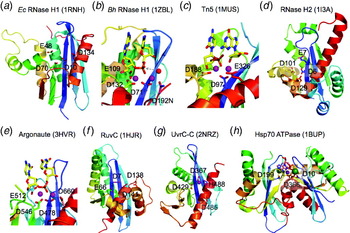
RNase H catalysis by the two-metal-ion mechanism has been established in a series of enzyme–nucleic acid complexes along the reaction pathway by X-ray crystallography (Nowotny et al. Reference Nowotny, Gaidamakov, Crouch and Yang2005; Nowotny & Yang, Reference Nowotny and Yang2006). Four carboxylates (D10, E48, D70 and D134 according to E. coli RNase H) form the catalytic core (Fig. 7a). The first and third carboxylates coordinate the two metal ions (Fig. 7b). The second carboxylate, Glu located on helix αA, is unique to RNase H1 and binds both the 2′-OH of RNA substrate and metal ion B, thereby enhancing the catalysis specificity (Nowotny et al. Reference Nowotny, Gaidamakov, Crouch and Yang2005). The fourth carboxylate coordinates metal ion A only. Although its replacement by Asn significantly reduces catalytic activity, it can be restored by the substitution of Mn2+ for Mg2+ (Nowotny & Yang, Reference Nowotny and Yang2006; Nowotny et al. Reference Nowotny, Gaidamakov, Crouch and Yang2005). In addition, the substrate actively participates in cleavage. The pro-Sp oxygen of the scissile phosphate coordinates both metal ions and the pro-Rp oxygen of the 3′ neighboring phosphate participates in nucleophile formation (Haruki et al. Reference Haruki, Tsunaka, Morikawa, Iwai and Kanaya2000; Nowotny et al. Reference Nowotny, Gaidamakov, Crouch and Yang2005).
The RNase H superfamily consists of various endonucleases. (a) E. coli RNase H1 is the prototype of this superfamily. (b) The active site of Bacillus halodurans RNase H complexed with an RNA/DNA hybrid substrate. (c) Tn5 transposase (complexed with a cleaved DNA product) represents the HIV integrase-like recombinases with the DDE motif. (d) RNase H2 has the same topology as RNase H1 but the second and fourth conserved carboxylates are differently located. (e) The active site of Tth Argonaute, essential for RNAi pathways is similar to that of RNase H1. (f) RuvC is a HJ resolvase. (g) The C-terminal nuclease domain of UvrC may use two conserved carboxylates and a His for metal-ion coordination and catalysis. (h) Monomeric Hsp70 ATPases contain two RNase H-like domains. The duplicated domains are both shown in blue- to red-colored gradients. The Asp residues in the first β-strand (D10 and D199) are conserved throughout the Hsp70 family and essential for the ATPase activity. Proteins, nucleic acids and divalent cations are shown in the same scheme as in Fig. 6. The green spheres represent the K+ ions.

6.2.2 Transposase and retroviral integrase with the DDE motif
When the structure of HIV integrase and bacterial Mu transposase were determined, it was a surprise to find that these DNA endonucleases bear clear similarity to RNase H1 (Dyda et al. Reference Dyda, Hickman, Jenkins, Engelman, Craigie and Davies1994; Rice & Mizuuchi, Reference Rice and Mizuuchi1995). Sequence alignment predicts that many cellular and retroviral enzymes catalyzing cut-and-paste sequence-specific transposition/recombination have the RNase H fold and a DDE motif, which is approximately equivalent to the 1st, 3rd and 4th carboxylates of RNase H1 (Fig. 7a–c). They include the human RAG1 and RAG2 proteins essential for immunoglobulin gene rearrangement (Kim et al. Reference Kim, Dai, Mundy, Yang and Oettinger1999; Landree et al. Reference Landree, Wibbenmeyer and Roth1999), and eukaryotic Mariner and Hermes transposases (Hickman et al. Reference Hickman, Perez, Zhou, Musingarimi, Ghirlando, Hinshaw, Craig and Dyda2005; Richardson et al. Reference Richardson, Colloms, Finnegan and Walkinshaw2009). Integrases and transposases are at least dimeric when associated with two donor-DNA ends and engaged in recombination. As revealed by the Tn5 transposase–DNA complex structures, the three carboxylates in these integrases and transposases are symmetrically arranged around the two metal ions (Davies et al. Reference Davies, Goryshin, Reznikoff and Rayment2000; Steiniger-White et al. Reference Steiniger-White, Rayment and Reznikoff2004). The first Asp coordinates both and the remaining two coordinate one metal ion each (Fig. 7c). The recently reported structure of a retroviral integrase complexed with two donor DNA ends, which are nearly perpendicular to each other rather than antiparallel as in the case of Tn5, confirms the active site configuration (Hare et al. Reference Hare, Gupta, Valkov, Engelman and Cherepanov2010). The symmetry surrounding the two metal ions has implications for the DNA transposition reaction. These integrase and transposase often catalyze two or more consecutive phosphoryltransfer reactions and the 3′-OH product of the first cleavage reaction can be used as a nucleophile for the second strand transfer reaction (Kennedy et al. Reference Kennedy, Haniford and Mizuuchi2000). It has thus been proposed that the two metal ions switch their roles in coordinating the nucleophile and the leaving group in the two consecutive steps (Nowotny, Reference Nowotny2009; Nowotny et al. Reference Nowotny, Gaidamakov, Crouch and Yang2005; Steitz & Steitz, Reference Steitz and Steitz1993).
6.2.3 RNase H2
RNase H2 is the second cellular RNase H found from bacteria to humans (Cerritelli & Crouch, Reference Cerritelli and Crouch2009). Eukaryotic RNase H2 consists of two auxiliary subunits, and all three subunits of human RNase H2 have been implicated in the Aicardi–Goutieres syndrome, a heritable autoimmune disease caused by either TREX1 or RNase H2 deficiency due to the accumulation of residual RNA and DNA (Crow & Rehwinkel, Reference Crow and Rehwinkel2009; Crow et al. Reference Crow, Leitch, Hayward, Garner, Parmar, Griffith, Ali, Semple, Aicardi, Babul-Hirji, Baumann, Baxter, Bertini, Chandler, Chitayat, Cau, Dery, Fazzi, Goizet, King, Klepper, Lacombe, Lanzi, Lyall, Martinez-Frias, Mathieu, Mckeown, Monier, Oade, Quarrell, Rittey, Rogers, Sanchis, Stephenson, Tacke, Till, Tolmie, Tomlin, Voit, Weschke, Woods, Lebon, Bonthron, Ponting and Jackson2006b). RNase H2 differs from RNase H1 in that it can cleave a single ribonucleotide embedded in a DNA duplex. Crystal structures of bacterial RNase H2 reveal an RNase H1-like structure with altered locations of catalytic carboxylates (Fig. 7d). Only the first and third of the four catalytic carboxylates are superimposable with those of RNase H1. The second carboxylate (E) is immediately located next to the first one (D) instead of 40 residues away. The reaction coordinate is yet to be determined.
6.2.4 Argonaute and PIWI
Argonautes (Ago) and PIWI are the key players in the RNAi pathway and the RNase activity cleaves target mRNA when it forms base pairs with guide RNA (Joshua-Tor, Reference Joshua-Tor2006; Nowotny & Yang, Reference Nowotny and Yang2009). But the catalytic residues are not always conserved, and some Ago and PIWI without catalytic activity function in RNAi pathways by binding complementary guide and target RNA duplexes (Farazi et al. Reference Farazi, Juranek and Tuschl2008; Joshua-Tor, Reference Joshua-Tor2006). For those having RNase activity, the three catalytic residues, either DDD or DDH, are approximately equivalent to the DDE in the integrase family and the 1st, 3rd and 4th carboxylates of RNase H1 (Joshua-Tor, Reference Joshua-Tor2006). The catalytic mechanism is revealed in a series of elegant crystallographic studies of Ago–nucleic acid complexes (Wang et al. Reference Wang, Juranek, Li, Sheng, Tuschl and Patel2008, Reference Wang, Juranek, Li, Sheng, Wardle, Tuschl and Patel2009). Similar to transposases, the first carboxylate coordinates the two metal ions and the remaining two coordinate one metal ion each (Fig. 7e).
6.2.5 RuvC and Ydc2
Bacterial RuvC and mitochondrial Cce1/Ydc2 are the HJ resolvases that have the RNase H fold (Ariyoshi et al. Reference Ariyoshi, Vassylyev, Iwasaki, Nakamura, Shinagawa and Morikawa1994; Ceschini et al. Reference Ceschini, Keeley, Mcalister, Oram, Phelan, Pearl, Tsaneva and Barrett2001; Iwasaki et al. Reference Iwasaki, Takahagi, Shiba, Nakata and Shinagawa1991; West & Connolly, Reference West and Connolly1992; White & Lilley, Reference White and Lilley1997). These proteins are obligatory dimers (Bennett et al. Reference Bennett, Dunderdale and West1993). The active site consists of four carboxylates, and the first two appear to be equivalent to the 1st and 3rd carboxylates of RNase H1, but the carboxylate at the end of β4 is Glu instead of Asp (Fig. 7f). The last two carboxylates are located on the C-terminal α-helix two residues apart. Based on biochemical analyses, a protein–DNA complex model has been proposed (Fogg et al. Reference Fogg, Kvaratskhelia, White and Lilley2001; Yoshikawa et al. Reference Yoshikawa, Iwasaki and Shinagawa2001). How these carboxylates together with the DNA substrate coordinate the metal ions is yet to be elucidated.
6.2.6 UvrC (C-terminal domain)
UvrC is a bacterial endonuclease involved in nucleotide excision repair. It makes two incisions, one on each side of a lesion (Verhoeven et al. Reference Verhoeven, Van Kesteren, Moolenaar, Visse and Goosen2000). UvrC has two nuclease domains in one polypeptide chain, one for each incision. The C-terminal domain of UvrC, which makes the incision 5′ to a lesion, is homologous to RNase H and shares two catalytic carboxylates with RNase H1 (1st and 3rd) (Karakas et al. Reference Karakas, Truglio, Croteau, Rhau, Wang, Van Houten and Kisker2007). But the third catalytic residue in UvrC may be a His that participates in single metal-ion coordination in the absence of DNA substrate (Fig. 7g). Interestingly, replacement of this conserved His with Ala only moderately reduces the enzymatic activity (Karakas et al. Reference Karakas, Truglio, Croteau, Rhau, Wang, Van Houten and Kisker2007). How the active site is formed upon substrate binding and whether the conserved Asp adjacent to the His is involved in catalysis awaits future analysis.
6.2.7 Structural roles and the ATPase catalytic core of Hsp70
The RNase H-like fold has been found in many proteins, where it plays a structural role, e.g. in T7 DNA polymerase and mismatch recognition protein MutS (Obmolova et al. Reference Obmolova, Ban, Hsieh and Yang2000) and RNase E (Callaghan et al. Reference Callaghan, Marcaida, Stead, Mcdowall, Scott and Luisi2005). Intriguingly, it is found in pre-mRNA splicing protein Prp8 (Pena et al. Reference Pena, Rozov, Fabrizio, Luhrmann and Wahl2008; Ritchie et al. Reference Ritchie, Schellenberg, Gesner, Raithatha, Stuart and Macmillan2008), where the first carboxylate is conserved and may play an essential structural rather than a catalytic role (Yang et al. Reference Yang, Zhang, Xu, Heroux and Zhao2008). Finally, two RNase H-like domains are found in actin and Hsp70 proteins and form the ATPase catalytic core (Flaherty et al. Reference Flaherty, Mckay, Kabsch and Holmes1991). Most interestingly, a conserved Asp is found in the first β-strand of each RNase H domain in Hsp70 (D10 and D199) just as in the nucleases of the RNase H superfamily and both are essential for cation binding (K+ and Mg2+) and ATPase activity (Johnson & McKay, Reference Johnson and Mckay1999) (Fig. 7h).
6.3 The major REase fold with the D-(D/E)XK motif
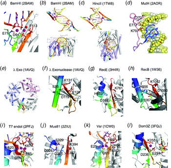
Many commonly used type-II restriction endonucleases (REases) share the conserved motif PD-(D/E)XK, or shortened as DEK (Ban & Yang, Reference Ban and Yang1998b; Orlowski & Bujnicki, Reference Orlowski and Bujnicki2008). The three catalytic residues are located close to each other on an uneven β-hairpin (Fig. 8a). The first D is located at the beginning of the first and shorter strand, and the E and K, separated by a hydrophobic residue x, are located in the middle of the second and longer strand. The first D is most conserved and coordinates both metal ions, whereas the second E can be replaced by Q, D, N, H or S, and the third K can be replaced E, Q, D, S, N or T (Lee et al. Reference Lee, Chang, Joseph, Ghirlando, Rao and Yang2005; Orlowski & Bujnicki, Reference Orlowski and Bujnicki2008). For instance, the third catalytic residue in BamHI is Glu (E113) rather than Lys (Fig. 8a). Members of this superfamily have a very diverse primary sequence and thus different structures surrounding the catalytic core. They can be monomeric, dimeric or even tetrameric and are involved in diverse processes, from restriction digestion, DNA repair and homologous recombination to the resolution of the HJ. In addition to endonucleases, members in this superfamily can also be 5′ or 3′ exonucleases. Although not every member with a crystal structure determined is shown to bind two metal ions, the two-metal ion mechanism is consistent with all biochemical and structural observations (Horton & Perona, Reference Horton and Perona2004; Lee et al. Reference Lee, Chang, Joseph, Ghirlando, Rao and Yang2005; Newman et al. Reference Newman, Lunnen, Wilson, Greci, Schildkraut and Phillips1998; Pingoud et al. Reference Pingoud, Fuxreiter, Pingoud and Wende2005; Viadiu & Aggarwal, Reference Viadiu and Aggarwal1998, Reference Viadiu and Aggarwal2000).
The DEK superfamily consists of sequence- or structure-specific endonucleases and processive 5′–3′ exonucleases. (a) The active site of BamHI contains (E)DEE, a variation of DEK. Except for (d), the region containing the catalytic residues is shown in rainbow colored ribbon diagrams. The rest of the protein is colored gray. Divalent cations and the nucleophilic water are shown in purple and red spheres, respectively. (b) Orthogonal views of BamHI–DNA interactions. The two BamHI subunits are shown in gray and light purple. The uneven β-hairpins containing the DEK motif are highlighted in the rainbow colors. Only the β-hairpins are shown when looking into the DNA major groove. BamHI cleaves DNA to form 4 nt 5′ overhangs. The nucleotides containing the scissile phosphate are highlighted in pink sticks. (c) Orthogonal views of HincII–DNA interactions. HincII cleaves DNA to blunt ends. The dimerization interface of HincII is nearly perpendicular to that of BamHI when looking down the DNA helical axis. (d) E. coli MutH is a sequence- and methyl-specific endonuclease with the DEK motif. The conserved Lys (K79) bridges the DNA sequence-specific binding in the major groove (shown in blue) and catalytic center approaching the minor groove (shown in pink). The divalent cations (Ca2+) are shown as green spheres. (e) The trimeric phage λ exonuclease has a toroidal shape and degrades ssDNA processively. The three subunits are shown in grey, pink and blue ribbon diagrams. The active site region of the grey subunit is highlighted in rainbow colors. (f) A zoom-in view of the active site with the (E)DEK motif. (g) RecE, which is a processive exonuclease and promotes DNA recombination, is shown in the same color scheme as BamHI. (h) The active site of RecB has the DEK motif with a His at its N-terminus like RecE. (i) The HJ resolvase T7 endonuclease I has the (E)DEK motif-like MutH and binds two metal ions. (j) The structure-specific endonuclease Mus81 and XPF also contain the DEK motif with two conserved carboxylates N-terminal to it. (k) Vsr is a sequence-specific endonuclease. The DEK motif may be replaced by DHH, and the two metal ions in the active site are coordinated by the carbonyl oxygen (of T63) in addition to the conserved Asp and scissile phosphate. (l) Dom3Z and Rai1 have the conserved DEK motif and can hydrolyze GTP.

6.3.1 Restriction endonucleases (BamHI, HincII, MutH, EcoR124I, etc.)
Nearly 70% REases belong to this superfamily (Orlowski & Bujnicki, Reference Orlowski and Bujnicki2008). Many REases in this family have a Pro immediately before the conserved Asp making the PD-EXK motif and a conserved N-terminal Glu, which contributes to the metal-ion coordination as observed in EcoRV, BglI, BamHI and MutH of type-II REase family (Horton & Perona, Reference Horton and Perona2004; Lee et al. Reference Lee, Chang, Joseph, Ghirlando, Rao and Yang2005; Newman et al. Reference Newman, Lunnen, Wilson, Greci, Schildkraut and Phillips1998; Viadiu & Aggarwal, Reference Viadiu and Aggarwal1998; Winkler et al. Reference Winkler, Banner, Oefner, Tsernoglou, Brown, Heathman, Bryan, Martin, Petratos and Wilson1993). These REases are most often dimeric (Bitinaite et al. Reference Bitinaite, Wah, Aggarwal and Schildkraut1998), but monomeric REase also exist (Lee et al. Reference Lee, Chang, Joseph, Ghirlando, Rao and Yang2005; Sokolowska et al. Reference Sokolowska, Kaus-Drobek, Czapinska, Tamulaitis, Szczepanowski, Urbanke, Siksnys and Bochtler2007; Xu et al. Reference Xu, Roberts and Guo2005). By varying dimeric interfaces and thus the relative positions of the two catalytic centers, dimeric REases can cleave DNA to generate blunt ends or staggered ends with various 5′ or 3′ overhangs (Fig. 8b and c) (Ban & Yang, Reference Ban and Yang1998b; Newman et al. Reference Newman, Lunnen, Wilson, Greci, Schildkraut and Phillips1998). The catalytic module invariably approaches DNA from the minor groove side, and the sequence-specific binding is conducted by a separate module/subdomain in the major groove (Fig. 8d). The first two carboxylates of the DEK motif coordinate the metal ions. The third, which usually is hydrogen bonded with both the nucleophilic water and the DNA-binding module in the major groove (Fig. 8d), has been attributed to couple DNA sequence recognition with the cleavage reaction (Lee et al. Reference Lee, Chang, Joseph, Ghirlando, Rao and Yang2005).
Type-I and type-III restriction/modification systems differ from type-II in the requirement of nucleotide triphosphate and DNA translocation (Dryden et al. Reference Dryden, Murray and Rao2001; Murray, Reference Murray2000), but their endonucleases have a similar active site composed of D and EXK motifs (Niv et al. Reference Niv, Ripoll, Vila, Liwo, Vanamee, Aggarwal, Weinstein and Scheraga2007). The crystal structure of EcoR124I of type-I motor-endonuclease subunit shows that the conserved DEK motif indeed forms the catalytic center although the Pro before the first conserved D is absent (Lapkouski et al. Reference Lapkouski, Panjikar, Janscak, Smatanova, Carey, Ettrich and Csefalvay2009).
6.3.2 Phage λ and RecE 5′–3′ exonuclease
The active site of the PD-(D/E)XK motif is found among bacterial and phage 5′–3′ exonucleases, which act on dsDNA and produce a protruding 3′ ssDNA for strand annealing or invasion in homologous recombination (Kolodner et al. Reference Kolodner, Hall and Luisi-Deluca1994; Stahl et al. Reference Stahl, Thomason, Poteete, Tarkowski, Kuzminov and Stahl1997). RecE together with the ssDNA annealing protein RecT has been exploited for enhanced homologous recombination for in vivo gene manipulation (Muyrers et al. Reference Muyrers, Zhang and Stewart2000; Zhang et al. Reference Zhang, Buchholz, Muyrers and Stewart1998). Phage λ exonuclease (aka Red recombinase) and E. coli RecE are trimeric and tetrameric, respectively, and both form a toroidal structure, whose central hole is proposed to bind DNA for processive degradation (Kovall & Matthews, Reference Kovall and Matthews1997; Zhang et al. Reference Zhang, Xing, Herr and Bell2009) (Fig. 8e). A conserved Glu precedes the DEK motif in Phage λ exonuclease (Fig. 8f). In RecE, the DEK motif becomes DDK and it is preceeded by a conserved His (Fig. 8g). Whether the multiple active sites in these highly processive exonucleases work sequentially or stochastically is not known yet.
6.3.3 RecB and AdnAB ssDNA nuclease
The nuclease domains of the bacterial motor-nuclease machines RecBCD and AdnAB are closely related to RecE and REases (Singleton et al. Reference Singleton, Dillingham, Gaudier, Kowalczykowski and Wigley2004; Sinha et al. Reference Sinha, Unciuleac, Glickman and Shuman2009). They have both exonuclease and endonuclease activities on ssDNA (Yu et al. Reference Yu, Souaya and Julin1998). Although RecB contains a single nuclease domain, it is one of the three subunits in the helicase/nuclease RecBCD complex. RecBCD forms a cylindrical molecule and DNA threads through the central hole similar to the cases of RecE and phage λ exonuclease. But the DNA becomes unwound by the helicase activity carried by RecB and RecD of RecBCD or by AdnA and AdnB (Unciuleac & Shuman, Reference Unciuleac and Shuman2009). The nucleases then nick the ssDNA and ultimately generate 3′ ssDNA overhangs for homologous recombination (Dillingham & Kowalczykowski, Reference Dillingham and Kowalczykowski2008; Sinha et al. Reference Sinha, Unciuleac, Glickman and Shuman2009). Deviating from the canonical DEK motif, the Glu is replaced by Asp in RecB as in RecE. RecB does not have the Pro preceding the DDK motif. A conserved His N-terminal to the DDK is likely to participatein catalysis (Fig. 8h).
Dna2 ubiquitous in all eukaryotes has a helicase domain and a RecB-like nuclease domain containing the DEK motif (Budd et al. Reference Budd, Choe and Campbell2000). Dna2 works with ssDNA-binding protein during DNA replication and removes excessively long primers (Kim et al. Reference Kim, Kim, Ryu, Kim, Hurwitz and Seo2006; Masuda-Sasa et al. Reference Masuda-Sasa, Imamura and Campbell2006).
6.3.4 HJ resolvase: T7 endonuclease I
Phage T7 endonuclease I and archaeal Hjc are HJ resolvases, whose active sites contain the REase-like (E)-PD-EXK motif (Lilley & White, Reference Lilley and White2001). These HJ resolvases are homodimeric and recognize four-way junctions in a distorted stacked-X form as shown in the crystal structures of enzyme–DNA complexes (PDB: 2PFJ and 2WJ0) (Hadden et al. Reference Hadden, Déclais, Carr, Lilley and Phillips2007). Recognition and resolution of HJ are recently reviewed (Declais & Lilley, Reference Declais and Lilley2008). T7 endonuclease I cleaves DNA by the two-metal-ion mechanism with the two most conserved carboxylates coordinating two metal ions (Hadden et al. Reference Hadden, Déclais, Carr, Lilley and Phillips2007) (Fig. 8i).
6.3.5 Structure-specific endonuclease: XPF, Hef, Rad1 and Mus81
The archaeal and eukaryotic structure-specific nucleases, Hef, Rad1, XPF and Mus81 all share the conserved nuclease domain, known as ERCC4 domain. The catalytic motif of D-ERK is homologous to that of REases (Chang et al. Reference Chang, Kim, Choi, Lee and Cho2008; Newman et al. Reference Newman, Murray-Rust, Lally, Rudolf, Fadden, Knowles, White and Mcdonald2005; Nishino et al. Reference Nishino, Komori, Ishino and Morikawa2003) (Fig. 8j). These nucleases are required for DNA repair, processing of a stalled replication fork, and repair-associated recombination (for an in-depth review, read Ciccia et al. Reference Ciccia, Mcdonald and West2008). In addition to the DEK motif, two conserved carboxylates, Asp and Glu, N-terminal to it are located in the active site (Fig. 8j). DNA substrates can vary from a 3′ flap, replication fork, to a HJ, all of which have the common feature of a 3′ flap, and cleavage occurs at the single- and double-strand junction. Although archaeal Hef nuclease is homodimeric, all eukaryotic nucleases in this family, for example, Rad1, XPF and Mus81, form heterodimers (Rad1/Rad10, XPF/ERCC1 and Mus81/Eme1, Mus81/Eme2, or Mus81/Mms4) with only one active subunit. Interestingly, even with homodimeric nucleases it is likely that only one subunit is engaged in catalysis at a given time (Newman et al. Reference Newman, Murray-Rust, Lally, Rudolf, Fadden, Knowles, White and Mcdonald2005). Based on the conservation of the active site, two-metal-ion catalysis is predicted.
6.3.6 DNA mismatch-repair endonuclease very short-patch repair (Vsr)
E. coli Vsr endonuclease specifically cleave 5′ to the T of mismatched T:G base pair, which results from the deamination of 5-methyl-cytosine in a specific sequence (Hennecke et al. Reference Hennecke, Kolmar, Brundl and Fritz1991). The crystal structures of Vsr and Vsr complexed with DNA cleavage products reveal the REase fold in spite of the absence of the last two conserved catalytic residues of the DEK motif (Bunting et al. Reference Bunting, Roe, Headley, Brown, Savva and Pearl2003; Tsutakawa et al. Reference Tsutakawa, Jingami and Morikawa1999). A conserved Glu preceding the central uneven β-hairpin and two His residues C-terminal to it are in the catalytic center and may be involved in metal-ion coordination and nucleophile activation (Fig. 8k). In addition to the absolutely conserved Asp at the beginning of the uneven β-hairpin, a backbone carbonyl oxygen in the second β-strand also contributes to the metal-ion coordination in the Vsr–DNA cleavage product complex (Fig. 8k). The presence of two Mg2+ ions in the enzyme–product complex structure is highly suggestive of the two-metal-ion mechanism as observed for the other members of this superfamily.
6.3.7 Rai1/Dom3Z for mRNA degradation and processing
Human Dom3Z and yeast Rai1 are the newest enzymes found to have the conserved REase fold and the (E)-D-EXK motif (Xiang et al. Reference Xiang, Cooper-Morgan, Jiao, Kiledjian, Manley and Tong2009) (Fig. 8l). Before the crystal structure of Rat1–Rai1 complex was determined, Rai1 was thought to activate the 5′ to 3′ exoribonuclease Rat1 in the processing of RNA (Xue et al. Reference Xue, Bai, Lee, Kallstrom, Ho, Brown, Stevens and Johnson2000). But the structure reveals a divalent cation-binding site in Rai1. The authors proceed to show that the Rai1 homologue Dom3Z binds GDP, possesses a pyrophosphatase activity and can remove pyrophosphate from a 5′ triphosphorylated RNA. However, the GDP in the crystal structure is too far from the (E)-D-EXK motif to be either a substrate or product (Fig. 8l). Whether Rai1 has nuclease activity is yet to be determined.
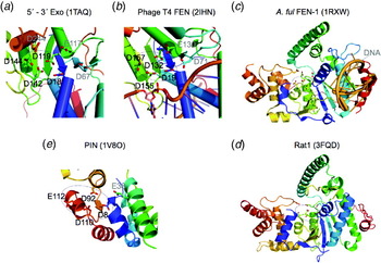
6.4 FEN1-like 5′ exo- and endo-nucleases
The nucleases in this family share a conserved βα Rossmann-like fold consisting of a central parallel five-stranded β-sheet (in the order of 54 123) surrounded by α-helices. The catalytic residues are located between the ends of strands (β1, β3 and β4) and the beginning of the following helices. They are almost exclusively Asp and Glu residues, and the number of conserved carboxylates ranges from five to eight, four of which may directly participate in phosphodiester bond cleavage (Fig. 9a). Although the crystal structures of Archaeoglobus fulgidus FEN1 and T4 RNase H were determined in complex with DNA (Chapados et al. Reference Chapados, Hosfield, Han, Qiu, Yelent, Shen and Tainer2004; Devos et al. Reference Devos, Tomanicek, Jones, Nossal and Mueser2007), the DNA is either far away from the active site (Fig. 9c) or approximately in the active site but with the scissile phosphate displaced in the absence of metal ions (Fig. 9b). However, two divalent cations have been observed in several apo-FEN1 structures and biochemical analyses suggest that these nucleases use the two-metal-ion mechanism like other nucleases containing an absolutely conserved Asp (Liu et al. Reference Liu, Kao and Bambara2004; Tomlinson et al. Reference Tomlinson, Atack, Chapados, Tainer and Grasby2010 and reference therein). Many members of this superfamily have dual 5′-exo and 5′ flap-endonuclease activities.
The FEN1 and 5′–3′ exonuclease superfamily. (a) The active site of the 5′–3′ exonuclease domain of Taq DNA polymerase. Four of the seven conserved carboxylates (encircled) are likely to participate in metal-ion binding and catalysis. (b) Phage T4 FEN1, previously known as T4 RNase H. The structure was determined in the complex with DNA substrate, but the scissile phosphate is too far away from the catalytic residues in the absence of divalent cations. (c) The archaeal FEN1 has the same active site composition as the bacterial and phage orthologs. The bound DNA is far away from the nuclease active site. (d) Rat1, which is an RNase, has the same overall structure and conserved active site as archaeal FEN1. (e) The PIN domain represents the minimal catalytic core of the nucleases in this superfamily. In all panels, the protein topology is shown in the color gradient from the blue N- to red C-terminus.

6.4.1 5′ FENs: T4 RNase H, yeast Rad27 and human FEN1
The 5′–3′ exonuclease intrinsic to E. coli DNA Pol I is the founding member of the superfamily (Klett et al. Reference Klett, Cerami and Reich1968; Setlow & Kornberg, Reference Setlow and Kornberg1972), which includes Phage T4 RNase H, yeast Rad27 and human FEN1. The exonuclease is active on either RNA or DNA and its main function is to remove RNA primers in lagging strand synthesis (Bhagwat et al. Reference Bhagwat, Hobbs and Nossal1997; Liu et al. Reference Liu, Kao and Bambara2004). These nucleases also contain the structure-specific endonuclease activity and can cleave the 5′ displaced ssDNA flap, such as the displaced primers in lagging strand DNA synthesis. The latter activity gives rise to the name of flap endonuclease, FEN1 (Harrington & Lieber, Reference Harrington and Lieber1994). In bacteriophages, archaea and mammals, the 5′–3′ exo and FENs can exist as an independent protein, for example, T4 RNase H (a mis-nomenclature; Bhagwat et al. Reference Bhagwat, Hobbs and Nossal1997). In fact, T4 RNase H has the same activities and structure as the 5′–3′ exonuclease of E. coli Pol I (Fig. 9a, b). To avoid confusion, phage T4 RNase H has been renamed as T4FEN (Tomlinson et al. Reference Tomlinson, Atack, Chapados, Tainer and Grasby2010). FEN activity is not only involved in DNA replication but also in long-patch base excision and DSB repair (Shen et al. Reference Shen, Singh, Liu, Qiu, Zheng, Finger and Alas2005).
To date, crystal structures of several apo-nucleases in this family and phage T4 and archaeal FEN in complex with DNA have been determined (Ceska et al. Reference Ceska, Sayers, Stier and Suck1996; Chapados et al. Reference Chapados, Hosfield, Han, Qiu, Yelent, Shen and Tainer2004; Devos et al. Reference Devos, Tomanicek, Jones, Nossal and Mueser2007; Hwang et al. Reference Hwang, Baek, Kim and Cho1998; Kim et al. Reference Kim, Eom, Wang, Lee, Suh and Steitz1995). The catalytic center often contains 6–7 highly conserved Asp and Glu residues (Fig. 9a–c). The four most conserved carboxylates located after β1, β3 and β4 are reminiscent of the catalytic carboxylates in RNase H (Fig. 7a). The remaining negatively charged residues in the catalytic center is likely to be involved in binding of a third metal ion, which may facilitate substrate binding and thus indirectly contribute to the catalysis (Syson et al. Reference Syson, Tomlinson, Chapados, Sayers, Tainer, Williams and Grasby2008). However, in the absence of knowledge of the active site complete with scissile phosphate and metal ions, the reaction coordinates and chemistry for bond breakage remain uncertain.
6.4.2 XPG and HJ resolvases Gen1 and Yen1
XPG is required for nucleotide excision repair to incise at the 3′ side of a lesion and shares sequence homology with FEN1 (Habraken et al. Reference Habraken, Sung, Prakash and Prakash1993; O'Donovan et al. Reference O'DONOVAN, Davies, Moggs, West and Wood1994; Scherly et al. Reference Scherly, Nouspikel, Corlet, Ucla, Bairoch and Clarkson1993). Recently isolated eukaryotic HJ resolvases Gen1 and Yen1 are also found to be homologous to FEN1 (Ip et al. Reference Ip, Rass, Blanco, Flynn, Skehel and West2008). Interestingly, like FEN1, human XPG and its yeast homolog Rad2 both exhibit 5′-exonuclease activity (Habraken et al. Reference Habraken, Sung, Prakash and Prakash1994). Although no crystal structure has been reported for these endonucleases, the conserved active site residues and functional assay indicate that they all recognize a 5′ flap or branch structure and endonucleolytically cleave DNA at or adjacent to a branch point (including a ss–ds junction), likely by the two-metal-ion mechanism.
6.4.3 Human and yeast 5′ exonuclease 1 (Exo1)
It is perhaps not surprising to find that human and yeast 5′ exonuclease I, which are implicated in mismatch and DSB repair (Genschel et al. Reference Genschel, Bazemore and Modrich2002; Szankasi & Smith, Reference Szankasi and Smith1995), are homologous to FEN1 and XPG and share the same conserved active site. Like its homologues, Exo1 also has the FEN activity (Lee & Wilson, Reference Lee and Wilson1999; Tran et al. Reference Tran, Erdeniz, Dudley and Liskay2002). This FEN activity of ExoI is reported to be stimulated by Wrn protein (Sharma et al. Reference Sharma, Sommers, Driscoll, Uzdilla, Wilson and Brosh2003). Exo1 also uses the two-metal-ion mechanism.
6.4.4 5′ exoribonuclease Rat1 (XRN2) and XRN1
Recently, the structure of mouse Rat1 or XRN2, the 5′ exoribonuclease for RNA processing, has been reported (Xiang et al. Reference Xiang, Cooper-Morgan, Jiao, Kiledjian, Manley and Tong2009). Rat1 is highly similar to the structure of archaeal FEN1 with a large N-terminal loop wrapping around the protein and a helical tower (Fig. 9c, and d). The highly conserved carboxylate-rich active site implicates a metal-ion-dependent mechanism of catalysis by these XRNs. It is unclear what makes them specific for RNA degradation and whether XRNs may have endonuclease activities if a proper substrate-binding partner is present. Eukaryotic XRN1, the major 5′–3′ exonuclease of RNAs, shares the conserved catalytic residues of XRN2 (Szankasi & Smith, Reference Szankasi and Smith1996) and is predicted to have a FEN1-like structure and active site.
6.4.5 PIN domain RNA exonuclease
PIN (PilT N-terminal domain) nuclease is a recent addition to the FEN1 superfamily. It is a component of the exosome for RNA turnover and is also involved in nonsense mediated mRNA decay (Arcus et al. Reference Arcus, Backbro, Roos, Daniel and Baker2004; Clissold & Ponting, Reference Clissold and Ponting2000; Eberle et al. Reference Eberle, Lykke-Andersen, Muhlemann and Jensen2009). Crystal structures of PIN nucleases reveal a stripped down minimal core of FEN1 (Glavan et al. Reference Glavan, Behm-Ansmant, Izaurralde and Conti2006) (Fig. 9e). Like many members of this superfamily, PIN has both 5′-exo and endonucleolytic activities (Eberle et al. Reference Eberle, Lykke-Andersen, Muhlemann and Jensen2009; Schneider et al. Reference Schneider, Leung, Brown and Tollervey2009). Three Asp residues after β1, β3 and β4 are highly conserved among PIN nucleases as well as all FEN1 and XPG superfamily members. The fourth (two residues away from the third) can be Asp, Glu, Asn, or His (Arcus et al. Reference Arcus, Backbro, Roos, Daniel and Baker2004; Glavan et al. Reference Glavan, Behm-Ansmant, Izaurralde and Conti2006). These four are superimposable with the core catalytic residues of all nucleases in the FEN1 superfamily (Fig. 9).
6.5 RecJ and DHH family
RecJ is a 5′–3′ single-strand exonuclease involved in DNA repair and recombination (Kowalczykowski et al. Reference Kowalczykowski, Dixon, Eggleston, Lauder and Rehrauer1994). It contains the DHH motif as does the drosophila prune nuclease (Aravind & Koonin, Reference Aravind and Koonin1998). Like the FEN1 superfamily, the nuclease core domain of RecJ consists of βα repeats with a central parallel β-sheet surrounded by α-helices. However, the order of β-strands is 54 312 in RecJ instead of 54 123 as in FEN1 (Fig. 10a, and b). The catalytically essential residues are located between the end of central three adjacent parallel β-strands (β1, β3 and β4) and the beginning of the following α-helices (Yamagata et al. Reference Yamagata, Kakuta, Masui and Fukuyama2002) (Fig. 10a). The consecutive DHH residues, which give rise to the name of this nuclease family (Aravind & Koonin, Reference Aravind and Koonin1998), are located at the end of the 4th strand just like the last two catalytically essential Asp residues in FEN1 (Figs 9 and 10b). Mn2+ is found in the active site coordinated by the two conserved His residues. In addition to the DHH, four conserved carboxylates are located in the active site and are likely to be involved in metal-ion binding and catalysis (Fig. 10b).
Two nuclease families related to FEN1. (a) RecJ is a representative member of the DHH family. The catalytic and DNA-binding domain of RecJ are shown in ribbon diagrams. The catalytic domain containing the DHH motif is shown in rainbow colors with the gradient of blue N- to red C-terminus. The rest of RecJ is shown in gray. (b) A divalent cation is found in the carboxylate-rich active site in the absence of substrate. (c) TOPRIM M5 nuclease family has a βα fold of a central parallel 4-stranded β-sheet surrounded by α-helices. The catalytic carboxylates are located on the loops linking secondary structures. (d) The active site of M5 nuclease. The four carboxylates are shown as sticks.

6.6 TOPRIM nucleases: RNase M5
TOPRIM is a metal-binding module found among TOpoisomerases (type IA and type II) as well as DnaG-like PRIMases (Aravind et al. Reference Aravind, Leipe and Koonin1998), giving rise to the name of this superfamily. In addition to the topoisomerases and primases, 5S rRNA maturases RNase M5 (Allemand et al. Reference Allemand, Mathy, Brechemier-Baey and Condon2005), and bacterial ATP-dependent OLD-family nucleases also contain TOPRIM and are thus members of this superfamily (Aravind et al. Reference Aravind, Leipe and Koonin1998). The TOPRIM module has a Rossmann-like βα fold quite similar to the nuclease core domain of RecJ. It consists of a central parallel four-stranded β-sheet in the order of 4312 surrounded by α-helices (Fig. 10c, d). TOPRIM is also marked by two conserved sequence motifs, E and DXD at the end of 1st and 3rd β-strand, respectively, which are proposed to coordinate divalent cations. Not all TOPRIM domains are involved in phosphoryl transfer reactions. RecR, which is an essential component of RecFOR for recombination, has the TOPRIM structure but is devoid of the metal-binding motifs and nuclease activity (Lee et al. Reference Lee, Kim, Park, Eom, Song and Suh2004).
Bacterial RNase M5 endonucleolytically cleaves precursors of 5S rRNA in the double-stranded region and produces the mature form in the context of ribosome (Condon et al. Reference Condon, Brechemier-Baey, Beltchev, Grunberg-Manago and Putzer2001). The cleavage products are 5′-phosphate and 3′-OH. The crystal structure of RNase M5 has been determined (PDB: 1T6 T) by the Midwest Structure Genomic group (Fig. 10c). In addition to the conserved E and DXD common to the TOPRIM, RNase M5 also contains an additional conserved Asp located three residues after the first E (Fig. 10d). These four carboxylates are conserved in the catalytic center of the DnaG-like primases (Keck et al. Reference Keck, Roche, Lynch and Berger2000). All DNA and RNA polymerases known to date use the two-metal-ion mechanism (Steitz, Reference Steitz1998; Yang et al. Reference Yang, Lee and Nowotny2006). Consistent with the two-metal-ion mechanism, three partially occupied metal ion sites are found in the active site of E. coli primase in the absence of nucleic acid substrate (Keck et al. Reference Keck, Roche, Lynch and Berger2000). Even though RNase M5 is a nuclease and catalyzes the reverse reaction of polymerization, its close relationship to primase and its similarities to RecJ and FEN1 suggest that RNase M5 and related OLD nucleases probably catalyze DNA and RNA cleavage by the two-metal-ion mechanism.
Interestingly, this fourth carboxylate is absent in the topoisomerases containing the TOPRIM module. The missing carboxylate and usage of a Tyr as the nucleophile may lead DNA cleavage by topoisomerases to depend on a single metal ion for catalysis (see section 7.3).
6.7 DNase I-like endo and 3′ exonucleases
Pancreatic DNase I is among the first nucleases discovered and characterized (Hoard & Goad, Reference Hoard and Goad1968; Kunitz, Reference Kunitz1950; Suck et al. Reference Suck, Oefner and Kabsch1984). This family of nucleases now encompasses DNase and possibly RNase, and their functions vary greatly from DNA digestion, base excision repair, to RNA processing (Table 1). Most of the members in this superfamily are endonucleases, but the 3′–5′ exonuclease activity of APE1 has also been reported and is essential for the DNA repair functions (Castillo-Acosta et al. Reference Castillo-Acosta, Ruiz-Perez, Yang, Gonzalez-Pacanowska and Vidal2009; Chen et al. Reference Chen, Herman and Demple1991). The catalytic core of the DNase I superfamily is a four-layered αββα structure (Fig. 11a and b). The two mixed β-sheets in the center share a similar, but permutated folding topology (Suck et al. Reference Suck, Oefner and Kabsch1984), and the tertiary structures are related by a pseudodyad axis roughly parallel to the β-strands (Mol et al. Reference Mol, Kuo, Thayer, Cunningham and Tainer1995). The active site is composed of conserved Asp, Glu and His residues located at the end of the central β-sheets (Fig. 11a).
DNase I family. (a) The overall structure of DNase I. Directions of strands are indicated in the parenthesis at the foot of the panel and the strands contributing catalytic residues are highlighted in red. The active site is encircled. (b) DNase I with bound substrate. The view is roughly orthogonal to (a). The two β-sheets are related by a pseudo-dyad axis lying in the plane of the page and roughly parallel to the strands. (c) The active site of DNase I. The pro-Sp oxygens of the scissile phosphate and the neighboring phosphate are encircled in green. Note the distortion of the scissile phosphate. (d) Similar distortion of the scissile phosphate is also observed in the APE1–substrate complex. Most of the catalytic residues are conserved between DNase I and APE1 except for the alternative His and Tyr as indicated by the black arrowheads. (e) The catalytic domain of RNase E upon dimerization is proposed to be similar to DNase I. Both subunits are shown as rainbow-colored ribbon diagrams from blue N- to red C-terminus. The dyad-axis between the two subunits is perpendicular to the plane of the page. (f) The catalytic domain of a single RNase E subunit. The divalent cation is shown as a purple sphere.

6.7.1 DNase I
DNase I is most prevalent in mammals and is also found in bacteria, but not in low eukaryotes or plants. DNase I functions in apoptosis and has been implicated in the autoimmune disease SLE (systemic lupus erythematosus) (Martinez Valle et al. Reference Martinez Valle, Balada, Ordi-Ros and Vilardell-Tarres2008). The crystal structure of bovine pancreatic DNase I – DNA complex was determined nearly 20 years ago (Weston et al. Reference Weston, Lahm and Suck1992). In the crystal structures of DNase I without DNA, two Ca2+ ions were observed at a distance from the active site and thought to play structural and substrate-binding roles (Chen & Liao, Reference Chen and Liao2006). DNase I is most active in the presence of mixed Ca2+ and Mg2+ (Pan & Lazarus, Reference Pan and Lazarus1999). All the catalytic residues (four Asp and Glu, two His and one Asn) are located at the end of the central 4 β-strands (↓↑↑↓) of both β-sheets (Fig. 11a–c). Two conserved His residues surrounding the scissile phosphate have been shown to be important for the general acid-and-base catalysis, and the His mutations can be rescued by imidazole (Chen et al. Reference Chen, Lai, Lai, Huang, Lin and Liao2007a). The scissile phosphate is distorted and both pro-Rp and pro-Sp oxygens interact with DNase I (Figs 11c, 5c). Although divalent cation is chelated away by 20 mM EDTA and thus absent in the crystal structure of enzyme–substrate complexes (Weston et al. Reference Weston, Lahm and Suck1992), the requirement of two Asp residues (D168 and D212) and Mg2+ for DNA cleavage suggests that metal ions are likely to be essential for catalysis (Jones et al. Reference Jones, Worrall and Connolly1996).
6.7.2 AP endonuclease Exo III and APE1
Exonuclease III (Exo III), which is the major apurinic/apyrimidinic endonuclease in E. coli and its eukaryotic homologue APE1 share the conserved tertiary structure and active site with DNase I (Gorman et al. Reference Gorman, Morera, Rothwell, De La Fortelle, Mol, Tainer, Hickson and Freemont1997; Mol et al. Reference Mol, Kuo, Thayer, Cunningham and Tainer1995). These nucleases cleave the phosphodiester bond 5′ to an abasic (apurinic/apyrimidinic) site endonucleolytically and also cleave exonucleolytically to remove a 3′-dRP (dRP=phosphoglycolate, phosphate, or α,β-unsaturated aldehyde) (Demple & Harrison, Reference Demple and Harrison1994). Crystal structures of Exo III and APE1 alone (Gorman et al. Reference Gorman, Morera, Rothwell, De La Fortelle, Mol, Tainer, Hickson and Freemont1997; Mol et al. Reference Mol, Kuo, Thayer, Cunningham and Tainer1995) and APE1 complexed with an abasic DNA substrate without divalent cation and complexed with metal ion and cleavage product have been determined (Mol et al. Reference Mol, Izumi, Mitra and Tainer2000). The active site is highly similar to that of DNase I, except for the replacement of the H134 and E78 by Y171 on the 3′ leaving group side (Fig. 11c and d). Two Asp and the C-terminal His surrounding the scissile phosphate are superimposable between DNase I and APE1.
In the APE1–DNA complex structure, the abasic nucleotide is flipped out of the DNA double helix. Concomitantly, the backbone phosphate turns as well and the pro-Sp non-bridging oxygen points inward instead of outward (Fig. 11d). The distortion of the scissile phosphate is identical to that in the DNase I complex, where the pro-Sp oxygen rotates toward the minor groove (Fig. 11c and d). In RNase H and many other two-metal-ion-dependent nucleases, the pro-Sp oxygen coordinates both metal ions (Fig. 5). Here the pro-Sp oxygen is stabilized by the C-terminal His in APE1 and DNase I. Instead, it is likely that the pro-Rp oxygen coordinates two metal ions for the nucleophilic attack (see additional discussion in section 6.16). Consistent with this prediction, sulfur replacement of pro-Sp or pro-Rp oxygen of the scissile phosphate eliminates endonucleolytic cleavage by Exo III (Takeuchi et al. Reference Takeuchi, Lillis, Demple and Takeshita1994).
6.7.3 RNase E and G
RNase E is an endoribonuclease participating in the degradation of messenger and regulatory RNA and the processing of tRNA, rRNA and tmRNA precursors (Worrall & Luisi, Reference Worrall and Luisi2007). In bacteria, it often functions in a multienzyme assembly known as the RNA degradosome, which contains RNA helicase, enolase and exoribonuclease PNPase. RNase G and RNase E are paralogs and share the conserved active site. RNase E forms a homotetramer with D2 symmetry (Callaghan et al. Reference Callaghan, Grossmann, Redko, Ilag, Moncrieffe, Symmons, Robinson, Mcdowall and Luisi2003). Each active site is composite and composed of two polypeptide chains, each of which contributes a two-layered αβ structure (Fig. 11e) (Callaghan et al. Reference Callaghan, Marcaida, Stead, Mcdowall, Scott and Luisi2005). The catalytic domain of a single RNase E subunit (one αβ structure) is similar to half of DNase I (Fig. 11f). The dyad axis that relates the two RNase E subunits that form a composite active site, however, is approximately perpendicular to the β-sheet instead of parallel to it as in the DNase I structure (Fig. 11a and e). Dimerization of RNase E actually results in a cluster of conserved carboxylates on either end of the β-sheets and thus two active sites instead of one as in DNase I (Callaghan et al. Reference Callaghan, Marcaida, Stead, Mcdowall, Scott and Luisi2005). The catalytic residues of the RNase E and their arrangement bear similarity to DnaQ and RNase H (Figs. 6 and 7). The mechanism for RNase E catalysis is likely two-metal-ion dependent with details yet to be revealed.
6.8 β-CASP nucleases with the metallo-β-lactamase fold
The β-CASP nucleases are named after representative members [CPSF-73 (Cleavage and Polyadenylation Specificity Factor-73), Artemis, Snm1 and Pso2] adopting the metallo-β-lactmase fold (Aravind, Reference Aravind1999; Callebaut et al. Reference Callebaut, Moshous, Mornon and De Villartay2002; Daiyasu et al. Reference Daiyasu, Osaka, Ishino and Toh2001; Dominski, Reference Dominski2007), which is a four-layer αββα structure with a pseudodyad axis relating the two halves. Metallo-β-lactamase is an amidohydrolase and degrades β-lactam in a group of powerful antibiotics that inhibit bacterial cell wall biogenesis (Wang et al. Reference Wang, Fast, Valentine and Benkovic1999), whereas β-CASP nucleases hydrolyze the phosphodiester bond in RNA or DNA. The nuclease activity can be either endo or 5′ exo. The β-lactamase fold is reminiscent of the DNase I fold (Callebaut et al. Reference Callebaut, Moshous, Mornon and De Villartay2002) (Fig. 12a), but each β-sheet contains seven strands instead of six and the strand polarities (↓↑↓↑↑↑↑) are different from those in DNase I and APE1 (↑↓↑↑↓↑). The catalytic residues are at one end of the β-sheets (Fig. 12a), which is again reminiscent of DNase I and APE1. The β-lactamase family enzymes are unique in their tight binding of two Zn2+ ions independent of substrate. The many His residues in the catalytic center may contribute to the selectivity for Zn2+ (Baldwin et al. Reference Baldwin, Waley and Abraham1979).
Nucleases of the metallo-β-lactamase and protein phosphatase (PP2A) families. (a) The overall structure of tRNase Z of the metallo-β-lactamase family. Directions of strands in the two β-sheets are indicated by the array of arrows. Strands contributing the catalytic residues are highlighted in red. (b) The active site of tRNase Z consists of six His and two Asp residues. Two metal ions are bound independent of substrate. (c) The overall structure of Mre11. It bears significant similarity to tRNase Z in the topology, tertiary structure, active site composition and metal-ion binding. However, the composition and locations of the catalytic residues are different. (d) The active site of Mre11 consists of five His, one Asn and two Asp. (e) A Ser/Thr phosphatase structure is shown for comparison. Its topology has some differences from tRNase Z and Mre11. (f) The active site of the protein phosphatase is nearly superimposable with that of Mre11.

6.8.1 RNase Z for tRNA maturation
RNase Z is an endonuclease that processes the 3′ end of tRNA precursors (pre-tRNA) and is also known as the 3′ tRNase. It is widely spread from bacteria, archaea to eukaryotes (Schiffer et al. Reference Schiffer, Rosch and Marchfelder2002). RNase Z cleaves 3′ to the discriminator base, which is the last in the acceptor stem and primes the CCA addition. If CCA is already present, RNase Z can cleave after CCA, but cleavage before CCA is severely inhibited thereby avoiding futile cycles of CCA addition (Minagawa et al. Reference Minagawa, Takaku, Takagi and Nashimoto2004). The crystal structure of RNase Z (de la Sierra-Gallay et al. Reference De La Sierra-Gallay, Pellegrini and Condon2005; Ishii et al. Reference Ishii, Minagawa, Takaku, Takagi, Nashimoto and Yokoyama2005; Kostelecky et al. Reference Kostelecky, Pohl, Vogel, Schilling and Meyer-Klaucke2006) confirms the sequence-based prediction of its resemblance to β lactamase (Schiffer et al. Reference Schiffer, Rosch and Marchfelder2002) and the two Zn2+-based catalytic mechanism (Minagawa et al. Reference Minagawa, Takaku, Takagi and Nashimoto2004). Five conserved His and two conserved Asp residues are involved in metal-ion coordination (Fig. 12b).
6.8.2 CPSF-73 and RNase J1
Mammalian CPSF-73 was predicted to have the metallo-β-lactamase fold and endonuclease activity (Aravind, Reference Aravind1999). Mutagenesis of the yeast CPSF-73 homolog, Ysh1p, confirms the predicted catalytic nuclease activity and its in vivo function (Ryan et al. Reference Ryan, Calvo and Manley2004). CPSF-73 has both an endo and 5′-exo nuclease activity and is essential for processing the 3′ end of mRNA precursors (Chanfreau et al. Reference Chanfreau, Noble and Guthrie1996; Jenny et al. Reference Jenny, Minvielle-Sebastia, Preker and Keller1996; Marzluff et al. Reference Marzluff, Wagner and Duronio2008; Yang et al. Reference Yang, Sullivan, Marzluff and Dominski2009). Crystal structures of the human CPSF-73 (Mandel et al. Reference Mandel, Kaneko, Zhang, Gebauer, Vethantham, Manley and Tong2006) show that the catalytic domain is essentially identical to RNase Z. Like RNase Z, two Zn2+ ions are coordinated by five His and two Asp residues.
Bacillus subtilis RNase J1 is a 5′ exonuclease and functions in pre-rRNA maturation and mRNA decay (Bechhofer, Reference Bechhofer2009; Mathy et al. Reference Mathy, Benard, Pellegrini, Daou, Wen and Condon2007). RNase J1 also has the metallo-β-lactamase fold and shares the conserved catalytic residues of CPSF-73.
6.8.3 Artemis, Snm1 and Pso2
Artemis, which is required to open DNA hairpins in V(D)J recombination, also belongs to the metallo-β-lactamase nuclease family (Moshous et al. Reference Moshous, Callebaut, De Chasseval, Corneo, Cavazzana-Calvo, Le Deist, Tezcan, Sanal, Bertrand, Philippe, Fischer and De Villartay2001). Mutations in Artemis are linked to defective V(D)J recombination and immune deficiency SCID. Like CPSF-73, Artemis has both endonuclease and 5′ exonuclease activities. In addition, the DNA cross-link repair nucleases Snm1 and Pso2 have similar nuclease domains (Callebaut et al. Reference Callebaut, Moshous, Mornon and De Villartay2002; Dominski, Reference Dominski2007). These related RNase and DNase share an additional conserved structural domain, called the β-CASP domain, which is inserted near the C-terminus of the nuclease domain (Mandel et al. Reference Mandel, Kaneko, Zhang, Gebauer, Vethantham, Manley and Tong2006). Although crystal structures of Artemis, Snm1 and Pso2 are not yet determined, based on the sequence homology these nucleases are likely to bind two Zn2+ ions in the active site and catalyze DNA cleavage by the two-metal-ion mechanism.
6.9 Mre11 and protein phosphatase 2B fold
Mre11 is an essential component of Mre11–Rad50–Nbs1 complex (MRN), which detects DSBs and promotes repair (Mimitou & Symington, Reference Mimitou and Symington2009). Similar to APE1, Mre11 has both the 3′ to 5′ exonuclease activity and endonuclease activity (Paull & Gellert, Reference Paull and Gellert1998, Reference Paull and Gellert1999). Structurally Mre11 is homologous to protein phosphatases 1, 2A and 2B, for example, calcinurin A (Griffith et al. Reference Griffith, Kim, Kim, Sintchak, Thomson, Fitzgibbon, Fleming, Caron, Hsiao and Navia1995; Hopfner et al. Reference Hopfner, Karcher, Craig, Woo, Carney and Tainer2001; Reiter et al. Reference Reiter, Reiter and Rusnak2002) (Fig. 12c–f). Like the Ser/Thr phosphatases, Mre11 binds tightly two Mn2+ in its active site even in the absence of a DNA substrate (Hopfner et al. Reference Hopfner, Karcher, Craig, Woo, Carney and Tainer2001).
The crystal structures of archaeal Mre11 alone and in complex with DNA reveal a homodimer with the tertiary structure and active sites not unlike that of β-CASP nucleases (Hopfner et al. Reference Hopfner, Karcher, Craig, Woo, Carney and Tainer2001; Williams et al. Reference Williams, Moncalian, Williams, Yamada, Limbo, Shin, Groocock, Cahill, Hitomi, Guenther, Moiani, Carney, Russell and Tainer2008) (Fig. 12). The core of each β-sheet is composed of six strands with the polarity of (↑↓↑↑↑↑), and the active site consists of multiple conserved His residues in addition to conserved carboxylates. The two manganese ions in the active site are also reminiscent of the Zn2+ ions in the β-CASP nucleases. Although the DNA in the co-crystal structure is located outside the active site, structural and mutagenic analyses indicate that one conserved His residue specifically orients DNA substrate for the exonuclease cleavage and the second His residue is involved in either metal-ion binding or catalysis directly (Williams et al. Reference Williams, Moncalian, Williams, Yamada, Limbo, Shin, Groocock, Cahill, Hitomi, Guenther, Moiani, Carney, Russell and Tainer2008).
The similarities in the tertiary structure, active site composition and tight binding of metal ions in addition to the shared exo- and endonuclease activities among Mre11 and the β-CASP family Snm1 and Pso2 may explain why their functions partially overlap (Lam et al. Reference Lam, Krogh and Symington2008). The high degree of homology between Mre11 and the Ser/Thr phosphatases (Voegtli et al. Reference Voegtli, White, Reiter, Rusnak and Rosenzweig2000) (Fig. 12c–f) raises the possibility that Mre11 may function as a phosphatase in addition to being a nuclease during DSB repair.
6.10 LAGLIDADG homing endonucleases
Homing endonucleases are encoded by introns or inteins and recognize long DNA sequences for specific cleavage (for a comprehensive review, see Stoddard, Reference Stoddard2005). They are found in single-cell organisms, fungal and protozoan mitochondria and chroloplasts and cleave dsDNA to enhance the mobility of introns and inteins, to which they belong. LAGLIDADG is one of four families of homing endonucleases, which is named after the conserved sequence motif. Despite the name, the sequence motif has many variations including the last D (Asp) that is critical for coordinating the metal ions (Spiegel et al. Reference Spiegel, Chevalier, Sussman, Turmel, Lemieux and Stoddard2006). The LAGLIDADG motif can occur once per subunit if a homing endonuclease is a homodimer, for example, I-CeuI (Spiegel et al. Reference Spiegel, Chevalier, Sussman, Turmel, Lemieux and Stoddard2006) (Fig. 13a). Alternatively, internal sequence duplication allows monomeric LAGLIDADG nucleases to function as a pseudodimer, for example, I-SceI (Duan et al. Reference Duan, Gimble and Quiocho1997; Moure et al. Reference Moure, Gimble and Quiocho2008) (Fig. 13b). The two active sites of a LAGLIDADG nuclease are fused together. Each contributes a conserved Asp and the two together coordinate three Mg2+ ions, which form two pairs. Each Mg2+ pair forms the catalytic center to cleave one DNA strand (Stoddard, Reference Stoddard2005) (Fig. 13c). Yeast mating type switch endonuclease (HO) is also a member of this family (Jin et al. Reference Jin, Binkowski, Simon and Norris1997).
The LAGLIDADG homing endonucleases. (a) The structure of homodimeric I–CeuI in complex with DNA substrate. Each subunit is shown in a rainbow color gradient from blue N- to red C-terminus. The DNA is shown in hot pink ribbon diagrams. The divalent cation bound in the active site is shown as a purple sphere. (b) Structure of the monomeric I–SceI with two homologous domains in one polypeptide chain. Each domain is colored in blue to red gradient from N- to C-terminus. The green and purple spheres represent the divalent and monovalent cations, respectively. (c) The active site of I–SceI consists of three cations and two overlapping catalytic centers, which are encircled in gray and yellow. The two scissile phosphates and surrounding nucleotides are colored pink (not cleaved) and white (cleaved). The most conserved Asp residues of the LAGLIDADG motif are shown as blue and red sticks.

6.11 RNase II and Rrp44
Bacterial RNase II and its eukaryotic homolog Rrp44 (also called Dis3 and the catalytic subunit of exosome) are 3′ exoribonucleases for ssRNA processing and degradation (Andrade et al. Reference Andrade, Pobre, Silva, Domingues and Arraiano2009; Lykke-Andersen et al. Reference Lykke-Andersen, Brodersen and Jensen2009). RNase R is a paralog of RNase II capable of degrading dsRNA. The active site of this conserved RNase family is composed of four closely spaced Asp residues situated on the loop (inner end) of an uneven β-hairpin structure (Fig. 14). The β-hairpin structure is reminiscent of the active site of the REases, but the DEK motif of the REase family is located at the outer end of the uneven β hairpin (Fig. 8). The active site of RNase II is invariably embedded near the end of a narrow channel, which may prevent RNA substrate from dissociation and promote processivity (Frazao et al. Reference Frazao, Mcvey, Amblar, Barbas, Vonrhein, Arraiano and Carrondo2006; Lorentzen et al. Reference Lorentzen, Basquin, Tomecki, Dziembowski and Conti2008; Zuo et al. Reference Zuo, Vincent, Zhang, Wang, Deutscher and Malhotra2006) (Fig. 14a). A divalent metal ion is observed in the active site of RNase II and Rrp44 complexed with a ssRNA substrate (Fig. 14b). Its position is close to the leaving group (3′-OH) and thus similar to the metal ion B. RNA cleavage by RNase II is most likely by the two-metal-ion mechanism. The absence of the second metal ion in the crystal structures may be due to the mutations of Asp to Asn, a metal-ion ligand (Frazao et al. Reference Frazao, Mcvey, Amblar, Barbas, Vonrhein, Arraiano and Carrondo2006; Lorentzen et al. Reference Lorentzen, Basquin, Tomecki, Dziembowski and Conti2008) (Fig. 14b).
Bacterial and eukaryotic RNase II. (a) E. coli RNase II. The catalytic domain is highlighted in rainbow colors and the remaining domains are shown in gray. The active site is marked by four conserved Asp residues and a bound metal ion (purple sphere). (b) The active site of Rrp44 in complex with ssRNA. Rrp44 is the yeast homolog of RNase II and is a part of exosome. The four Asp residues are all located on the loop of a β hairpin.

6.12 RNase III and Dicer
Bacterial RNase III and eukaryotic Dicer and Drosha are homologous dsRNA endonucleases, and Dicer and Drosha generate guide RNAs in the processes of RNAi (Ji, Reference Ji2008; MacRae & Doudna, Reference Macrae and Doudna2007; Nowotny, Reference Nowotny2009). These enzymes are either homodimers or pseudodimeric monomers that have two active sites for dsRNA cleavage. Crystal structures of a bacterial RNase III and eukaryotic dicer have been determined (Gan et al. Reference Gan, Tropea, Austin, Court, Waugh and Ji2006; MacRae & Doudna, Reference Macrae and Doudna2007). The active sites are situated such that they cleave dsRNA symmetrically and produce a 2-nt 3′ overhang on each cleaved end (Fig. 15a, b). Each active site is composed of two Asp and Glu pairs located on two closely placed α-helices, one pair of carboxylates a piece (Fig. 15c). RNase III has a dsRNA-binding domain C-terminal to the catalytic domain (Fig. 15a) to facilitate substrate binding and sequence-non-specific cleavage. Eukaryotic dicers generate dsRNA of defined length, often ~20 bp in length. This is because they contain additional dsRNA and 3′ end binding domains to confer the length and structure specificity (MacRae et al. Reference Macrae, Zhou, Li, Repic, Brooks, Cande, Adams and Doudna2006, Reference Macrae, Zhou and Doudna2007). Kinetic analysis of RNase III clearly shows that two closely placed metal ions are required for the catalysis of cleavage (Sun et al. Reference Sun, Pertzev and Nicholson2005). The crystal structures of RNase III complexed with dsRNA substrate and product provide atomic details of the binding of two metal ions and a catalytic mechanism similar to that of RNase H (Fig. 15c) (Gan et al. Reference Gan, Tropea, Austin, Court, Waugh and Ji2006, Reference Garcin, Hosfield, Desai, Haas, Bjoras, Cunningham and Tainer2008).
RNase III and dicers. (a) The orthogonal views of a homodimeric RNase III. Each subunit consists of a catalytic domain (rainbow color) and the RNA-binding domain (light blue). The two active sites are marked by the bound divalent cations (purple spheres). The red arrowheads point at the scissile bonds. The catalytic residues and nucleotides flanking the scissile phosphates are shown in sticks. (b) A eukaryotic dicer is shown in a similar orientation as in (a). It contains two RNase III-like domains. The region of the duplicated catalytic domain is shown in rainbow color from the blue N- to red C-terminus, and the two active sites are encircled in yellow. (c) The active site of the bacterial RNase III and the cleaved RNA.

6.13 RNase PH, PNPase and exosome
Bacterial RNase PH is a 3′ phosphorolytic exoribonuclease that carries out RNA processing and degradation (Deutscher & Li, Reference Deutscher and Li2001) (Fig. 2f). It uses inorganic phosphate as the nucleophile and releases nucleoside-5′-diphosphates as cleavage products. Its functional form is a hexameric ring formed as a trimer of dimers (Fig. 16a). Bacterial polynucleotide phosphorylase (PNPase) is a paralog of RNase PH and turns over mRNA and processes tRNA by phosphorolysis. PNPases are ring-shaped homo-trimers. Each subunit containing two RNase PH domains, but only one such domain is catalytically active. Archaeal exosomes are homologous to PNPases and contain six RNase PH-like chains, three Rrp41 and three Rrp42 (Buttner et al. Reference Buttner, Wenig and Hopfner2005; Lorentzen et al. Reference Lorentzen, Walter, Fribourg, Evguenieva-Hackenberg, Klug and Conti2005). Only Rrp41 subunits have the catalytic activity. Interestingly in eukaryotic exosomes, RNase PH homologues form the hexameric ring-shaped structure as well, but have no catalytic activity for RNA degradation (Dziembowski et al. Reference Dziembowski, Lorentzen, Conti and Seraphin2007; Liu et al. Reference Liu, Greimann and Lima2006).
Archaeal and eukaryotic RNase PH (PNPase). (a) The RNase PH core of an archaeal exosome consists of a trimer of Rrp41–Rrp42 heterodimer. The red arrowheads indicate the active sites. (b) The catalytically active Rrp41 is shown as rainbow-colored ribbon diagrams. The pink sphere represents the Cl− observed in the crystal structure. The two 3′ nucleotides of ssRNA and the residues in the active site are shown as sticks. (c) A zoom-in view of the catalytic center as boxed in (b). The Cl− mimicking the nucleophile Pi is poised for nucleophilic attack to produce a fresh 3′-OH and NDP.

A single RNase PH domain is a four-layered βαβα structure (Fig. 16b). The active site, composed of both basic residues and carboxylates, is located at one end of the βαβ sandwich. Two such domains tightly associate to form a dimer and ssRNA substrate is bound at the dimer interface. A Cl− ion in the co-crystal structure of archaeal exosome–ssRNA complex is suggested to mimic the phosphate for nucleophilic attack (Fig. 16c). The locations of two conserved carboxylates resemble many two-metal-ion-dependent nucleases. However, a metal ion is found outside of the active site of the archeal exosome and shown to play a structural role (Lorentzen et al. Reference Lorentzen, Dziembowski, Lindner, Seraphin and Conti2007). Although a catalytic metal-ion binding site is not evident in the crystal structures, it was reported that the phosphorolytic activity of RNase PH and PNPase is Mg2+ dependent (Deutscher et al. Reference Deutscher, Marshall and Cudny1988; Gorchakova, Reference Gorchakova1981). Whether and how many Mg2+ ions are needed for phosphorolysis by RNase PH and PNPase are yet to be determined.
6.14 RusA
RusA is a HJ resolvase encoded by a defective prophage in E. coli, and its homologues are found in many bacteria (Sharples et al. Reference Sharples, Chan, Mahdi, Whitby and Lloyd1994). RusA is a homodimer in solution and each active site appears to be composite and consists of two Asp from one subunit and a third Asp from the second subunit (Fig. 17) (Macmaster et al. Reference Macmaster, Sedelnikova, Baker, Bolt, Lloyd and Rafferty2006). DNA-junction cleavage by RusA is Mg2+ dependent (Sharples et al. Reference Sharples, Chan, Mahdi, Whitby and Lloyd1994), and the carboxylate-rich active site is suggestive of the two-metal-ion mechanism. The crystal structures of RusA alone and RusA–DNA duplex complex have been determined. However, the required Mg2+ has not been located (Macmaster et al. Reference Macmaster, Sedelnikova, Baker, Bolt, Lloyd and Rafferty2006; Rafferty et al. Reference Rafferty, Bolt, Muranova, Sedelnikova, Leonard, Pasquo, Baker, Rice, Sharples and Lloyd2003). One possible reason for the absence of metal ions is that the DNA co-crystallized with RusA is a duplex rather than a four-way junction and hence not a true substrate. The other possible reason is that a RusA mutant D70N was used in DNA co-crystallization and the mutation is likely to reduce Mg2+ binding (Fig. 17b).
HJ resolvase RusA. (a) An overall view of RusA homodimer. Each subunit is shown in rainbow colors. The active site residues and bound DNA near one active site are depicted in sticks. (b) A close-up view of the active site. Each active site consists of residues from both subunits (one in rainbow color and the other in silver). Hence, it is composite. The catalytic side chains and two nucleotides surrounding the scissile phosphate are shown as sticks.

6.15 Ribozymes
To date, two types of ribozymes are confirmed to cleave RNA in a metal-ion-dependent manner. They are self-splicing group I and group II introns (Kruger et al. Reference Kruger, Grabowski, Zaug, Sands, Gottschling and Cech1982; Peebles et al. Reference Peebles, Perlman, Mecklenburg, Petrillo, Tabor, Jarrell and Cheng1986) and the universal tRNA 5′-processing RNase P (Guerrier-Takada et al. Reference Guerrier-Takada, Gardiner, Marsh, Pace and Altman1983; Kirsebom, Reference Kirsebom2007). These metal-ion-dependent ribozyme nucleases generate 5′-phosphate and 3′-OH cleavage products. In fact, the two-metal-ion mechanism was first proposed to explain how group I and II introns catalyze RNA cleavage (Steitz & Steitz, Reference Steitz and Steitz1993).
6.15.1 Group I intron
Self-splicing group I introns catalyze two consecutive phosphoryl transfer reactions (Fig. 18a) and are the best characterized example of two Mg2+-dependent ribozymes (Stahley & Strobel, Reference Stahley and Strobel2006). The nucleophile in the first step (cleavage reaction of the 5′ exon–intron juction) is the 3′-OH of a free guanosine (or GMP, GTP), which becomes covalently linked to the 5′-end of the intron. In the second step, the 3′-OH of the cleaved exon (product of the first cleavage reaction) serves as the nucleophile and attacks the 3′ intron–exon junction to produce the ligated exons and a free intron (Vicens & Cech, Reference Vicens and Cech2006) (Fig. 18a). Cleavage substrates, the intron–exon junctions, are structured and the bases surrounding each scissile phosphate are paired and stacked in a double helix (Fig. 18b) (Adams et al. Reference Adams, Stahley, Kosek, Wang and Strobel2004). Two divalent cations are coordinated between a non-bridging oxygen (pro-Sp) of the scissile phosphate and a backbone phosphate of the ribozyme, which acts exactly like the most conserved Asp in the active site of many two-metal-ion-dependent protein enzymes (Yang et al. Reference Yang, Lee and Nowotny2006). The two metal ions are likely to switch their roles of activating nucleophiles in the consecutive reaction steps, similar to DNA transposition by retroviral integrase and bacterial transposases (section 6.2.2, Fig. 7c) (Nowotny et al. Reference Nowotny, Gaidamakov, Crouch and Yang2005; Steitz & Steitz, Reference Steitz and Steitz1993).
Self-splicing introns. (a) A schematic drawing of phosphoryl transfer reactions catalyzed by group I introns. G–O: represents a free guanosine with its 2′-O serving as the nucleophile. (b) The structure of the catalytic center of group I introns. The exons to be ligated are shown in yellow and the nucleotides of the intron involved in catalyses are highlighted in pink and hot pink. Two Mg2+ (purple spheres) are jointly coordinated by the scissile phosphate and a backbone phosphate that mimics the side chain of Asp in protein enzymes. (c) A schematic drawing of phosphoryl transfer reactions catalyzed by group II introns. A–O: represents an internal adenosine with its 2′-O serving as the nucleophile. (d) The structure of the catalytic center of a group II intron in complex with ligated exons as the product of splicing. Two metal ions (purple spheres) again are found in the active site.

6.15.2 Group II intron
Group II introns also catalyze their own excision and function in retro-transposition in addition (Aizawa et al. Reference Aizawa, Xiang, Lambowitz and Pyle2003; Peebles et al. Reference Peebles, Perlman, Mecklenburg, Petrillo, Tabor, Jarrell and Cheng1986). The splicing reaction consists of two steps like group I introns, but group II introns use the 2′-OH of an internal Ade to initiate self-splicing (Doudna & Cech, Reference Doudna and Cech2002). The intron is excised in a lariat form of a 2′–5′ cyclic diester bond (Fig. 18c), which is similar to mRNA splicing by spliceosomes in eukaryotes (Michel et al. Reference Michel, Costa and Westhof2009). The crystal structure of the product form of a group II intron was recently determined, and two metal ions are found in the catalytic center (Toor et al. Reference Toor, Rajashankar, Keating and Pyle2008) (Fig. 18d). A two-metal-ion-dependent catalysis mechanism similar to that of group I introns has been postulated for self-splicing group II introns and spliceosomes, in general, for mRNA splicing (Toor et al. Reference Toor, Keating and Pyle2009).
6.15.3 RNase P
RNase P is one of the first ribozymes identified and the universally conserved nuclease that endonucleolytically cleaves pre-tRNA to generate the mature 5′ end (Guerrier-Takada et al. Reference Guerrier-Takada, Gardiner, Marsh, Pace and Altman1983). In addition to the catalytic RNA subunit, RNase P also includes a variable number of protein subunits (Kirsebom, Reference Kirsebom2007). Crystal structures of two types of bacterial RNase P have been determined (Torres-Larios et al. Reference Torres-Larios, Swinger, Pan and Mondragon2006). These structures reveal a conserved architecture of the catalytic core. But, in the absence of an RNase P-tRNA ribozyme-substrate complex, details of the active site are uncertain. The catalytic mechanism is likely to be similar to that of group I introns in spite of the uncertainty of which ribonucleotide coordinates the Mg2+ ions.
6.16 Three-Zn2+-dependent nucleases
Three metal ions, more specifically three Zn2+, have been found in the active site of two structurally unrelated endonucleases: AP endonuclease IV (Endo IV) and nuclease P1/S1 (Garcin et al. Reference Garcin, Hosfield, Desai, Haas, Bjoras, Cunningham and Tainer2008; Romier et al. Reference Romier, Dominguez, Lahm, Dahl and Suck1998). Both result in 5′ phosphate and 3′ hydroxyl cleavage products. Nuclease P1 cleaves ssDNA. Although Endo IV cleaves dsDNA endonucleolytically or as a 3′ exonuclease (Demple & Harrison, Reference Demple and Harrison1994), the nucleotide carrying the scissile phosphate (apurine or apyrimidine) is not base-paired and thereby effectively single-stranded with large backbone distortions. The crystal structures of Endo IV complexed with DNA substrate and product reveal that one Zn2+ is located in the minor groove of the unpaired scissile phosphate and two remaining Zn2+ are coordinated similarly to the two metal ions in RNase H1 and DnaQ (Figs 6, 7 and 19) (Garcin et al. Reference Garcin, Hosfield, Desai, Haas, Bjoras, Cunningham and Tainer2008). Therefore, two out of the three metal ions are likely to be involved in catalysis. This is reminiscent of the case of three metal ions in alkaline phosphatase with two participating in actual catalysis and the third for alignment of the nucleophile relative to the scissile phosphate (Coleman, Reference Coleman1992; Kim & Wyckoff, Reference Kim and Wyckoff1991; Stec et al. Reference Stec, Holtz and Kantrowitz2000; Steitz & Steitz, Reference Steitz and Steitz1993; Zalatan et al. Reference Zalatan, Fenn and Herschlag2008).
Nucleases using the three-metal-ion mechanism. (a) The structure of AP endonuclease IV (Endo IV) has a TIM barrel fold. The ribbon diagram of Endo IV is colored according to the secondary structures: red α-helices, yellow β-strands and green loops. A bound phosphate ion is shown as sticks and three Zn2+ ions are shown as purple spheres. (b) The structure of the Endo IV–DNA complex. The protein is shown in rainbow colors from blue N- to red C-terminus. DNA substrate is shown as sticks. The cleavage strand is shown in pink and the other strand in yellow. (c) A close-up view of the active site. The scissile phosphate is turned so that both pro-Rp and pro-Sp oxygen atoms are coordinated by metal ions. (d) Nuclease P1 consists of mostly α-helices and a β-hairpin. The structure represents an enzyme–ssDNA product complex. (e) A close-up view of the nuclease P1 active site as boxed in (d). In spite of the unrelated structures, the three Zn2+ ions and the coordination ligands are reminiscent of Endo IV.

6.16.1 E. coli Endo IV (AP endonuclease)
Endo IV is the class II and minor AP endonuclease in E. coli (second to Exo III, see section 6.7.2) and is known for its EDTA-resistant activity (Demple & Harrison, Reference Demple and Harrison1994). Its yeast homologue is known as Apn1. Endo IV has the TIM-barrel fold, and the active site is located at one end of the β barrel, where the three Zn2+ ions bind (Fig. 19a). The crystal structures of the enzyme complexed with substrate and product were also determined at high resolutions (Garcin et al. Reference Garcin, Hosfield, Desai, Haas, Bjoras, Cunningham and Tainer2008) (Fig. 19b). Two out of three metal ions, Zn2 and Zn3, are reminiscent of the metal ions A and B, respectively, in the two-metal-ion mechanism (Fig. 19c). However, instead of the pro-Sp non-bridging oxygen of the scissile phosphate jointly coordinating these two metal ions, the scissile phosphate in the Endo IV complex turns toward the minor groove and the pro-Rp oxygen coordinates Zn2 and Zn3. This is very similar to the cases of DNase I and APE1 (Fig. 11c, d). In Endo IV, Zn1 (instead of a His as in APE1 and DNase I) stabilizes the pro-Sp oxygen and facilitates the distortion of the scissile phosphate (Fig. 19c).
In Endo IV, the Zn3 metal ion is coordinated by both the pro-Rp and the 3′ leaving oxygens just like the B site ion in the two-metal-ion mechanism (Fig. 5a and c). Mutational studies also confirm that coordination of Zn3 is more sensitive to amino acid substitution than the other two-metal-ion-binding sites and that the Zn3 metal ion plays a more important role in catalysis (Garcin et al. Reference Garcin, Hosfield, Desai, Haas, Bjoras, Cunningham and Tainer2008). Moreover, for Endo IV to be active, the Zn3 site also has a strong preference for Mn2+ over Zn2+ (Demple & Harrison, Reference Demple and Harrison1994; Garcin et al. Reference Garcin, Hosfield, Desai, Haas, Bjoras, Cunningham and Tainer2008). Supporting the crystallographic observation, sulfur replacement of the pro-Sp oxygens at the scissile phosphate reduces the Endo IV cleavage activity, but sulfur substitution of the pro-Rp oxygen eliminates the nuclease activity (Takeuchi et al. Reference Takeuchi, Lillis, Demple and Takeshita1994). Five His and four carboxylates contribute to tight binding of the three metal ions (Fig. 19c), which may explain why Endo IV is EDTA resistant. E261, which coordinates the Zn2 ion (equivalent to metal ion A), is suggested to be essential for nucleophile activation and cleavage (Garcin et al. Reference Garcin, Hosfield, Desai, Haas, Bjoras, Cunningham and Tainer2008).
6.16.2 Nuclease P1 and S1
Nuclease P1 and S1 are single-strand-specific nucleases that degrade DNA or RNA either endonucleolytically or as a 3′ exonuclease (Desai & Shankar, Reference Desai and Shankar2003). P1 nuclease is all α-helical and resembles bacterial phospholipase C (Hough et al. Reference Hough, Hansen, Birknes, Jynge, Hansen, Hordvik, Little, Dodson and Derewenda1989; Romier et al. Reference Romier, Dominguez, Lahm, Dahl and Suck1998) (Fig. 19d). Although P1 nuclease and Endo IV are unrelated in amino acid sequence or tertiary structure, they both use carboxylates and histidines to tightly bind three Zn2+ ions even in the absence of substrate (Fig. 19). In P1 nuclease, two Zn2+ ions are jointly coordinated by two conserved Asp (Fig. 19e). Similar to Endo IV, the pro-Rp rather than the pro-Sp non-bridging oxygen of the scissile phosphate is intolerant of thio-replacement (Romier et al. Reference Romier, Dominguez, Lahm, Dahl and Suck1998). Although a P1-substrate complex structure is not available, the sulfur replacement experiment is in agreement with the metal ion coordination observed in Endo IV. It is likely that the pro-Rp oxygen of the scissile phosphate together with the two conserved Asp residues coordinates the two catalytic metal ions.
6.17 Nucleases that may use two metal ions for catalysis
6.17.1 Staphylococcal nuclease
Staphylococcal nuclease (SNase) is a Ca2+ dependent extracellular phosphodiesterase that cleaves ssRNA and ssDNA to 3′-phospho mono- and di-nucleotides (Tucker et al. Reference Tucker, Hazen and Cotton1978). The enzyme contains two antiparallel β-sheets and three α-helices (Fig. 20a). The active site is composed of four carboxylates at the end of the large β-sheet. High-resolution crystal structures of its complex with nucleotide analog/inhibitor pdTp and metal ions Ca2+ or Co2+ (inhibitor) reveal a single but mobile metal ion (Loll et al. Reference Loll, Quirk, Lattman and Garavito1995). Because of the lack of a confirmed general base, the metal ion is postulated to deprotonate and to activate the nucleophilic water (Hale et al. Reference Hale, Poole and Gerlt1993; Loll et al. Reference Loll, Quirk, Lattman and Garavito1995). In the absence of an enzyme–substrate complex, the catalytic mechanism remains undefined. It is possible that SNase uses a single metal ion as observed in the crystal structure, or the two-metal-ion mechanism given the carboxylate-rich active site, or even three metal ions similar to Endo IV and DNase P1/S1 because SNase prefers ssDNA and ssRNA. Since the cleavage product of SNase is 3′-phosphate, which is radically different from all the confirmed two-metal-ion-dependent nucleases, it is likely that it has a different catalytic mechanism.
Nucleases that may use the two-metal-ion mechanism. (a) Staphylococcal nuclease (SNase) is shown in a rainbow-colored ribbon diagram. Thymidine diphosphate (pdTp, THP) co-crystallized and the catalytic residues are shown as sticks. A Ca2+ ion captured in the structure is shown as a purple sphere. (b) The MutL endonuclease activity resides in its C-terminal domain (CTD). The crystal structure of E. coli MutL CTD (known as LC20) is shown in a rainbow-colored ribbon diagram. Conserved sequence motifs for the metal-ion-binding site are indicated in matching colors of the ribbon diagram and modeled onto the E. coli structure. The potential metal ion ligands are shown as sticks. (c) Cas1 is a CRISPR nuclease. One catalytic domain is shown in rainbow-colored ribbon diagrams with helices shown as rods. The active site is composed of three carboxylates and one His. A bound Mn2+ is shown as a purple sphere. The dimerization domain is not shown here. (d) The structure of Cas2. Cas2 is a CRISPR RNase with the ferredoxin fold. Both subunits of the homodimer Cas2 are shown in rainbow color from the blue N- to the red C-terminus. The active site is located at the dimer interface with the conserved and catalytically essential Asp shown as sticks and labeled.

6.17.2 MutL
MutL is an essential enzyme for DNA mismatch repair in bacteria and eukaryotes, and inactivation of MutL in humans leads to dramatically increased susceptibility to hereditary colon cancers (Li, Reference Li2008). All MutL homologues contain a highly conserved N-terminal ATPase domain (Ban & Yang, Reference Ban and Yang1998a). Four years ago, it was discovered that some MutL homologues also contain metal-ion-dependent weak endonuclease activity in the C-terminal dimerization domain (Kadyrov et al., Reference Kadyrov, Dzantiev, Constantin and Modrich2006, Reference Kadyrov, Holmes, Arana, Lukianova, O'Donnell, Kunkel and Modrich2007). A conserved metal-binding site in the C-terminal domain, which is essential for the MutL endonuclease activity (Duppatla et al., Reference Duppatla, Bodda, Urbanke, Friedhoff and Rao2009), is modeled based on the crystal structure of E. coli MutL (Guarne et al., Reference Guarne, Ramon-Maiques, Wolff, Ghirlando, Hu, Miller and Yang2004; Kosinski et al., Reference Kosinski, Plotz, Guarne, Bujnicki and Friedhoff2008; Yang, Reference Yang2007) (Fig. 20b). Mutations of the metal ion-coodinating residues lead to the loss of the MutL endonuclease activity in vitro and mismatch repair capacity in vivo (Fukui et al., Reference Fukui, Nishida, Nakagawa, Masui and Kuramitsu2008).
6.17.3 Cas1
CRISPR (clustered regularly interspaced short palindromic repeats) is a bacterial and archaeal adaptive immune system to resist phage infection (Sorek et al. Reference Sorek, Kunin and Hugenholtz2008). Phage DNA is cleaved to short oligonucleotides and incorporated into host CRISPR between repetitive DNA sequences and the transcription products are used for target degradation if the same phage infection is encountered again. Cas1 and Cas2 are CRISPR-associated proteins essential for CRISPR (Brouns et al. Reference Brouns, Jore, Lundgren, Westra, Slijkhuis, Snijders, Dickman, Makarova, Koonin and Van Der Oost2008). Cas1 is the universally conserved protein in CRISPRs and is a dimeric metal-dependent DNA endonuclease. The crystal structure reveals a novel fold and a bound Mn2+ in the absence of DNA substrate (Wiedenheft et al. Reference Wiedenheft, Zhou, Jinek, Coyle, Ma and Doudna2009). The structure contains a helical catalytic domain (Fig. 20b) and an αβ dimerization domain. The helical structure of the Cas1 catalytic domain is reminiscent of DNase P1/S1. With the active site composed of EHDD residues and at least one conserved Asp indispensable for the nuclease activity (Wiedenheft et al. Reference Wiedenheft, Zhou, Jinek, Coyle, Ma and Doudna2009), it is possible that DNA cleavage by Cas1 is by the two-metal-ion mechanism.
6.17.4 Cas2
Cas2 is ferredoxin-like and metal-dependent single-strand endoribonuclease (Beloglazova et al. Reference Beloglazova, Brown, Zimmerman, Proudfoot, Makarova, Kudritska, Kochinyan, Wang, Chruszcz, Minor, Koonin, Edwards, Savchenko and Yakunin2008). It is dimeric, prefers U-rich regions and produces 3′ -OH and 5′-phosphate oligonucleotides. Given its metal-ion dependence and requirement for a conserved Asp residue, a two-metal-ion-dependent catalysis mechanism has been proposed. Interestingly, in the crystal structure of Cas2 protein, the catalytically essential Asp (D10) is situated at the dimeric interface and two symmetry-related Asp residues are 6·5 Å apart (Beloglazova et al. Reference Beloglazova, Brown, Zimmerman, Proudfoot, Makarova, Kudritska, Kochinyan, Wang, Chruszcz, Minor, Koonin, Edwards, Savchenko and Yakunin2008). These two Asp side chains may belong to a single active site (Fig. 20c).
7. One-metal-ion-dependent nuclease superfamilies
The one-metal-ion mechanism can be viewed as a variation on the two-metal-ion theme as the location and function of the single metal ion is equivalent to the B-site metal ion in the two-metal-ion mechanism (Yang, Reference Yang2008) (Fig. 5b). The role of this single metal ion appears to be destabilizing the scissile phosphodiester bond and neutralizing the highly negatively charged transition state. A large variety of endonucleases belonging to the ββα-Me and HUH superfamilies (see detailed description below) have been unequivocally shown to cleave DNA or RNA using the one-metal-ion mechanism. The A metal ion in the two-metal-ion mechanism appears to be replaced by positively charged protein side chains. Dependence on the metal ion to activate the nucleophile is alleviated by using His as the general base, or using Tyr as a nucleophile, which is more readily deprotonated than a water molecule at the neutral pH. All nucleases using the one-metal-ion mechanism generate 5′ phosphate and 3′ OH products.
7.1 The ββα-Me superfamily
All nucleases in this superfamily share the conserved structural motif, a β hairpin followed by an α-helix (Fig. 21a). Within the consecutive ββα structures, an invariable His at the end of the first β-strand serves as the general base to deprotonate and activate the nucleophilic water molecule (Fig. 21). Additional conserved His (H) and Asn (N) in the active site are most often involved in coordinating the single metal ion (Me). Nucleases in the ββα-Me family have very diverse functions. The HNH and His-Cys homing endonucleases, DNases involved in cell death, many non-sequence and non-sugar-specific nucleases, colicin E nucleases, certain restriction endonucleases, and a HJ resolvase, all belong to this superfamily (Hsia et al. Reference Hsia, Li and Yuan2005; Stoddard, Reference Stoddard2005). Not surprisingly, the overall structures and DNA-binding modes are as varied as the functions. A number of nucleases have large helical insertions between the β and β elements. Nevertheless, the catalytic core structure and the metal-ion-binding sites are superimposable.
The ββα-Me nucleases. (a) The overall structure of homing endonuclease I–PpoI. It is homodimeric and the identical protein subunits are shown in the same color schemes. The catalytic domain is shown in green, the DNA recognition domain in blue and the connection region in yellow. The cations in the active sites are shown as purple spheres and structurally important Zn2+ ions are shown as orange spheres. DNA is shown in hot pink. (b) A close-up of the active site. The ββα structural motif is shown in rainbow colors. The scissile phosphate and surrounding nucleotides are shown as yellow sticks. A Na+ mimicking the catalytic divalent cation in this structure is shown in purple. The catalytic residues are shown in sticks and labeled. The potential nucleophilic water is shown as a red sphere. (c) T4 endonuclease VII (Endo VII) resolves HJ. One of the two subunits is shown in rainbow color (from blue N- to red C-terminus) and the other in silver. The lime-green DNA strands are to be cleaved. The Mg2+ ions in the active site are shown as purple spheres and structural Zn2+ ions as orange spheres. One ββα motif in this panel is colored cyan. (d) A close-up view of the active site of Endo VII. The presentation scheme is same as in (b). (e) The structure of restriction endonuclease Hpy99I in complex with DNA substrate. The two subunits of Hpy99I are shown as pale blue and yellow ribbon diagrams. The DNA-binding modules of both subunits are highlighted in blue and the two ββα-Me motifs in green and cyan. The catalytically essential Mg2+ ions are substituted by Na+ ions and shown as purple spheres. Zn2+ ions (orange spheres) play a structural role. DNA is colored pink and the scissile phosphates are indicated in light pink. (f) The active site of Hpy99I. (g, h) The overall structures of NucA/EndoG and Vvn. The ββα-Me motif in each structure is highlighted in rainbow colors and the metal ion shown as a purple sphere. The remaining protein structures are shown in silver grey. (i) The active site (ββα-Me motif) of colicin E7 in complex with DNA substrate. The catalytic Zn2+ is coordinated by three His side chains and two oxygens of the scissile phosphate. The general base H545 is mutated to Q to stop the cleavage reaction. (j) The catalytic domain of CAD. The ββα-Me motif is highlighted as in (f, g).

7.1.1 HNH and His-Cys homing endonucleases
Although HNH and His-Cys were initially classified into two separate families of homing endonucleases based on conserved sequence motifs, close scrutiny of amino acid sequences has led to the proposal that they derived from a common ancestor and share the same ββα-Me catalytic core (Friedhoff et al. Reference Friedhoff, Franke, Meiss, Wende, Krause and Pingoud1999; Kuhlmann et al. Reference Kuhlmann, Moore, James, Kleanthous and Hemmings1999). The HNH motif is involved in the catalytic metal coordination, whereas the His-Cys motif coordinates additional metal ions to stabilize the tertiary structure (Fig. 21a) (Stoddard, Reference Stoddard2005). The catalytic metal ion can be Mg2+, Zn2+, Ca2+ or other divalent cations, which are often coordinated by His, Asn and Asp (Fig. 21b). Most noticeably, the ββα-Me motif is nearly a stand-alone domain and separate from the DNA sequence-specific binding domains (Fig. 21a). In the His-Cys homing endonuclease I-PpoI, two structural metal ions (Zn) tie the DNA-binding and ββα-Me catalytic domains together in each subunit (Flick et al. Reference Flick, Jurica, Monnat and Stoddard1998).
7.1.2 HJ resolvase: phage T4 endonuclease VII
T4 endo VII is the only HJ resolvase that contains the ββα-Me motif for catalysis. It forms a homodimer and recognizes an H-shaped four-way junction (Biertümpfel et al. Reference Biertümpfel, Yang and Suck2007) (Fig. 21c and d). The two strands that make a U-turn (aka exchanged strands) in the four-way junction are cleaved. Mg2+ is the preferred metal ion for DNA cleavage. Like His-Cys homing endonuclease, a Zn2+ is also tightly bound by each T4 endo VII subunit and stabilizes the tertiary structure. Relative to T7 endonuclease I, which has the REase-type active site and uses the two-metal-ion mechanism, T4 endo VII is not as specific for four-way junctions and can cleave 3-way junctions and mismatched DNA heteroduplexes, all of which are characterized by distorted double-helix structures (Jensch & Kemper, Reference Jensch and Kemper1986; Solaro et al. Reference Solaro, Birkenkamp, Pfeiffer and Kemper1993).
7.1.3 Type-II restriction endonucleases
Several type-II restriction endonucleases also contain the ββα-Me motif for DNA cleavage. They include HphI (Cymerman et al. Reference Cymerman, Obarska, Skowronek, Lubys and Bujnicki2006), Eco311 (Jakubauskas et al. Reference Jakubauskas, Giedriene, Bujnicki and Janulaitis2007), KpnI (Chatterjee et al. Reference Chatterjee, Hammond, Blakesley, Adams and Gerard1991; Vasu et al. Reference Vasu, Saravanan, Bujnicki and Nagaraja2008) and Hpy99I (Sokolowska et al. Reference Sokolowska, Czapinska and Bochtler2009). The active site of these sequence-specific endonucleases contains the conserved DH and N residues just like T4 endo VII (Fig. 21d and e). Additional DNA-binding domains in these restriction enzymes target cleavage to a specific sequence. The crystal structure of Hpy99I complexed with its cognate DNA substrate reveals an interlocked homodimer, which bears remarkable similarity to T4 endo VII (Fig. 21c and e). In addition to the catalytic motifs approaching the cognate DNA from the minor groove side, Hpy99I embraces the DNA with sequence-specific binding in the major groove. Different from I-PpoI (Fig. 21a), the catalytic motifs, DNA-binding modules and the dimeric interface in Hpy99I are rolled into one. Concommitantly, the recognition sequence of Hpy99I is much shorter (5 bp) than I-PpoI (14 bp) and the cleavage sites border the major-groove recognition rather than being at a distance as with I-PpoI (Fig. 21a and e).
7.1.4 Sugar-non-specific endonucleases
Sugar-non-specific nucleases degrade ssDNA and ssRNA or dsDNA and dsRNA in a sequence-independent manner (Friedhoff et al. Reference Friedhoff, Kolmes, Gimadutdinow, Wende, Krause and Pingoud1996). Members of this family have very diverse functions and can take part in bacterial defense, invasion, nutrient scavenging and eukaryotic programmed cell death. Periplasmic Vvn (Hsia et al. Reference Hsia, Li and Yuan2005) defends host cells by degrading foreign nucleic acids. The secreted serratia and cyanobacterium NucA (Muro-Pastor et al. Reference Muro-Pastor, Flores, Herrero and Wolk1992) nucleases may be regulated depending on growth phases (Chen et al. Reference Chen, Shipley, Ball and Benedik1992). The other secreted nucleases, colicins E7 and E9 (Hsia et al. Reference Hsia, Li and Yuan2005), kill non-self-target cells and thereby enhance the chance of host cell survival under stress. Eukaryotic mitochondrial endonuclease G also belongs to this family and is a key enzyme in cell death (Buttner et al. Reference Buttner, Eisenberg, Carmona-Gutierrez, Ruli, Knauer, Ruckenstuhl, Sigrist, Wissing, Kollroser, Frohlich, Sigrist and Madeo2007). These non-specific nucleases are very efficient and non-discriminative in nucleic acid degradation. Host cells often have specific inhibitors, known as immunity proteins, to avoid self-destruction.
7.1.4.1 Serratia, NucA and Endo G
The Serratia family of nucleases includes bacteria nucleases (NucA) and eukaryotic mitochondrial endonuclease G (Endo G), which is also known as Nuc1p in yeast. The nuclease from Serratia marcescens encoded by the nucA gene is the first example described in the family (Benedik & Strych, Reference Benedik and Strych1998). Its possible role is scavenging for nutrients. Other bacterial NucA may act as virulence factors in invasion or establishment of a colony. Bacterial hosts, which secrete the nuclease, often have an inhibitor to protect themselves. In eukaryotes, mitochondrial Endo G may play a role in mitochondrial DNA metabolism. Endo G, however, can migrate to the nucleus and function in Caspase-independent cell death (Buttner et al. Reference Buttner, Eisenberg, Carmona-Gutierrez, Ruli, Knauer, Ruckenstuhl, Sigrist, Wissing, Kollroser, Frohlich, Sigrist and Madeo2007).
These nucleases are structurally very similar. The catalytic ββα motif forms a structural subdomain and is packed against one face of a six-stranded antiparallel β-sheet (Fig. 21g). The active site is marked by a conserved sequence motif RG immediately preceding the general base H in the first β-strand of the ββα motif (Ghosh et al. Reference Ghosh, Meiss, Pingoud, London and Pedersen2007b). They usually prefer Mg2+ for catalysis, and the metal ion is coordinated by the conserved N and E on the α-helix. The catalytic metal ion may be directly chelated by an immunity protein and prevented from substrate binding (Ghosh et al. Reference Ghosh, Meiss, Pingoud, London and Pedersen2007b).
7.1.4.2 Vvn and periplasmic nucleases
Vvn (from Vibrio vulnificus) represents the bacterial periplasmic endonuclease I family and is likely to serve a defensive role against uptake of foreign DNA (Wu et al. Reference Wu, Lo, Shao, Tsai and Hor2001). Its tertiary structure is unique and has long helical insertions between the β-strand of the ββa-Me motif, but the active site is conserved (Hsia et al. Reference Hsia, Li and Yuan2005) (Fig. 21h). Homologous proteins are found in bacteria only, which include endonuclease I from Vibrio cholerae and Vibrio Salmonicida (Altermark et al. Reference Altermark, Smalas, Willassen and Helland2006; Niiranen et al. Reference Niiranen, Altermark, Brandsdal, Leiros, Helland, Smalas and Willassen2008). The residues for Mg2+ coordination are (W)EH and N(GDRG)N within the ββα-Me motif.
7.1.4.3 Colicins E7 and E9
Colicins are toxins produced by E. coli and other enterobacteriaceae to kill unprotected competitive organisms as part of the SOS response. Colicins E2, E7, E8 and E9 are E-group toxins containing the ββα-Me motif, which kills by the highly efficient and non-specific DNase activity (James et al. Reference James, Kleanthous and Moore1996). E. coli and host cells protect themselves using immunity proteins, which bind and inhibit the DNase (Kolade et al. Reference Kolade, Carr, Kuhlmann, Pommer, Kleanthous, Bouchcinsky and Hemmings2002). The crystal structures of colicins E7 and E9 in complex with DNA substrate and their respective immunity protein have been determined (Kleanthous et al. Reference Kleanthous, Kuhlmann, Pommer, Ferguson, Radford, Moore, James and Hemmings1999; Ko et al. Reference Ko, Liao, Ku, Chak and Yuan1999; Mate & Kleanthous, Reference Mate and Kleanthous2004; Wang et al. Reference Wang, Yang, Li, Doudeva and Yuan2007). The active site of Colicins E7 and E9 consists of four conserved His. Three appear to coordinate a single metal ion, for which Mg2+ and Zn2+ are preferred (Mate & Kleanthous, Reference Mate and Kleanthous2004; Wang et al. Reference Wang, Yang, Li, Doudeva and Yuan2007). The remaining His (the second in sequence and also most conserved) functions as the general base to deprotonate and activate the nucleophile water (Wang et al. Reference Wang, Yang, Li, Doudeva and Yuan2007) (H545Q in Fig. 21i).
7.1.4.4 Cell death and caspase-activated DNase (CAD)
Caspase-activated DNase (CAD), also known as DFF40 (DNA fragmentation factor 40 kDa), is responsible for degradation of genomic DNA in apoptosis when its specific inhibitor ICAD (or DFF45) is removed (Enari et al. Reference Enari, Sakahira, Yokoyama, Okawa, Iwamatsu and Nagata1998; Liu et al. Reference Liu, Zou, Slaughter and Wang1997; Widlak & Garrard, Reference Widlak and Garrard2005). The crystal structure of the CAD reveals a Zn2+-dependent dimer shaped like a pair of scissors (Woo et al. Reference Woo, Kim, Kim, Han, Shin, Robinson, Park and Oh2004) (Fig. 21j). The active site consists of the ββα-Me motif and is very similar to the nuclease domain of colicin E7 and E9. CAD requires Mg2+ for the DNase activity (Widlak, Reference Widlak2000). The putative Mg2+ in the active site is coordinated by two His and one Asp (Fig. 21i).
7.2 GIY-YIG endonucleases
GIY-YIG endonucleases are named after their conserved sequence motifs first found among homing endonucleases (Paquin et al. Reference Paquin, O'KELLY and Lang1995; Van Roey et al. Reference Van Roey, Meehan, Kowalski, Belfort and Derbyshire2002). The family also includes the DNA repair endonuclease UvrC (the N-terminal domain), phage T4 EndoII and type-II restriction endonucleases (Kaminska et al. Reference Kaminska, Kawai, Boniecki, Kobayashi and Bujnicki2008; Lagerback et al. Reference Lagerback, Andersson, Malmberg and Carlson2009; Orlowski & Bujnicki, Reference Orlowski and Bujnicki2008; Truglio et al. Reference Truglio, Rhau, Croteau, Wang, Skorvaga, Karakas, Dellavecchia, Wang, Van Houten and Kisker2005). Recognition of a specific sequence or DNA lesion is usually accomplished by DNA-binding modules outside of the nuclease core (Stoddard, Reference Stoddard2005). To date no nuclease-substrate complex structure has been determined for the GIY-YIG nucleases. The structures of the conserved GIY-YIG catalytic core of a homing endonuclease I-TevI and UvrC have been reported (Truglio et al. Reference Truglio, Rhau, Croteau, Wang, Skorvaga, Karakas, Dellavecchia, Wang, Van Houten and Kisker2005; Van Roey et al. Reference Van Roey, Meehan, Kowalski, Belfort and Derbyshire2002). The catalytic core consists of a three-stranded antiparallel β-sheet, which is surrounded by helices on either side (Fig. 22a). The active site is marked by two Tyr residues of the GIY-YIG motif. The GIY-YIG motif is located on a β-hairpin structure, which is followed by an α-helix (Fig. 22b). Thus, the structure of GIY-YIG motif is reminiscent of the ββα module. However, the active site includes residues (Arg, His or Tyr, Glu and Asn) outside of the GIY-YIG motif (Fig. 22a and c). A divalent metal ion is found in the UvrC-N structure and is bound to the conserved Glu (Fig. 22c). The catalysis is metal ion dependent and is likely by the one-metal-ion mechanism.
GIY-YIG nucleases. (a) I-TevI homing endonuclease. The catalytic domain is shown in a rainbow-colored ribbon diagram. (b) A close-up view of the GIY-YIG motif, which forms a β-hairpin. The conserved Tyr are labeled. With the following α-helix, the structure resembles the ββα-Me motif, but the GIY-YIG motif does not bind metal ion. (c) The N-terminal domain of UvrC has the GIY-YIG motif. A divalent cation is coordinated by E76 and probably marks the active site. The catalytic residues Y (of YIG), E and N near the C-terminus are conserved in both and are encircled in yellow.

7.3 The HUH superfamily
HUH, which stands for His, hydrophobic residue and His, is the motif shared among endonucleolytic DNases involved in rolling-circle replication. The initiation of rolling-circle replication (RCR) of circular DNA requires a free 3′-OH generated by an endonuclease. HUH endonucleases were initially divided into two families, ‘rep’ indicating the association with DNA replication and ‘mob’ indicating the association with DNA mobility via conjugation or transposition (Koonin & Ilyina, Reference Koonin and Ilyina1993). Recently, the HUH motif has been found in ssDNA transposases of the ISHp608 family (Ton-Hoang et al. Reference Ton-Hoang, Guynet, Ronning, Cointin-Marty, Dyda and Chandler2005). The two conserved His residues are involved in the metal ion (most often Mg2+) binding. All HUH endonucleases use a Tyr as the nucleophile to catalyze the DNA cleavage and ligation reactions via a 5′-phosphotyrosyl covalent intermediate. DNA cleavage by these nucleases is necessarily rejoined and the phosphotyrosyl bond reverts to a phosphodiester bond after DNA replication or rearrangement. This process of cleavage–religation is similar to the process of topoisomerization and site-specific recombination.
7.3.1 Rep nucleases
Members of the rep family are found in ssDNA and dsDNA bacterial phages, plasmids, plant geminiviruses and animal parvoviruses, such as Adeno-associated virus (Aav) (Hickman et al. Reference Hickman, Ronning, Kotin and Dyda2002). By forming 5′-phosphotyrosyl linkage, these nucleases generate a free 3′-OH to initiate DNA synthesis. In addition to the HUH motif for metal-ion coordination, this superfamily also contains the YxxxY motif with two conserved Tyr residues. The crystal structure of Aav5 Rep reveals a nuclease core consisting of a five-stranded antiparallel β-sheet and a number of surrounding α-helices (Hickman et al. Reference Hickman, Ronning, Perez, Kotin and Dyda2004) (Fig. 23a). Most catalytic residues including HUH are located on a pair of adjacent central β-strands, and the nucleophile Tyr is on a nearby α-helix (Fig. 23a). As predicted, an Mg2+ is coordinated by the two absolutely conserved His and a conserved Glu (Fig. 23a). The catalytic mechanism has not been verified by a crystal structure of enzyme-substrate complexes, but the nuclease alone structure resembles that of mob nucleases (see below) and hence the catalysis is likely by the one-metal-ion mechanism.
The HuH nucleases. (a) The nuclease domain of Adeno-associated virus 5 (Aav5) Rep shown in rainbow-colored ribbon diagrams. The associated metal ion is shown in purple. (b) The structure of TraI relaxase (Y16F mutant) representing the mob family. Only two nucleotides surrounding the scissile phosphate of the ssDNA substrate are shown as sticks. (c) A close-up view of the TraI active site. Three His side chains coordinate the Mg2+, which in turn aligns the scissile phosphate for nucleophilic attach by Y16. (d) Ribbon diagrams of the DNA transposase (TnpA) of IS608 complexed with a 3′-end cleavage product. TnpA is homodimeric. The two domain-swapped protein subunits are shown in yellow and cyan and the two DNAs in hot pink. (e) A close-up view of the composite TnpA active site. The last nucleotide at the 3′ end is also shown. The metal-chelating His residues (of the HUH motif) are from one subunit. The third metal ligand (Q131) and the nucleophile Y127 are from the other subunit.

7.3.2 Mob nucleases (TraI and TrwC)
Mobility of bacterial integrative conjugative elements (ICEs, including conjugative F plasmids and integrative transposons) and ssDNA phage (ϕx174) depends on DNA nicking by mob nucleases, which are also known as relaxases (Khan, Reference Khan2005; Llosa et al. Reference Llosa, Gomis-Ruth, Coll and De La Cruz Fd2002). Even though some mob nucleases recognize ssDNA and make a single-stranded nick, the DNA substrate is actually folded into a base-paired hairpin structure and the scissile phosphate bond is embedded in the double helix. TraI encoded by E. coli F plasmid and TrwC by R388 conjugation plasmid are characterized by two consecutive Tyr residues near the very N-terminus in addition to the HUH motif (Gomis-Ruth & Coll, Reference Gomis-Ruth and Coll2006; Larkin et al. Reference Larkin, Haft, Harley, Traxler and Schildbach2007). The first Tyr serves as the nucleophile. Because the cleavage–ligation reactions catalyzed by these nucleases relax supercoiled DNA, they are called relaxases, very much like topoisomerases (Fekete & Frost, Reference Fekete and Frost2000). In the crystal structure of TraI, the nucleophile Tyr is mutated to Phe and the relaxase is co-crystallized with its specific DNA substrate (Fig. 23b) (Datta et al. Reference Datta, Larkin and Schildbach2003; Larkin et al. Reference Larkin, Datta, Harley, Anderson, Ebie, Hargreaves and Schildbach2005). Several positively charged Lys and Arg side chains facilitate substrate DNA binding. The catalytically essential Mg2+ is coordinated by the scissile phosphate, the two absolutely conserved His, and a third His only conserved within the mob family (Fig. 23c). The active site is superimposable with the metal-ion center in colicin E7 (Yang, Reference Yang2008), indicating the conserved metal-ion function and catalytic mechanism.
7.3.3 TnpA DNA transposase of IS200/IS605 family
Nucleases with the HUH motif are also involved in DNA transposition of IS elements (Ton-Hoang et al. Reference Ton-Hoang, Guynet, Ronning, Cointin-Marty, Dyda and Chandler2005). They differ from rep and mob nucleases by possessing only one instead of two conserved Tyr residues besides the HUH motif. These small and dimeric nucleases (<200 aa) are represented by the IS200/IS605 family of DNA transposases (Tnp). The ssDNA transposase (TnpA) from transposon ISHp608 is the best-studied example. TnpA recognizes specific ssDNA sequence and utilizes it for substrate recognition by forming base-paired duplex structures for cleavage (Barabas et al. Reference Barabas, Ronning, Guynet, Hickman, Ton-Hoang, Chandler and Dyda2008). The tertiary structure and catalytic mechanism of TnpA is reminiscent of TraI and TrwC relaxases (Fig. 23), but the nucleophile Tyr is permutated to be near the C-terminus. Each active site is composite with two metal-chelating His residues from one subunit and the nucleophile Tyr from the second subunit by domain swapping (Fig. 23d and e). A third metal-ion ligand, Gln, is located near the Tyr and supplied in trans as well. Interestingly, the domain swapping and subsequent active site formation are entirely dependent on the DNA substrate binding, which induces a gross conformational change (Barabas et al. Reference Barabas, Ronning, Guynet, Hickman, Ton-Hoang, Chandler and Dyda2008).
8. TOPRIM topoisomerase and Ser recombinase: partial metal-ion dependence
TOPRIM topoisomerases and Ser recombinases both cleave DNA by forming 5′ phospho-protein covalent bonds (via Tyr and Ser, respectively) and after topological change or rearrangement of DNA they catalyze DNA religation and restore phosphodiester bonds (Grindley et al. Reference Grindley, Whiteson and Rice2006; Schoeffler & Berger, Reference Schoeffler and Berger2008). In both cases, the strand cleavage step may be metal ion independent but is stimulated by Mg2+ ion, whereas the religation step is metal ion dependent (Bhat et al. Reference Bhat, Leelaram, Hegde and Nagaraja2009; Domanico & Tse-Dinh, Reference Domanico and Tse-Dinh1991; Reed, Reference Reed1981; Zhu & Tse-Dinh, Reference Zhu and Tse-Dinh2000). When complexed with either a type-II topoisomerase or Ser recombinase for the cleavage reaction, the DNA substrate is severely bent around the scissile phosphate, suggesting that distortion and stretching of phosphodiester bonds may facilitate the bond breakage. The number, location and mechanism of metal ions involved in the recombination are still unclear. Recent structural analyses suggest that type II topoisomerases use the one-metal-ion mechanism for DNA cleavage and rejoining.
8.1 TOPRIM topoisomerases
Type IA, IIA and IIB topoisomerases are members of the TOPRIM superfamily, an established metal-ion-binding module (Aravind et al. Reference Aravind, Leipe and Koonin1998). Three conserved carboxylates are found in every member of the TOPRIM family. Two differences divide nucleases containing a TOPRIM domain. The OLD nucleases and RNase M5 (like primases) have an additional carboxylate in the first sequence motif (EGxxD or EGxxE) (see section 6.6), whereas in topoisomerases it is a Lys instead (EK or ESxxK). Secondly, the topoisomerases use a Tyr instead of a water molecule as the nucleophile for DNA cleavage and form a phosphotyrosyl covalent intermediate. These differences may divide the TOPRIM nucleases and topoisomerases into families that use different numbers of metal ions for the DNA cleavage reaction. Spo11 nucleases, which introduce DSB to initiate meiotic DNA recombination, are highly homologous to type IIB topoisomerases and likewise have three instead of four conserved carboxylates (Keeney, Reference Keeney2001). Mutation of the carboxylates in the TOPRIM domain abolishes the nuclease activity of Spo11 (Diaz et al. Reference Diaz, Alcid, Berger and Keeney2002). The cleavage products of TOPRIM topoisomerases and Spo11 are 5′-phosphate and 3′-OH as for the other TOPRIM nucleases.
8.1.1 Type-II topoisomerases
Type-II topoisomerases are divided into distinct IIA and IIB families, both of which cleave dsDNA (Wang, Reference Wang2002). Type-II topoisomerases are also Mg2+-dependent ATPases, and Mg2+ cannot be omitted from the reaction. These enzymes are dimeric or hetero–tetrameric. Each active site is composite and consists of the nucleophile Tyr on the WHD domain of one subunit and carboxylates on the TOPRIM domain of another subunit (Schoeffler & Berger, Reference Schoeffler and Berger2008). A single Mg2+ coordinated by the conserved E and DxD motifs of TOPRIM was first found in the crystal structure of an archaeal type IIB topoisomerase in the absence of DNA substrate (Nichols et al. Reference Nichols, Deangelis, Keck and Berger1999). A Mg2+ ion was later found in the active site of yeast Topo II co-crystallized with a nicked DNA substrate (Dong & Berger, Reference Dong and Berger2007) (Fig. 24a).
Topoisomerases containing the TOPRIM motif. (a) The active site of S. cerevisiae Topo II in complex with dsDNA substrate. The TOPRIM domain is shown in rainbow-colored ribbon diagram. The nucleophile Y782 and its neighbor R781 are supplied in trans by the other subunit of the dimeric Topo II and shown as yellow sticks. The scissile phosphate and surrounding nucleotides are shown as pink sticks. Nitrogen and oxygen atoms are colored blue and red, respectively. A Mg2+ ion (purple sphere) is coordinated by the conserved carboxylates in the TOPRIM domain. (b) Ribbon diagrams of E. coli Topo III (Type IA) in complex with ssDNA substrate. The α-helices are represented by cylinders. The TOPRIM domain (aka domain I) is shown in rainbow colors and the catalytic domain (domain III) carrying the Tyr nucleophile is shown in yellow. The ssDNA substrate in shown as sticks. (c) A close-up view of the active site of Topo III. The scissile phosphate, the conserved carboxylates and the nucleophile Y328 are nearly superimposable with that of Topo II shown in (a). (d) A model of the reaction coordinates of DNA cleavage by type IA and type II topoisomerases. The active sites of the two types of TOPRIM topoisomerase are superimposed. The metal ion observed in Topo II is incorporated in the composite active site and shown as a lilac sphere. A Lys (K8) of Topo III approximately occupies the metal ion A position. The Arg next to the nucleophile (R781 of Topo II or R330 of Topo III) in the catalytic domain further stabilizes the scissile phosphate and neutralizes the developing negative charges.
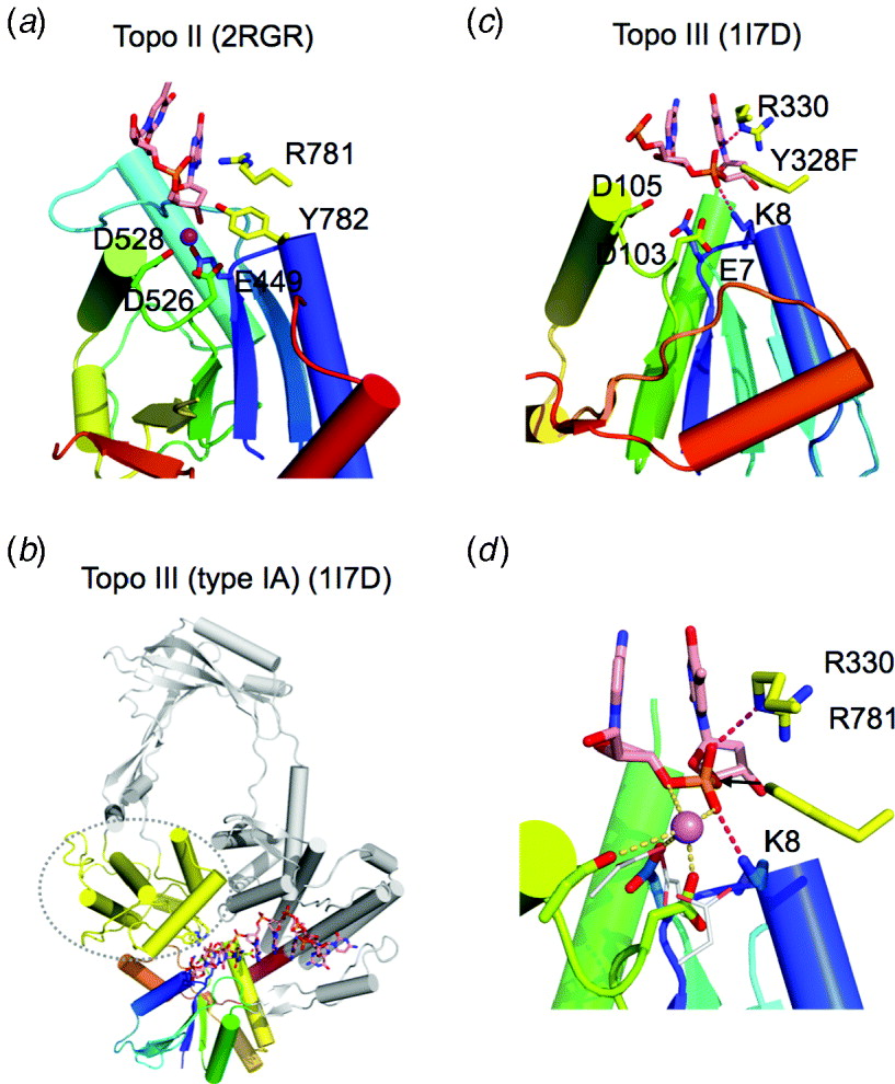
The location of this metal ion is close to the leaving group and is reminiscent of the B metal ion in RNase H1 and the single metal ion in TraI (Figs 7 and 23). The absence of the second metal ion (equivalent to metal ion A on the nucleophile side) is not a proof of a one-metal-ion mechanism, because the second metal ion may have escaped detection due to a crystallographic artifact. The arrangement of the Tyr and the metal ion relative to the DNA substrate in Topo II is rather similar to the mob nucleases, suggesting that the one-metal-ion mechanism may be applicable here. The absence of the fourth carboxylate in the active site and the presence of the Arg adjacent to the nucleophile Tyr to stabilize the scissile phosphate (Fig. 24a) also lend support to the one-metal-ion mechanism.
8.1.2 Type-IA topoisomerases
Type IA topoisomerases are monomeric and nick one strand of DNA (Schoeffler & Berger, Reference Schoeffler and Berger2008). In addition, type-IA enzymes are ATP-independent and relax negative supercoils (with the exception of reverse gyrase). Interestingly, the ssDNA co-crystallized with E. coli Topo III (type IA) adopts the double-helix-like conformation (Changela et al. Reference Changela, Digate and Mondragon2001) (Fig. 24b). Similar to Topo II, the active site of monomeric type IA is assembled between two domains and composed of ExxK and DxD from the TOPRIM domain (aka domain I) and the nucleophile Tyr from domain III (equivalent to the WHD of Topo II) (Fig. 24b and c). It was controversial as to whether metal ion is required for Topo IA. It is now clear that efficient DNA cleavage in addition to the religation by type-IA topoisomerases requires Mg2+ (Domanico & Tse-Dinh, Reference Domanico and Tse-Dinh1991). In support of the metal-ion requirement, amino-acid substitutions of the conserved carboxylates in TOPRIM render the mutant topoisomerases defective in DNA supercoil relaxation (Bhat et al. Reference Bhat, Leelaram, Hegde and Nagaraja2009; Zhu & Tse-Dinh, Reference Zhu and Tse-Dinh2000).
The crystal structure of a type-IA topoisomerase (Topo III) with the nucleophile Tyr replaced by Phe and in complex with ssDNA substrate provides the highest-resolution view of the active site of TOPRIM topoisomerases (Changela et al. Reference Changela, Digate and Mondragon2001). Relative to the scissile phosphate, the carboxylates and the Tyr of Topo III occupy positions similar to those in Topo II. In addition to the Arg next to the Tyr, a Lys conserved in type IA stabilizes the scissile phosphate (Fig. 24c). But due to the high salt concentration in the crystallization buffer (1·3 M (NH4)2SO4), soaking in 10 mM MgCl2 was not successful in detecting metal ion binding (Changela et al. Reference Changela, Digate and Mondragon2001). The superposition of the catalytic residues of E. coli Topo III and yeast Topo II show that the metal ion observed in the Topo II falls right between the conserved carboxylates and scissile phosphate in Topo III (Fig. 24d).
The striking similarities of metal ion coordination and nucleophilic attack of the scissile phosphate between TOPRIM topoisomerases and HUH nucleases TraI and TnpA imply a similar catalytic mechanism. The nuclease activity of Topo IA in the absence of Mg2+ may be explained by the conserved Lys and Arg in the active site, which can neutralize the developing negative charge on the scissile phosphate (Fig. 24c). A divalent cation on the leaving group side may not be necessary, but, in the strand ligation step, the metal ion is on the 3′-OH nucleophile side and may become essential for its deprotonation. It is most likely that type II and type IA topoisomerases with so much in common use the same metal-ion-dependent cleavage mechanisms.
The M5 and OLD family nucleases and type-IA and type-II topoisomerases of the TOPRIM superfamily share the conserved TOPRIM domain yet may use a two- versus one-metal-ion-dependent mechanism for DNA cleavage. The divergence appears to originate from different nucleophiles and different environments surrounding the respective scissile phosphate in their cleavage reactions. They may represent an early divergence from a primordial sequence motif and structural domain.
8.2 Ser recombinases
The Ser-recombinase family consists of a large number of bacterial and archaeal resolvases/invertases, transposases and DNA-partitioning enzymes, which use the conserved Ser as a nucleophile to cleave and ligate DNA by forming a 5′-phosphoserine covalent intermediate (Grindley et al. Reference Grindley, Whiteson and Rice2006). A pair of long DNA recognition sites is required for DNA recombination, and DNA rearrangement takes place between the steps of cleavage and religation of both sites. Although Ser recombinases are usually compared with DNA recombinases rather than topoisomerases, they can relax negative supercoils like topoisomerases if cleavage of four strands (two duplexes) are incomplete and the recombination of DNA is prevented (Falvey et al. Reference Falvey, Hatfull and Grindley1988).
Ser recombinases are bi-lobal and contain an N-terminal catalytic domain and a C-terminal sequence-specific DNA-binding domain (Yang & Steitz, Reference Yang and Steitz1995a) (Fig. 25a). Dimerization is mandatory for binding to a pseudo-palindromic recognition site and for double strand cleavage (Salvo & Grindley, Reference Salvo and Grindley1988). For recombination between two specific sites to take place, Ser recombinases have to form a synaptic dimer of dimers (Fig. 25a and b). After cleavage of both recombination sites, protein and DNA undergo dramatic conformational changes (Li et al. Reference Li, Kamtekar, Xiong, Sarkis, Grindley and Steitz2005). Unlike the Tyr recombinases (see section 10.2.2), the recombination intermediate of Ser recombinases is not a HJ. The DNA rearrangement catalyzed by a Ser recombinase is rather similar to the topological change catalyzed by type-II topoisomerases (Fig. 2d) except for that two dsDNA cleavages are required rather than one.
Metal-independent Ser recombinases. (a) The structure of γδ resolvase dimer complexed with a palindromic DNA cleavage site (site I). Each protein subunit and DNA are shown in different colors. The catalytic residues are shown as sticks. (b) The γδ resolvase tetramer complexed with two cleaved DNA site I. Each protein dimer and DNA site are extensively rearranged. (c) The catalytic domain of resolvase is shown in rainbow colors from blue N- to red C-terminus. The catalytic residues S10 and R68 are shown in sticks. (d) The catalytic domain of RecJ. The metal coordinating carboxylates and the DHH motif are shown as sticks. (e) Superposition of the catalytic residues of resolvase (blue) and RecJ (yellow).

The catalytic domain of Ser recombinases is composed of ~120 N-terminal residues, which form a mixed five-stranded central β-sheet (54 312, ↓↑↑↑↑) surrounded by α-helices (Fig. 25c). The nucleophile Ser is at the end of the 1st β-strand. A number of conserved basic residues are essential for DNA cleavage and religation. The catalytic domain of γδ resolvase was first suggested to be similar to T5-FEN, because they share the first four parallel strands (Artymiuk et al. Reference Artymiuk, Ceska, Suck and Sayers1997), but γδ resolvase appears even more similar to RecJ than FEN (Fig. 25c and d). Structural superposition of RecJ and resolvase reveals an uncanny similarity and sheds light on catalytic mechanisms of both nucleases. The Ser nucleophile of resolvase overlays with the two conserved carboxylates in RecJ, which are likely to coordinate a metal ion (A site) in the presence of the DNA substrate for the two-metal-ion-dependent hydrolysis. The metal ion observed in the RecJ structure, which is coordinated by two His residues of the DHH motif, overlays with the catalytically essential Arg (R68) in γδ resolvase. The overlay suggests that this metal ion may occupy the B site in RecJ and stabilize the transition state. The similarity is consistent with a catalytic mechanism in which Ser attacks the scissile phosphate and Arg68 stabilize the penta-covalent intermediate. How the metal ion comes into play in DNA religation awaits elucidation by future studies.
Interestingly, Ser recombinases bear structural and functional similarity to TOPRIM topoisomerases because of the resemblance between RecJ and TOPRIM OLD nucleases (Fig. 10). The passage of one DNA duplex through another in site-specific recombination by a Ser recombinase is not dissimilar to the dsDNA passing in topological changes by type-II topoisomerases. Ser recombinases may represent the transition from metal-independent to metal-dependent nucleic acid enzymes. Metal-ion-dependent mechanisms have been associated with (1) high specificity, (2) high efficiency and (3) reversibility of forward and reverse reactions (Yang et al. Reference Yang, Lee and Nowotny2006). Ser recombinases achieve their specificity through DNA sequence recognition by the C-terminal domain and reaction efficiency is not a concern as recombination is a rare event in the life of cells. The metal ion is indeed required for the reverse reaction of cleavage (ligation) by Ser recombinases. For topoisomerases, the efficiency of DNA cleavage and religation is in the center of topological change. Correlated with the demands of reaction efficiency and two consecutive phosphoryl transfer reactions, TOPRIM domain, a metal-ion-binding module, comes to being and plays a central role during topoisomerization. In contrast to the metal-independent type IB topoisomerases (see section 10.2), type-IA and type-II topoisomerases have little preferences for specific DNA sequences. The requirement for metal ion may also contribute to the tight coupling of DNA cleavage with topological changes, one (double or single) strand-passage event in each DNA cleavage-and-rejoining cycle.
9. Metal-independent RNase
A large number of RNases catalyze phosphodiester bond breakage without metal ions. This is largely due to the 2′-OH of ribose, which readily makes intramolecular nucleophilic attack on the adjacent 3′-phosphate, thus breaking the RNA backbone (Figs 2e, 3b, and c). RNase A, T1 and T2 are three large RNase families in this class (Deshpande & Shankar, Reference Deshpande and Shankar2002; Raines, Reference Raines1998; Yoshida, Reference Yoshida2001). Endonucleases for tRNA splicing, colicin D and E5, ferredoxin-like Cas6 and CasE and Ire1 are additional metal-independent RNases. Metal-independent RNases, whether protein enzymes or ribozymes, produce 5′-OH and 2′,3′-cyclic phosphate, which are subsequently converted to 3′ phosphate and occasionally to 2′ phosphate as during tRNA splicing (Abelson et al. Reference Abelson, Trotta and Li1998).
These RNases expose and distort the scissile phosphate by splaying the bases surrounding the scissile phosphate far apart (Fig. 3b and c). Despite secondary structures recognized by some of these RNases, the nucleotides placed in the active site are inevitably single-stranded and unpaired. For example, when bound to barnase the RNA substrate is in an extended conformation with the bases surrounding the scissile phosphate more than 9 Å apart as compared with the 3·2–3·4 Å in a double helix (Fig. 26a) (Buckle & Fersht, Reference Buckle and Fersht1994). The active site residues facilitate the splay of bases in the substrate and distortion of the scissile phosphate for the 2′-O to attack it. An efficient cleavage reaction requires a general base to deprotonate the 2′-OH, positive charges to neutralize the penta-covalent transition state, and a general acid to protonate the 5′-O leaving group. When a general base and acid with pK a close to neutral pH are absent, such as in ribozymes, the catalytic rate is slow (Cochrane & Strobel, Reference Cochrane and Strobel2008; Zamel et al. Reference Zamel, Poon, Jaikaran, Andersen, Olive, De Abreu and Collins2004). In contrast, many protein RNases have excellent general bases and acids and are thus highly active (Deshpande & Shankar, Reference Deshpande and Shankar2002; Raines, Reference Raines1998; Yoshida, Reference Yoshida2001) and usually acquire special inhibitors known as immunity proteins for host protection (Kobe & Deisenhofer, Reference Kobe and Deisenhofer1996; Kolade et al. Reference Kolade, Carr, Kuhlmann, Pommer, Kleanthous, Bouchcinsky and Hemmings2002; Lee et al. Reference Lee, Auld and Vallee1989).
Metal-independent RNases of T1 family and colicins. (a) Barnase in complex with a substrate mimic d(GGAC). The RNase is shown in rainbow-colored ribbon diagrams. The scissile phosphate and surrounding nucleotides are shown as magenta sticks. The active site residues are shown in sticks and labeled. Hydrogen bonds with the scissile phosphate and the surrounding bases are shown as dashed lines. (b) RNase T1 complexed with a phosphorothioate cleavage product. The sulfur atom in the 2′,3′ cyclophosphorothioate is shown in light yellow. (c) Sarcin homolog restrictocin in complex with the mimic of sarcin/ricin loop. Only the active site is shown. The RNA is not positioned for cleavage. (d) The ribonuclease domain of colicin E3. The potential active site residues are shown as sticks in the same colors as their Cα atoms. Those, whose mutations do not decrease the nuclease activity, are shown in silver. (e) The nuclease domain of colicin D. The active site residues are shown in sticks. (f) Colicin E5 complexed with d(GU). Sequence specificity is determined by the numerous hydrogen bonds between the RNase and the bases surrounding the scissile phosphate. The nucleophilic attack would take place as indicated by the black dashed arrowhead if 2′-OH is present.

9.1 RNase T1, Barnase and microbial RNases
Members of RNase T1 family are found in bacteria and fungi only. Related are the restrictocin/sarcin and colicins E3, E4 and E6. The active site consists of a Glu-His pair as general base and acid.
9.1.1 Barnase
Barnase, an extracellular ribonuclease of Bacillus amyloliquefaciens, has been extensively studied for its folding properties, interaction with its intracellular protein inhibitor barstar and its substrate recognition (Fersht & Daggett, Reference Fersht and Daggett2002; Urakubo et al. Reference Urakubo, Ikura and Ito2008). Homologous RNases are found in other bacillus species, e.g. Binase from Bacillus intermedius (Schulga et al. Reference Schulga, Nurkiyanova, Zakharyev, Kirpichnikov and Skryabin1992). Barnase has a characteristically twisted five-stranded contiguous antiparallel β-sheet with long loops surrounding it and short helices covering one side (Fig. 26a). The exposed face of the β-sheet binds nucleic acid substrate. Most of the catalytically essential residues are located on the loops except for the central Glu. The crystal structure of Barnase bound to a tetradeoxynucleotide (instead of RNA to prevent cleavage) reveals the mode of substrate recognition and a possible catalytic mechanism (Buckle & Fersht, Reference Buckle and Fersht1994). Barnase cleaves 3′ to a Gua, which is sandwiched between an Arg and Phe and forms double hydrogen bonds with a Glu (Fig. 26a). The scissile phosphate is neutralized by two Arg and one Lys. The catalytic residues Glu (E73) on the central β-strand and His (H102) on a long loop are on either side of the scissile phosphate and function as the general base and acid, respectively. The conformation of the deoxyribose linked to the scissile phosphate, however, is not in position for the in-line attack in this crystal structure, probably because of the absence of 2′-OH (Buckle & Fersht, Reference Buckle and Fersht1994). Y103, which stacks against the (deoxy)ribose, together with the Arg and Lys residues, may align and distort the substrate for the nucleophilic attack.
9.1.2 RNase T1
RNase T1, RNase Sa and fungal RNase U2, F1 and Ms belong to RNase T1 family and are homologous to Barnase in sequence and tertiary structures (Yoshida, Reference Yoshida2001). They too cleave ssRNA after Gua. The general base and acid pair, Glu and His, are conserved in these RNases. Several crystal structures of RNase T1 bound to various substrate analogs have been reported (Arni et al. Reference Arni, Watanabe, Ward, Kreitman, Kumar and Walz1999), and the complex with a cyclic thiophosphate substrate is particularly revealing (Zegers et al. Reference Zegers, Loris, Dehollander, Fattah Haikal, Poortmans, Steyaert and Wyns1998). RNase T1 can cleave the cyclic phosphodiester to 3′ phosphate, which is the second half of the normal hydrolysis reaction. The sulfur replacement (thiophosphate) greatly reduces the reaction rate. In the crystal structure, the cyclic phosphate is placed between the conserved Glu (E58) and His (H92) and hydrogen bonded to both (Fig. 26b). The ribose is flipped compared to that in Barnase (Fig. 26a) and well placed for the general acid–base catalysis. In addition, positively charged H40 and R77 interact and orient the scissile phosphate. The guanine base preceding the scissile phosphate is sandwiched between two Tyr residues and forms bifurcated hydrogen bonds with a glutamate (E46) in addition.
9.1.3 Restrictocin
The homologous restrictocin and sarcin are ribotoxins and specifically cleave 28S ribosome RNA (Olmo et al. Reference Olmo, Turnay, Gonzalez De Buitrago, Lopez De Silanes, Gavilanes and Lizarbe2001). Structurally these ribotoxins are related to Barnase and RNase T1 (Yang et al. Reference Yang, Gerczei, Glover and Correll2001), yet they specifically recognize a well-folded hairpin loop (sarcin/ricin loop) in the 28S rRNA. Because of the modified 2′-groups used in co-crystallization of restrictocin-RNA to prevent cleavage (Yang et al. Reference Yang, Gerczei, Glover and Correll2001), the RNA substrate is out of register by one nucleotide and the scissile-phosphate mimic is displaced from the catalytic residues. Nevertheless, the structure reveals a dramatically deformed hairpin RNA substrate and the unpaired and flipped-out base 5′ to the scissile phosphate (Correll et al. Reference Correll, Yang, Gerczei, Beneken and Plantinga2004) (Fig. 26c). Such distortions are necessary to align the 2′-OH nucleophile for the in-line attack. The bases surrounding the scissile phosphate are thus splayed much like in the cases of Barnase and RNase T1. The catalytic Glu-His pair is E95 and H136 in Ristrictocin. Like RNase T1, restrictocin also depends on aromatic and positively charged residues to bind and orient the scissile phosphate for the cleavage reaction (Fig. 26c).
9.1.4 Colicin E3, E4 and E6
Colicin E3, E4 and E6 are homologous and specifically cleave 16S rRNA. Bacteriocin Cloacin DF13 is a ribonuclease homologous to E3 and E6 colicins and it again cleaves 16S rRNA (Akutsu et al. Reference Akutsu, Masaki and Ohta1989). The crystal structure of E3 complexed with its inhibitor reveals the similarity to that of Barnase and RNase T1 (Carr et al. Reference Carr, Walker, James, Kleanthous and Hemmings2000) (Fig. 26d). The conserved E62 and H71 assume comparable positions as the catalytic Glu-His pair found in the T1 superfamily. But mutagenic studies show that H58 instead of H71 is critical for catalysis and that E62 and H58 are likely to be the general base and acid pair (Walker et al. Reference Walker, Lancaster, James and Kleanthous2004). Additionally charged residues, Asp, Glu, Lys and Arg, align the active site. Mutagenesis suggests that R40, D55, E60, Y64 and R90 are essential for catalysis (Walker et al. Reference Walker, Lancaster, James and Kleanthous2004). How these RNases recognize their substrate remains unsettled in the absence of a co-crystal structure and additional biochemical studies.
9.2 Colicin D and E5
Colicin D cleaves the anticodon loop of all four Arg tRNA (Kolade et al. Reference Kolade, Carr, Kuhlmann, Pommer, Kleanthous, Bouchcinsky and Hemmings2002). The crystal structure of colicin D complexed with its immunity protein has been determined (Graille et al. Reference Graille, Mora, Buckingham, Van Tilbeurgh and De Zamaroczy2004). Although colicin D consists of a contiguous antiparallel β-sheet supported by long α-helix on one side as does RNase T1, the topology is different with the strands (1234) going from right to left instead of left to right (Fig. 26e). The crystal structure also reveals that its active site lacks the Glu-His pair. Instead colicin D has a single His (H611), which is suspected to be the general base. In addition, the active site is marked by aromatic residues, which may determine the base specificity, and by positively charged residues, which may orient and neutralize the scissile phosphate (Graille et al. Reference Graille, Mora, Buckingham, Van Tilbeurgh and De Zamaroczy2004).
Colicin E5 is similar to colicin D and is also a tRNase. It recognizes the GU sequence in the anticodon loop of E. coli Tyr, His, Asn and Asp tRNA (James et al. Reference James, Kleanthous and Moore1996). The crystal structure of colicin E5 complexed with a substrate mimic (dGpdUp) reveals that GU bases are mainly specified by hydrogen bonds with the backbone atoms of the protein (Yajima et al. Reference Yajima, Inoue, Ogawa, Nonaka, Ohsawa and Masaki2006) (Fig. 26f). This structure reveals a near-active reaction coordination, where the scissile phosphate and the preceding (deoxy)ribose (to which the 2′-OH would belong to if present) are perfectly aligned for the nucleophile attack (compared with the reaction product of 2′,3′ cyclicphosphate shown in Fig. 25b). Colicin E5 has no Glu or His in its active site (Fig. 26f). Three basic residues (K25, R33 and K60) provide charge neutralization of the scissile phosphate. These positively charged residues may serve as the general acid and base for catalysis at high pH as proposed (Yajima et al. Reference Yajima, Inoue, Ogawa, Nonaka, Ohsawa and Masaki2006). Interestingly, two Gln (Q29 and Q93) are located near the scissile phosphate and are potentially involved in the cleavage reaction.
9.3 RNase A
Bovine pancreatic RNase A is perhaps the most studied ribonuclease, which has culminated in four Nobel prizes (Raines, Reference Raines1998). It is the protein that Chris Anfinsen used to show that information for disulfide formation and tertiary folding is intrinsically stored in the amino acid sequence (Anfinsen, Reference Anfinsen1973). Homologs of RNase A are widespread among animals. There are eight versions of RNase A in the human genome, six expressed in pancreas (RNase 1–6) and two in skin (RNase 7 and 8). Genes encoding these RNases tend to be in a cluster on one chromosome. Only homologs of RNase 5 have been reported outside the mammals. The crystal structures of pancreatic RNase A (Avey et al. Reference Avey, Boles, Carlisle, Evans, Morris, Palmer, Woolhouse and Shall1967; Kartha et al. Reference Kartha, Bello and Harker1967; Wyckoff et al. Reference Wyckoff, Tsernoglou, Hanson, Knox, Lee and Richards1970) and homologous seminal (Mazzarella et al. Reference Mazzarella, Capasso, Demasi, Di Lorenzo, Mattia and Zagari1993; Merlino et al. Reference Merlino, Ercole, Picone, Pizzo, Mazzarella and Sica2008) and eggwhite ribonucleases (PDB: 2ZPO) have been determined. Like Barnase, RNase A contains a central antiparallel β-sheet, but it is much more extended and twisted (Fig. 27a). The catalytic triad, two His (H12 and H119) and one Lys (K41) surround a central substrate-binding groove (Fig. 27a).
RNase A and related RNases. (a) RNase A in complex with d(ATAAG) substrate mimic. RNase A is shown in rainbow-colored ribbon diagrams with the catalytic triad (HKH) shown as sticks. The scissile phosphate is shown in yellow, the surrounding nucleotides in magenta and other nucleotides in pink sticks. (b) An example of RNase T2 family, RNase NT, complexed with two GMP. The structure is shown in the same scheme as in (a). (c) A close-up view of the substrate recognition by RNase NT. The two bases surrounding the putative scissile phosphate are sandwiched by aromatic side chains (shown in yellow). The HKH catalytic triad (shown in green sticks) appears to be conserved and the two His side chains serve as general acid and base. (d) The overall structure of a homodimeric tRNA splicing endonuclease. Each catalytic unit consists of a catalytic domain (shown in green and cyan) and an associated unique domain (shown in yellow and steel blue). Two structural units are shown in silver gray and are homologous to the catalytic units (green+yellow or cyan+light) by gene duplication. The four units can be independent polypeptide chains in other orthologs. The pre-tRNA mimic is shown in orange ribbon diagrams. The uncleaved and cleaved scissile phosphates are boxed and labeled as ‘e’ and ‘f’, respectively. (e) A close-up view of the active site (green subunit) with uncleaved RNA (substrate). Hydrogen bonds with the scissile phosphate are shown as hot pink dashed lines. RNA substrate is stabilized by cation-π-cation stacking with a neighboring subunit (cyan). (f) The active site (cyan subunit) with the cleavage product, 2′,3′-cyclic phosphate.

Pancreatic RNases recognize U or C for endonucleolytic cleavage to produce 3′-Up or 3′-Cp. The apo-protein structure of RNase A was among the first protein crystal structures determined (Kartha et al. Reference Kartha, Bello and Harker1967; Wyckoff et al. Reference Wyckoff, Hardman, Allewell, Inagami, Johnson and Richards1967), and the RNase complexed with ssDNA substrate mimics were determined in the 1990s (Raines, Reference Raines1998). The structure of RNase A complexed with d(ATAAG) with the dT mimicking a U is most revealing (Fontecilla-Camps et al. Reference Fontecilla-Camps, De Llorens, Le Du and Cuchillo1994). The preference for cleaving after a pyrimidine is due to steric exclusion of purines. The reaction coordinate is rather similar to the other known metal-independent RNases (Figs 26 and 27a). The conserved H12 (potentially the general base) is hydrogen bonded with the scissile phosphate on the 3′-O side (2′-OH is replaced by H), and H119 (potentially the general acid) is hydrogen bonded with the leaving group O5′. Studies by chemical perturbation of the catalytic triad and pH profiling indicate that indeed H12 is the general base, H119 the general acid and K41 specifically neutralizes the transition state (Raines, Reference Raines1998).
9.4 RNase T2
Members of RNase T2 family are widespread from E. coli (RNase I), fungi (Rd and DdI), plants (LE and NT) and mammals (Deshpande & Shankar, Reference Deshpande and Shankar2002; Irie & Ohgi, Reference Irie and Ohgi2001). RNase T2 are sequence non-specific. The structure of RNase T2 consists of a central 4-stranded antiparallel β-sheet surrounded by short strands and long helices on one face. The topology and tertiary structure, however, are different from RNase A and T1 (Fig. 27b). The active site is located on the exposed face of the β-sheet. The conserved Glu, Lys and three His residues in the active site are essential for the catalytic activity. Interestingly, mutation of individual amino acids only reduces, but not eliminates, the RNase activity (Tanaka et al. Reference Tanaka, Arai, Inokuchi, Koyama, Ohgi, Irie and Nakamura2000). The crystal structure of tobacco RNase T2 (NT) complexed with two 5′ GMP in the active site may mimic the enzyme–substrate complex (Kawano et al. Reference Kawano, Kakuta, Nakashima and Kimura2006). Each guanine bases is sandwiched by two aromatic side chains. The catalytic residues are within hydrogen bonding distance to the scissile phosphate mimics (Fig. 27c), a His and Lys coordinating the attacking nucleophile 2′-OH, and the remaining three coordinating the phosphate.
9.5 tRNA-splicing endonuclease
About 5–25% of archaeal and eukaryotic tRNA contain introns, which are recognized and excised by the tRNA-splicing endonuclease (Abelson et al. Reference Abelson, Trotta and Li1998). The tRNA-splicing endonucleases vary from homodimeric (archaea) to hetero-tetrameric (eukarya), but the overall structures are identical. Regardless of the number of peptides, each active molecule contains two catalytic and two structural units, all of which are structurally similar (Calvin & Li, Reference Calvin and Li2008). All tRNA-splicing endonucleases recognize the bulge-helix-bulge (BHB) structure in pre-tRNAs. The crystal structure of an archaeal homodimeric tRNA endonuclease complexed with RNA substrate with two bulges reveals two composite active sites (Xue et al. Reference Xue, Calvin and Li2006) (Fig. 27d). One active site is bound to a non-cleavable 2′-deoxy substrate, and the other is bound to the cleaved product (Fig. 27e and f).
The pre-tRNA substrate is mostly base-paired except for the cleavage sites in the 3-nt bulges. The bases flanking the scissile phosphate are unpaired and splayed as in substrate complexes of other metal-independent RNases. Each active site is composite. The catalytic residues are from one subunit, while the other subunit donates the Arg residues that sandwich the flipped out base flanking the scissile phosphate by the cation–π interactions (Fig. 27e, f). The catalytic unit contains a five-stranded mixed β-sheet not unlike metal-independent RNases. The conserved Tyr, His and Lys in the active site are confirmed to play catalytic roles. Double mutation of the Tyr and His eliminates the nuclease activity (Calvin et al. Reference Calvin, Xue, Ellis, Mitchell and Li2008). This Tyr–His pair appears to replace the Glu–His in Barnase and the His–His pair in RNase A and acts as the general acid and base in the RNA cleavage reaction. Interestingly, replacing the conserved Lys in the active site by Glu, Gln or Arg abolishes the cleavage activity too, but the K to E mutant enzyme alone can be rescued by a specific divalent cation (Calvin et al. Reference Calvin, Xue, Ellis, Mitchell and Li2008). This may be an in-test tube mimic of the evolution from metal-independent to metal-dependent nucleases.
Ligation of the cleavage products 5′-OH and 2′,3′-cyclic phosphate in tRNA splicing is rather baroque (Abelson et al. Reference Abelson, Trotta and Li1998). It requires the 5′ end to be phosphorylated and the 2′,3′ cyclic phosphate to be converted to the 3′-OH and 2′-phosphate by a special phosphodiesterase (Wang & Shuman, Reference Wang and Shuman2005). After the standard ligation, the 2′-phosphate needs to be removed by another enzyme. The archaic processing of such metal-ion-independent RNA cleavage products may suggest an ancient origin. The simpler metal-dependent 3′-OH and 5′-phosphate splicing and cleavage intermediates ready for ligation and replication may arise in parallel to this or after, and they are clearly favored by evolution as shown by their abundance in today's world.
9.6 Ferredoxin-fold Cas6
Cas6 (for CRISPR associated 6) and CasE (Cse3) are related endoribonucleases that specifically recognize and cleave CRISPR RNA repeats to generate guide RNA for antiviral defense in prokaryotes (Brouns et al. Reference Brouns, Jore, Lundgren, Westra, Slijkhuis, Snijders, Dickman, Makarova, Koonin and Van Der Oost2008). The cleavage by Cas6 occurs 21 or 22 nucleotide downstream of the first repeat nucleotide. It is metal ion independent and presumably generates 5′-OH and 2′,3′-cyclic phosphate (Carte et al. Reference Carte, Wang, Li, Terns and Terns2008). Cas6 and CasE belong to one of the repeat-associated mysterious protein (RAMP) families (Makarova et al. Reference Makarova, Grishin, Shabalina, Wolf and Koonin2006). The crystal structure of Cas6 reveals that it is composed of two ferredoxin domains in a direct repeat (Fig. 28a). Ferredoxin-like domains are often found as RNA-binding modules. In addition to Cas6, Cas2 is also an RNase with a ferredoxin-fold, but Cas2 is metal-ion dependent and generates 5′-phosphate and 3′-OH products (section 6.17.4).
Other metal-independent RNases. (a) Pyrococcus furiosus Cas6. The two ferredoxin-like domains arranged in a direct repeat are both shown in rainbow-colored ribbon diagrams, from blue N- to red C-terminus. Putative catalytic residues are shown as sticks. (b) Thermus thermophilus CasE is related to Cas6 and shown in the same color scheme. Only the Lys near the C-terminal is conserved between Cas6 and CasE. (c) Ribbon diagram of XendoU. The protein is in rainbow color from blue N- to red C-terminus. A pseudo-dyad symmetry relates the two 3-stranded β-sheets. A bound phosphate is shown as orange and red sticks. The conserved and catalytic essential residues are also shown and labeled. (d) The overall structure of IreI homodimer. The two subunits are shown in green and cyan, and the bound ADP in the kinase domains are shown in red sticks. The nuclease domains (top) are highlighted with the catalytic residues shown in sticks. (e) A close-up view of the putative active site of IreI. Each nuclease domain is shown in rainbow colors. The residues (in one subunit) with strong effects on catalysis are labeled in black, and those of with medium effects in gray.

The two Ferredoxin domains in Cas6 are arranged approximately side-by-side and a G(Gly)-rich region fills the gap between the domains (Carte et al. Reference Carte, Wang, Li, Terns and Terns2008) (Fig. 28a). The active site of Cas6 consists of highly conserved Tyr, His and Lys in the first ferredoxin domain and a conserved Lys in the G-rich region. The Tyr–His pair is reminiscent of the catalytic residues of tRNA endonucleases. The structure of archaeal protein TTHB192, a homolog of CasE (Cse3), was reported before its function was identified (Ebihara et al. Reference Ebihara, Yao, Masui, Tanaka, Yokoyama and Kuramitsu2006). Whether CasE is an RNase has not been confirmed. Interestingly, His, Glu and Arg from the first ferredoxin domain and Lys from the G-rich region of CasE occupy the same positions as the catalytic residues in Cas6 (Fig. 28a, b). A His–Glu pair may replace the Tyr–His pair for catalysis if CasE is an RNase.
9.7 XendoU
Xenopus laevis RNA endonuclease is involved in the processing of the intron-encoded boxC/D U16 snoRNA for ribosome biogenesis (Laneve et al. Reference Laneve, Altieri, Fiori, Scaloni, Bozzoni and Caffarelli2003). Cleavage is poly(U) specific and homologs of XendoU are found in most metazoa (Laneve et al. Reference Laneve, Gioia, Ragno, Altieri, Di Franco, Santini, Arceci, Bozzoni and Caffarelli2008). Although the catalysis was initially thought to be metal ion dependent, metal ions cannot be found in the active site in the crystal structure (Renzi et al. Reference Renzi, Caffarelli, Laneve, Bozzoni, Brunori and Vallone2006). Considering the cleavage products are 3′-phosphate and 5′-OH, the required metal ion was suggested to facilitate RNA substrate binding. The tertiary structure of XendoU is unique. It contains a bunch of α-helices at the N-terminus and a duplication of an α-helix and three-stranded β-sheet unit at the C-terminus (Fig. 28c). The catalytic residues are concentrated in the first repeat of the α/β structure and include two His, one Lys and one Arg (Renzi et al. Reference Renzi, Caffarelli, Laneve, Bozzoni, Brunori and Vallone2006).
9.8 Ire1 and RNase L
Ire1 is an ancient transmembrane sensor of ER (endoplasmic reticulum) stress and is conserved in all eukaryotes (Sidrauski & Walter, Reference Sidrauski and Walter1997). In addition to the N-terminal ER luminal domain (receptor of unfolded protein) and a transmembrane segment, Ire1 has both a C-terminal cytoplasmic kinase domain and a kinase-extension nuclease (KEN) domain. Ire1 catalyzes splicing of target mRNAs in a spliceosome-independent manner. The cleavage products are 2′,3′ cyclic phosphate and 5′-OH, and the cleavage mechanism is proposed to be similar to RNase A. Dimerization and auto-phosphorylation of Ire1 activate its ribonuclease activity, and binding ADP/ATP in the kinase domain enhances dimerization of the KEN and thereby the nuclease activity (Sidrauski & Walter, Reference Sidrauski and Walter1997). The crystal structure of the kinase and KEN domain has been recently determined (Lee et al. Reference Lee, Dey, Neculai, Cao, Dever and Sicheri2008) (Fig. 28d and e). The KEN nuclease is rather unusual and is entirely α-helical without any β-strand. The active site residues are concentrated along an α-helix at the dimer interface, including His, Asn, Tyr and several Arg side chains.
A kinase-like and KEN domains are also found in mammalian RNase L (Bork & Sander, Reference Bork and Sander1993; Dong et al. Reference Dong, Niwa, Walter and Silverman2001), which contains nine ankarin repeats in addition. In parallel to the nuclease activation of Ire1 by ER stress, RNase L is activated by 2′,5′ oligoadenylates (whose production is induced by viral infection and mediated by interferon) binding to the ankarin repeats (Silverman, Reference Silverman2007). RNase L cleaves ssRNA at the 3′ of UA or UU sequences and plays an essential role in breaking down viral RNA in innate immune response. Like all other metal-ion-independent RNases, the cleavage products of RNase L are 3′ phosphate and 5′-OH. Reminiscent of the correlated kinase and nuclease activities in Ire1, RNase L activity is dampened by an ATP-binding cassette protein ABCE1 (Silverman, Reference Silverman2007).
9.9 Hammerhead, hairpin, glmS, HDV and VS ribozymes
Metal-independent ribozymes cleave RNA very much like metal-independent protein enzymes by splaying bases surrounding the scissile phosphate and aligning the 2′-OH for in-line nucleophilic attack (see the recent comprehensive review by Cochrane & Strobel, Reference Cochrane and Strobel2008). Because ribozymes lack a general acid and base with pK a close to neutrality, the fastest ribozyme VS RNA has a turnover rate of 10 s−1 (Zamel et al. Reference Zamel, Poon, Jaikaran, Andersen, Olive, De Abreu and Collins2004), which is orders of magnitude slower than protein ribonucleases. Three-dimensional structures of the hammerhead, hairpin, glmS, HDV and VS ribozymes, which cleave RNA to generate 2′,3′ cyclic phosphate and 5′-OH, have been determined (Chi et al. Reference Chi, Martick, Lares, Kim, Scott and Kim2008; Ferre-D'Amare et al. Reference Ferre-D'amare, Zhou and Doudna1998; Klein & Ferre-D'Amare, Reference Klein and Ferre-D'amare2006; MacElrevey et al. Reference Macelrevey, Salter, Krucinska and Wedekind2008; Rupert et al. Reference Rupert, Massey, Sigurdsson and Ferre-D'amare2002; Scott et al. Reference Scott, Murray, Arnold, Stoddard and Klug1996). In most cases, the unprotonated N1 of a Gua base is placed adjacent to the 2′-OH nucleophile and may serve as a general base. This is evident in the high-resolution structures of hammerhead in the pre- and post-catalytic states and of the hairpin ribozyme along the reaction pathway (Chi et al. Reference Chi, Martick, Lares, Kim, Scott and Kim2008; Rupert et al. Reference Rupert, Massey, Sigurdsson and Ferre-D'amare2002) (Fig. 29). The general acid to donate a proton to the 5′ leaving group may also be a nucleobase (Cochrane & Strobel, Reference Cochrane and Strobel2008). Although Mg2+ is critical for the folding of ribozymes and may play a role in substrate binding, there is no evidence that Mg2+ directly participates in catalysis.
Metal-independent ribozymes. (a) Hammerhead ribozyme. The RNA is shown in silver ribbon diagrams. The scissile phosphate is shown in yellow, the surrounding nucleotides (C−1 and A+1) in magenta, and the catalytic nucleotides in cyan. A Mg2+ (purple sphere) happens to be coordinated by the scissile phosphate. (b) A close-up view of the catalytic center in the hammerhead. The Mg2+ is omitted for clarity. The reaction is stopped because of the G12A mutation, which reduces the effectiveness of the general base. The 2′-OH of G8 is proposed to be the general acid. (c) The overall structure of a catalytically active Hairpin ribozyme. The RNA substrate is shown in pink ribbon diagrams, and the others are shown in the same scheme as in (a). (d) Close-up views of the active site of the hairpin ribozyme complexed with a substrate analog (A-1 is 2′-O-methylated), transition state analog (vanadate) and product (2′,3′-cyclic phosphate).

10. Metal-ion-independent DNase
A number of DNases are known to be metal-ion independent. To date, these DNases have been shown to catalyze cleavage reactions in two steps and require a phosphoenzyme covalent intermediate. The classic examples are recombinases and topoisomerases, which use Tyr as a nucleophile and form a 3′-phosphotyrosine covalent intermediate (Grindley et al. Reference Grindley, Whiteson and Rice2006; Schoeffler & Berger, Reference Schoeffler and Berger2008). In addition, DNase II and BfiI restriction endonuclease represent members of the phospholipase D (PLD) family and cleave DNA via a phosphohistidine intermediate (Sasnauskas et al. Reference Sasnauskas, Zakrys, Zaremba, Cosstick, Gaynor, Halford and Siksnys2010). The cleavage products of DNase II and BfiI are 5′-phosphate and 3′-OH. The last example of metal-independent DNase is the newly identified halfpipe restriction endonuclease PabI (Miyazono et al. Reference Miyazono, Watanabe, Kosinski, Ishikawa, Kamo, Sawasaki, Nagata, Bujnicki, Endo, Tanokura and Kobayashi2007). A conserved Tyr is required for catalysis, but a phospho-Tyr intermediate is yet to be confirmed. The metal-ion-independent DNases use positively charged side chains to align the scissile phosphate for nucleophilic attack and to stabilize the penta-covalent intermediate.
10.1 DNase with PLD fold
DNases in the PLD superfamily contain the HXK(X)4D (or HKD in short) motif and cleave DNA via a two-step mechanism by forming an phosphohistidine intermediate (Gottlin et al. Reference Gottlin, Rudolph, Zhao, Matthews and Dixon1998). The conserved HKD motif is present twice in each PLD family member either by homodimerization or an internal duplication (Uesugi & Hatanaka, Reference Uesugi and Hatanaka2009). The His and Lys of the HKD motif are involved in catalysis, whereas the Asp appears to stabilize the structure by hydrogen bonding with the main chain amides. The catalytic mechanism appears to be highly conserved in the PLD family, whether the substrate is phospholipids, DNA or phosphorylated proteins (Davies et al. Reference Davies, Interthal, Champoux and Hol2004). The reaction coordinate is examplified by the crystal structure of human tyrosyl-DNA phosphodiesterase (TDP1) complexed with vanadate mimicking the transition state (Davies et al. Reference Davies, Interthal, Champoux and Hol2004) (Fig. 30a). One of the two conserved His residues acts as the nucleophile and the other as the general acid to protonate the leaving group (Fig. 30b).
Metal-independent DNase of the PLD family. (a) The overall structure of tyrosine dephosphatase (TDP1). TDP1 serves as a paradigm of phosphoryl transferases using His as the nucleophile. Two gene-duplicated domains are shown in green and cyan. The phosphorylated Tyr is shown in magenta, the scissile phosphate in yellow sticks and the remaining oligonucleotides in pink. The catalytic residues are shown in sticks and colored in green and cyan as the Cα atoms. (b) A close-up view of the catalytic center. A mimic of the covalent intermediate (vanadate) is shown in yellow. The duplicated His and Lys are directly involved in catalysis. (c) The overall structure of S. typ Nuc is shown in the orientation similar to TDP1 in (a). The two identical subunits are both shown in rainbow colors. (d) The catalytic domains of dimeric restriction endonuclease BfiI. The catalytic domains of two subunits are shown in green and cyan and oriented similarly as TDP1 and Nuc. Sequence-specific DNA-binding domains are shown in silver gray.

10.1.1 Nuc and DNase II
Nuc of Salmonella typhimurium provided the first glimpse of a PLD family enzyme. It is a homodimer. The conserved His residues from two subunits (H94) align a narrow groove forming one active site and most probably act as the nucleophile and general acid in the trans-esterification reaction (Stuckey & Dixon, Reference Stuckey and Dixon1999) (Fig. 30c). The reaction intermediate may be stabilized by the conserved Lys residues (K96) much as in cleavage by RNase A. The structure and metal-ion-independent catalysis indicate that the DNA substrate is recognized in the single-stranded form. Like all PLD-like DNases, Nuc generates 5′-phosphate and 3′-OH. Metazoa DNase II are homologs of bacterial Nuc and belong to the PLD family. DNase II in lysosomes of macrophages plays an essential role in degrading DNA downstream from CAD/DFF40 after engulfment of apoptotic cells (Nagata, Reference Nagata2007). Without DNase II, residual DNA fragments lead to autoimmune diseases. The Nuc and DNase II are non-sequence specific.
10.1.2 BfiI restriction endonuclease
BfiI restriction endonuclease presents the first crystal structure of Mg2+ independent type-II restriction endonucleases (Grazulis et al. Reference Grazulis, Manakova, Roessle, Bochtler, Tamulaitiene, Huber and Siksnys2005; Vitkute et al. Reference Vitkute, Maneliene, Petrusyte and Janulaitis1998). It is a homodimer in solution and the two PLD-like domains form one active site (Lagunavicius et al. Reference Lagunavicius, Sasnauskas, Halford and Siksnys2003) (Fig. 30d). Like all PLD-family enzymes, BfiI cleaves DNA via a two-step trans-esterification mechanism (Sasnauskas et al. Reference Sasnauskas, Connolly, Halford and Siksnys2007). In the first step, it uses the conserved His as a nucleophile to cleave DNA forming a 5′ phosphoenzyme. In the second step, it transfers the phosphodiester bond to a water or alcohol molecule to regenerate the enzyme. The transition intermediate is likely to be stabilized by the conserved Lys residues as in TDP1 (Davies et al. Reference Davies, Interthal, Champoux and Hol2004). Although BfiI cleaves dsDNA, it cleaves one strand first and then the second (Sasnauskas et al. Reference Sasnauskas, Zakrys, Zaremba, Cosstick, Gaynor, Halford and Siksnys2010). Sequence-specific recognition is achieved by the DNA-binding domains that are separate from the catalytic domains (Fig. 30d) and occurs several base pairs away from the cleavage site (Grazulis et al. Reference Grazulis, Manakova, Roessle, Bochtler, Tamulaitiene, Huber and Siksnys2005; Vitkute et al. Reference Vitkute, Maneliene, Petrusyte and Janulaitis1998).
10.2 TopIB and Tyr recombinase
TopIB and Tyr recombinases share a conserved catalytic core and similar reaction mechanism (Cheng et al. Reference Cheng, Kussie, Pavletich and Shuman1998; Hickman et al. Reference Hickman, Waninger, Scocca and Dyda1997). These recombinases and topoisomerases contain one or more DNA-binding domains in addition to the catalytic domain and often have a preference for certain DNA sequences. They all have an absolutely conserved Tyr, which serves as the nucleophile and forms a phosphotyrosyl DNA–protein bond. The cleavage reaction produces a 3′-phosphotyrosine and 5′-OH. The DNA–enzyme covalent bond is cleaved by the 5′-OH of DNA in the religation step of recombination or topoisomerization (Grindley et al. Reference Grindley, Whiteson and Rice2006; Schoeffler & Berger, Reference Schoeffler and Berger2008). If Topo IB fails to religate the DNA, the covalently linked DNA and protein intermediate is the substrate of TDP1 (Fig. 30a and b) (Pouliot et al. Reference Pouliot, Yao, Robertson and Nash1999). In the absence of a DNA substrate, the Tyr is disordered or in a conformation unsuitable for catalysis (Biswas et al. Reference Biswas, Aihara, Radman-Livaja, Filman, Landy and Ellenberger2005; Cheng et al. Reference Cheng, Kussie, Pavletich and Shuman1998; Kwon et al. Reference Kwon, Tirumalai, Landy and Ellenberger1997; Perry et al. Reference Perry, Hwang, Bushman and Van Duyne2006b; Yang & Mizuuchi, Reference Yang and Mizuuchi1997). In the assembled active site, the Tyr is surrounded by a handful of basic residues which participate in the chemistry of cleavage and religation.
10.2.1 TopIBs
TopIBs relax supercoiled DNA. They can be sequence-specific like viral topoisomerases (Cheng et al. Reference Cheng, Kussie, Pavletich and Shuman1998) or have weak preference for certain DNA sequences like eukaryotic cellular TopIB (Redinbo et al. Reference Redinbo, Stewart, Kuhn, Champoux and Hol1998). Many crystal structures of Topo 1B, from protein alone (Cheng et al. Reference Cheng, Kussie, Pavletich and Shuman1998), protein–DNA complexes (Perry et al. Reference Perry, Hwang, Bushman and Van Duyne2006b; Redinbo et al. Reference Redinbo, Stewart, Kuhn, Champoux and Hol1998) to a cleavage transition mimic (Davies et al. Reference Davies, Mushtaq, Interthal, Champoux and Hol2006), have been determined. The active site contains two Arg, one Lys, one His and the nucleophile Tyr (Fig. 31a). The basic residues orient and neutralize the DNA backbone. One conserved Arg that is hydrogen bonded to both scissile phosphate and the nucleophile Tyr (Fig. 31b) is postulated to be the general base to deprotonate the Tyr (Davies et al. Reference Davies, Mushtaq, Interthal, Champoux and Hol2006; Yakovleva et al. Reference Yakovleva, Chen, Hecht and Shuman2008). The remaining Arg is proposed to work together with the Lys to act as the general acid and protonate the 5′ leaving group (Hwang et al. Reference Hwang, Baek, Kim and Cho1998; Yakovleva et al. Reference Yakovleva, Chen, Hecht and Shuman2008).
Metal-independent topoisomerases and Tyr recombinases. (a) The overall structure of Human Topo IB. The protein is shown in rainbow-colored ribbon diagrams. The DNA duplex is shown in pink/blue ribbons. The active site is highlighted with the catalytic residues shown in sticks. The nucleophile Y723 (mutated to Phe) is labeled. (b) A close-up view of the active site of Topo IB. The reaction is stopped because of the Y723F (shown in pink) mutation. The scissile phosphate is neutralized by Arg and Lys side chains. (c) The overall structure of the Cre recombinase complexed with substrate LoxP site. The tetrameric Cre is shown in four colors and the two LoxP in pink and yellow. The DNA-binding domains are above the DNA and the catalytic domains are below. (d) A close-up view of the active site of Cre-LoxP. The scissile phosphate is already pre-nicked. (e) Superposition of the catalytic residues of Topo IB and Cre.

10.2.2 Tyr recombinases: Cre, Flp, Xer and Phage integrase
The representative members of the Tyr recombinase superfamily include phage integrases (λ and HP1) (Biswas et al. Reference Biswas, Aihara, Radman-Livaja, Filman, Landy and Ellenberger2005; Hickman et al. Reference Hickman, Waninger, Scocca and Dyda1997), the Cre–loxP recombination system (Baldwin et al. Reference Baldwin, Martin, Abel, Gelato, Kim, Schultz and Santoro2003; Guo et al. Reference Guo, Gopaul and Van Duyne1997) widely used in the gene knockout techniques, XerC/D for bacterial DNA segregation (Subramanya et al. Reference Subramanya, Arciszewska, Baker, Bird, Sherratt and Wigley1997), and yeast Flp recombinase (Chen et al. Reference Chen, Narendra, Iype, Cox and Rice2000). They are all site-specific recombinases and share the conserved catalytic motif RHR in addition to the Tyr nucleophile. These recombinases are most often bi-lobal with a sequence-specific DNA-binding domain at the N-terminus and catalytic domain at the C-terminus (Fig. 31c). Many crystal structures have been determined along the reaction pathway (Grindley et al. Reference Grindley, Whiteson and Rice2006), and the active site and the surrounding structure of these recombinases can be superimposed with that of Topo IB (Fig. 31d and e). The Tyr nucleophile is located on the third helix in a three-helix bundle, which resembles a classic HTH domain (Yang & Steitz, Reference Yang and Steitz1995a) (Fig. 31d). Positioning the Tyr for nucleophilic attack requires the synapsis of four protein subunits and two palindromic DNA recombination sites (Ghosh et al. Reference Ghosh, Guo and Van Duyne2007a) (Fig. 31c). A HJ is a necessary intermediate in recombination (Voziyanov et al. Reference Voziyanov, Pathania and Jayaram1999). A catalytically competent active site may consist of residues from two adjacent subunits (so-called trans-configuration) (Chen et al. Reference Chen, Narendra, Iype, Cox and Rice2000). The Tyr nucleophile is likely to be deprotonated by one of the two conserved Arg residues as with human and vaccinia Topo IB (Fig. 31e) (Redinbo et al. Reference Redinbo, Champoux and Hol2000; Yakovleva et al. Reference Yakovleva, Chen, Hecht and Shuman2008). The other conserved Arg and Lys residues in the active site orient the scissile phosphate, stabilize the transition state, and may also protonate the 5′-O leaving group.
10.3 Halfpipe restriction endonuclease
The last group of metal-ion-independent nucleases is represented by the newly identified restriction endonucleases PabI encoded by transposable elements in archaea (Ishikawa et al. Reference Ishikawa, Watanabe, Kuroita, Uchiyama, Bujnicki, Kawakami, Tanokura and Kobayashi2005). Homologs of PabI possibly exist in bacteria as well. They are called ‘halfpipe’, because the quaternary structure of the homodimeric enzyme resembles a halfpipe with a deep groove in the center (Fig. 32) (Miyazono et al. Reference Miyazono, Watanabe, Kosinski, Ishikawa, Kamo, Sawasaki, Nagata, Bujnicki, Endo, Tanokura and Kobayashi2007). The nuclease is highly toxic to cells, presumably because of its nuclease activity. Overexpression was achieved in a cell-free system for the biochemical and structural studies. The crystal structure is determined without DNA substrate, and the catalytic mechanism is unclear. Mutagenesis indicates that the conserved Tyr (Y134), Glu and Arg are indispensable for the catalytic activity (Miyazono et al. Reference Miyazono, Watanabe, Kosinski, Ishikawa, Kamo, Sawasaki, Nagata, Bujnicki, Endo, Tanokura and Kobayashi2007). It is not known whether a DNA–tyrosyl covalent bond is formed in the process of DNA cleavage.
PabI family of restriction endonuclease. (a) The overall structure of homodimeric PabI. Two subunits are both shown in rainbow colors. The shape is likened to a halfpipe. The catalytically essential Y134 from both subunits are close together at the bottom of the central groove. (b) An orthogonal view of PabI. The catalytically essential residues are shown in sticks and labeled.

11. Concluding remarks and future directions
11.1 Metal ions and nucleases
Classification of nucleases by metal-ion dependence highlights the following general properties. Firstly, when metal ions are not directly involved in catalysis, RNase and DNase do not (and perhaps cannot) use water molecules as nucleophiles to attack a phosphodiester bond. The 2′-OH immediately adjacent to the scissile phosphate is universally used by metal-independent RNase for nucleophilic attack, whereas the hydroxyl group of Tyr or Ser or the imidazole of His is used by metal-independent DNase. Each of these ribose and side-chain nucleophiles has a lower pK a than water and is more readily deprotonated at physiological pH. In these metal-independent nucleolytic reactions, cleavage requires a second step to hydrolyze the cyclic diphosphate or covalent enzyme–substrate intermediate (Figs 1b, 2d and e). Incorporation of one or two metal ions into an active site, however, allows water to be a nucleophile and cleavage to occur in one step. Interestingly, the metal-ion site in common between the one and two metal-ion-dependent mechanisms is situated on the leaving group side (B site) (Yang, Reference Yang2008) and is not directly involved in deprotonating and activating the nucleophilic water.
Secondly, metal-ion-dependent nucleases cleave nucleic acid in the base-stacked helical conformation whether single- or double-stranded, whereas metal-independent nucleases prefer a base unpaired and unstacked single-stranded substrate, for example, all metal-independent RNase and PLD-like DNase. TopIBs and Tyr recombinases are exceptional and cleave a scissile phosphate embedded in the double helix with assistances of Arg and Lys side chains reaching into the DNA minor groove (Fig. 31a). Interestingly, these DNases cleave one strand only. This is likely because two such active sites cannot be easily configured in the minor groove adjacent to each other.
Thirdly, cleavage products of metal-ion-dependent reactions are a 5′-phosphate and 3′-OH groups, and products of metal-ion-independent reactions are most often a 5′-OH and 3′-phosphate. This division is observed among all RNase, but among DNase there are a couple of exceptions. SNase (section 6.17.1) is metal dependent but produces 3′-phosphate products. Ser recombinases (section 8.2) and DNase II and BfiII of the PLD family (section 10.1) cleave DNA without metal ions and produce 5′-phosphoserine and 5′-phosphohistidine intermediates, respectively.
Fourthly, nucleases that require no or one metal ion for catalysis most often require specific inhibitors to keep their activity in check. This requirement for inhibition is evident for eukaryotic RNase A and homologs (section 9.3), death-associated DNase (Endo G, Nuc, CAD and DNase II, sections 7.1.4 and 10.1.1), and microbial colicins (sections 7.1.4.3, 9.1.4 and 9.2). The process of inhibition can be quite cumbersome, and each nuclease often requires a specific inhibitor (Ghosh et al. Reference Ghosh, Meiss, Pingoud, London and Pedersen2007b; Kolade et al. Reference Kolade, Carr, Kuhlmann, Pommer, Kleanthous, Bouchcinsky and Hemmings2002; Rutkoski & Raines, Reference Rutkoski and Raines2008; Widlak & Garrard, Reference Widlak and Garrard2005). In contrast, two-metal-ion-dependent nucleases rarely require specific inhibitors to regulate their cleavage activity. The most prevalent protection is by DNA methylation in the restriction–modification systems (Bujnicki, Reference Bujnicki2001). An addition of methyl groups to a DNA recognition sequence is often sufficient to stop nuclease digestion. The reason that restriction–modification systems can work may be a result of the improved coupling between substrate recognition and catalysis of bond cleavage by the two-metal-ion mechanism (Yang et al. Reference Yang, Lee and Nowotny2006). Although one-metal- and no-metal-dependent nucleases can be sequence specific, substrate recognition and the cleavage reaction are less tightly coupled for these enzymes than for two-metal-ion-dependent nucleases, and specificity is achieved by the recognition of longer DNA sequences (e.g. homing endonucleases), additional separate DNA-binding domains (KpnI and BfiI), and formation of high-order protein–DNA complexes (e.g. site-specific recombination).
11.2 Diversity and convergence
Two themes emerge from analyzing the diversity and convergence of nucleases and their biological functions. At one end of the spectrum, a wide range of nucleases of different structures and catalytic mechanisms are employed to catalyze a similar biological reaction. At the other end of the spectrum, a conserved structural module and catalytic sequence motif may function in divergent or completely unrelated biological pathways.
11.2.1 Diversity of nucleases in a similar cleavage reaction
In the first category, the divergence of restriction endonucleases, HJ resolvases, site-specific recombinases, homing endonuclease and AP endonucleases stand out. As noted by Darwin, greater variation is found closer to the evolutionary source of a species. This principal is exemplified in the recent genomic sequencing of African Bushmen (Schuster et al. Reference Schuster, Miller, Ratan, Tomsho, Giardine, Kasson, Harris, Petersen, Zhao, Qi, Alkan, Kidd, Sun, Drautz, Bouffard, Muzny, Reid, Nazareth, Wang, Burhans, Riemer, Wittekindt, Moorjani, Tindall, Danko, Teo, Buboltz, Zhang, Ma, Oosthuysen, Steenkamp, Oostuisen, Venter, Gajewski, Zhang, Pugh, Makova, Nekrutenko, Mardis, Patterson, Pringle, Chiaromonte, Mullikin, Eichler, Hardison, Gibbs, Harkins and Hayes2010), which reveals that the genetic differences between any two Bushmen is greater than that between a European and an Asian. The diversity of nucleases may indicate that repair of an abasic lesion, homologous and site-specific DNA recombination and restrictive digestion of foreign DNA are ancient processes. The different nucleases that result in a shared outcome may also provide hints of a convergent relationship. For example, two AP endonucleases, Endo IV and Exo III (APE1) (sections 6.7.2 and 6.1.6.1), recognize the abasic site in a very similar way and use the pro-Rp oxygen to coordinate the catalytic two metal ions despite their unrelated tertiary structure and catalytic motifs.
Two cases of diverse nucleases for one biological process are worth noting. HJ or four-way DNA junctions are resolved to two duplexes by nucleases with different structure and mechanisms. Even among bacterial and single-cell eukaryotes there are more than four different HJ resolvases (Table 1). In higher eukaryotes, HJ resolvases exhibit additional diversity both in catalytic motifs and nuclease organization (Fekairi et al. Reference Fekairi, Scaglione, Chahwan, Taylor, Tissier, Coulon, Dong, Ruse, Yates, Russell, Fuchs, Mcgowan and Gaillard2009; Ip et al. Reference Ip, Rass, Blanco, Flynn, Skehel and West2008). HJ resolvases in lower organisms are homodimeric regardless of differences in catalytic mechanisms and symmetrically cleave fully base-paired four-way junctions in antiparallel or square–planar configurations (Biertümpfel et al. Reference Biertümpfel, Yang and Suck2007; Hadden et al. Reference Hadden, Déclais, Carr, Lilley and Phillips2007; Lilley & White, Reference Lilley and White2001). Eukaryotic HJ resolvase SLX1–SLX4 and Mus81-Eme1, however, are heterodimeric, and only one subunit contains the active site (Blanco et al. Reference Blanco, Matos, Rass, Ip and West2010; Ciccia et al. Reference Ciccia, Mcdonald and West2008). Instead of symmetric and fully base-paired junctions, eukaryotic HJ resolvases can recognize and cleave asymmetric ssDNA flaps. In bacteria, HJ often occur due to localized short palindromic sequences, which can be easily arranged in symmetric antiparallel or open-square conformation to be cleaved by homodimeric HJ resolvases. But in eukaryotes, four-way junctions often arise during homologous recombination between two distal sites or sister chromatids, where a symmetric HJ in antiparallel conformation is not easily achievable. The increasing diversity in HJ resolvases may correlate with the increased genomic complexity from bacteria to eukaryotes.
At least four different types of endonucleases are involved in site-specific DNA recombination: two kinds of Tyr recombinases (sections 7.3 and 10.2), Ser recombinases (section 8.2) and many two-metal-ion-dependent transposases (section 6.2.2). The catalytic mechanisms of these nucleases range from no metal, one-metal to two-metal dependent. Ser and Tyr recombinases are rarely found in eukaryotes and are absent in plants and metazoa. In contrast, two-metal-ion-dependent transposases/recombinases are widespread in bacteria, retroviruses and lower and higher eukaryotes. Usage of two-metal-ion catalysis is also associated with the cut-and-paste mechanism and a widened selection of target DNA for recombination.
11.2.2 Adaptation of a catalytic motif in various nucleases
Several catalytic modules of conserved structure and mechanism are found in many DNases and RNases (see sections 6.1–6.4 and 7.1). The wide distribution of a particular catalytic module is likely to reflect its catalytic efficiency. For example, the DEDD motif is associated with a large number of 3′ exonucleases, regardless of whether the substrate is DNA or RNA, and FEN1-like nucleases containing both a 5′ exo and endonuclease activity are found to remove primers in DNA replication and to cleave nucleic acid in HJ resolution, DNA repair and RNA processing.
Two catalytic motifs have the widest adaptation and are associated with the most diverse functions. They are the REase fold with the DEK motif (section 6.3) and the ββα-Me motif (section 7.1). These two catalytic motifs share the following features, which may allow them to be incorporated into different nucleases and different pathways. Firstly, the conserved catalytic motif is modular and small in size. Both the DEK and ββα-Me motifs are often contained within a stretch of 20–30 residues. The DEK motif occurs in a β-hairpin structure (Fig. 8), and the ββα-Me motif consists of a β-hairpin followed by an α-helix (Fig. 21). Thus, the DEK and ββα-Me motifs are readily incorporated into diverse surrounding tertiary structures and adapted to different nuclease activities and pathways. Secondly, both the DEK and ββα-Me motif are extremely tolerant of amino acid substitutions. Except for the first D in the DEK motif and a His serving as the general base in ββα-Me, other catalytic residues can vary greatly (see sections 6.3 and 7.1). Thirdly, the REase-like nucleases are further aided by the two-metal-ion mechanism in substrate specificity and catalytic efficiency.
It is worth noting that metal-ion-dependent mechanisms appear most adaptable. This observation may be explained by the advantages of specificity, efficiency and diversity endowed by metal ions (Yang et al. Reference Yang, Lee and Nowotny2006). Multiple metal-independent mechanisms and varied catalytic motifs are found among RNases, and this is likely due to efficient catalysis using a 2′-OH as the nucleophile. Adaptation of metal-independent mechanisms among DNase is rather limited and with few alterations, e.g. DNase II versus BfiI and TopIBs versus Tyr recombinases. The small number of metal-independent DNase may be a result of the limited types of reactions catalyzed and additional requirements for regulation and inhibition of cleavage activity.
11.3 Future directions
Although nucleases are the oldest nucleic acid enzymes characterized, new nucleases are being discovered every year and it is safe to predict that this trend will continue. Among the more recently discovered nucleases, several have novel sequence motifs, and their structures cannot be modeled using existing nucleases. For example, Sae2 in meiotic DNA break repair and nucleases in CRISPR pathways have unique catalytic motifs and unknown structures. For the well-studied processes of mRNA splicing and processing, the mechanism of the spliceosome and involvement of exon junction complex and nonsense-mediated decay are still being fleshed out. Furthermore, for many nucleases whose structures are known or are homologous to nucleases of known structure the catalytic mechanism remains unclear without additional atomic structures of functional enzyme–substrate complexes. Last but not the least many nucleases are linked to genetic or infectious diseases and are potential therapeutic targets. It is clear that exciting discoveries will continue to expand our understanding of these diverse and essential enzymes.
12. Acknowledgment
W.Y. is supported by NIH, NIDDK intramural research fund. The author thanks Dr C. Biertuempfel for initially collecting nucleases deposited with PDB database, and Drs R. Craigie and D. Leahy for reading and editing the manuscript.