Introduction
Multisystem inflammatory syndrome in children is a systemic disorder that involves incessant fever, extreme inflammation, and organ dysfunction, which is at first associated with exposure to COVID-19. 1,2 Multisystem inflammatory syndrome in children may manifest several weeks after the initial infection (typically 2–6 weeks later, often after 3–4 weeks), during which time symptoms may be inconspicuous. The pathogenesis of multisystem inflammatory syndrome in children was mediated by acquired immunity rather than direct viral injury. The antibody tests may be positive in cases of multisystem inflammatory syndrome in children, but COVID-19 polymerase chain reaction (PCR) results were negative in most cases. The clinical manifestations of multisystem inflammatory syndrome in children typically include gastrointestinal symptoms, such as diarrhoea, vomiting, intense abdominal pain, and extremity pain, rather than respiratory symptoms. Other clinical symptoms are enlarged lymph nodes, red or swollen hands and feet, red eyes (conjunctivitis without pus), widespread rash (consistent with the aetiology of leukocytoclastic vasculitis), or some Kawasaki-like symptoms that may be present, including mucosal alterations around the mouth (strawberry tongue). Reference Henderson, Canna and Friedman3 According to the literature, only five different types of clinical presentations of multisystem inflammatory syndrome in children have been reported. These manifestations are refractory vasodilatory shock (toxic shock syndrome-like) with normal cardiac function, septic or cardiogenic shock with impaired cardiac function, Kawasaki-like illness (both typical and atypical presentation), children with elevated inflammatory markers and tachycardia 3–4 weeks after COVID-19 infections, the different combination of the above. Reference Erika4–Reference Recalcati6 Kawasaki-like illness is more frequently detected in young children, though cardiogenic shock is seen in older children who require noradrenaline and milrinone for haemodynamic support. Skin findings related to Kawasaki-like illness are widely featured visually in the literature. To the best of our knowledge, this is the first report of the dermatological manifestation of cardiogenic shock with impaired cardiac function requiring noradrenaline and milrinone for haemodynamic support in a COVID-19 paediatric patient with multisystem inflammatory syndrome in children.
Case presentation
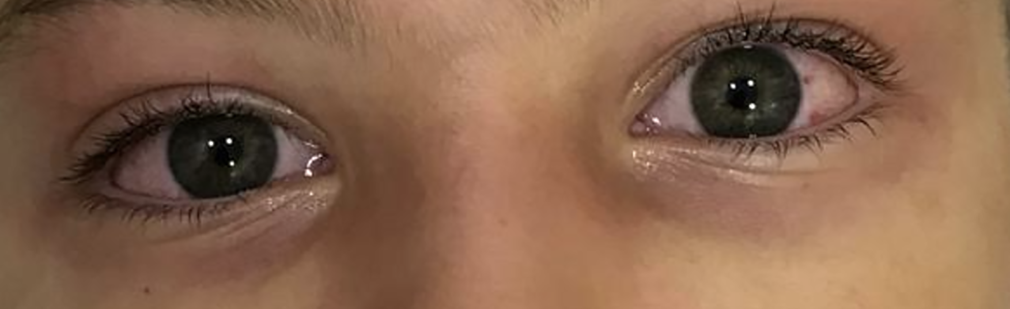
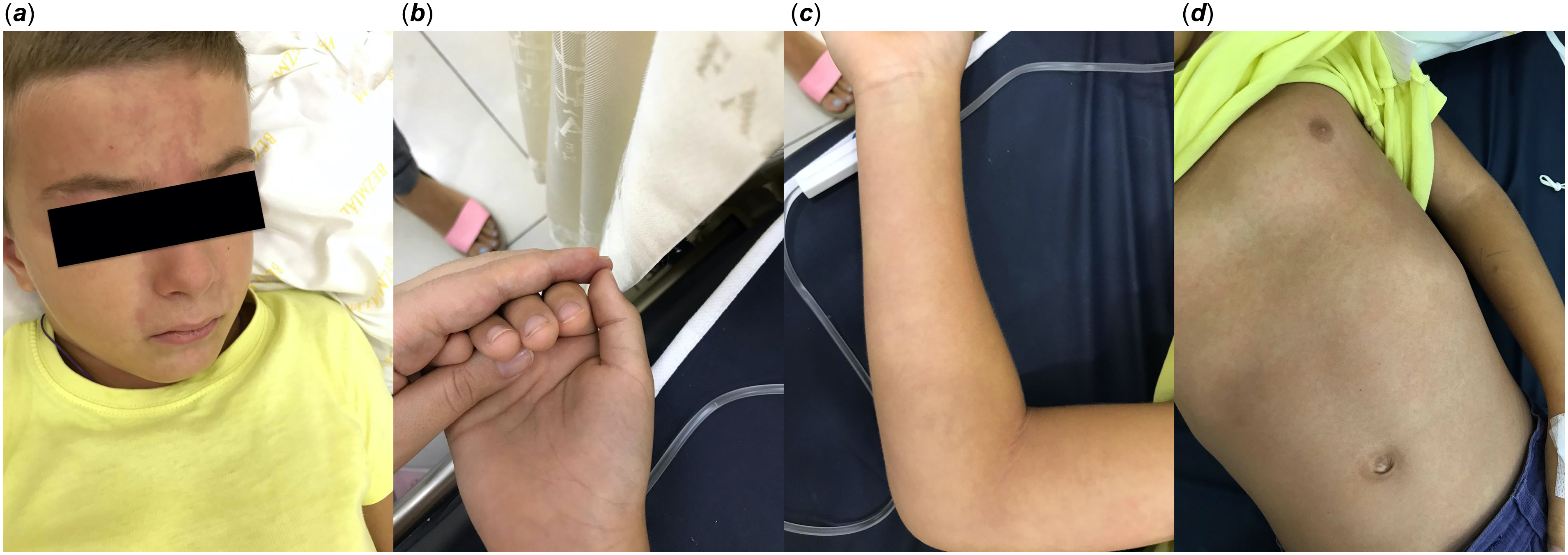
Early intervention played a crucial role in the successful management of this case—a 7-year-old boy presented to our institution’s emergency department with a fever and rash. The fever started seven days ago, and he was prescribed co-amoxicillin at another hospital; one day before presenting to the emergency department, a rash developed, which initially appeared on the trunk and subsequently spread to the extremities. On admission, the patient’s body temperature was 40.3°C. The patient had non-purulent conjunctivitis (Figure 1). There was a haemangioma and erythematous rash on the patient’s forehead (Figure 2A). A polymorphous rash, varying from macular to maculopapular and cutis marmorata-like in appearance, especially on the extremities, was detected (Figures 2B, 2C, 2D). There was no significant change on the patient’s lips, but circumoral pallor was noted. No rales were heard on auscultation, and cardiac sounds were normal. Lymph nodes were not enlarged, and the liver and spleen were not palpable. Initial blood chemistry results showed a normal count of leukocytes (5000/μl), lymphopenia (700/μl), a haemoglobin level of 12.2 g/dL, thrombocytopenia (97 000/mm3), a high C-reactive protein level of 177 mg/dL, average erythrocyte sedimentation rate (17 mm/h), a high D-dimer level of 1368 ng/mL, a high ferritin level of 749 ng/mL, a troponin level of 32 ng/mL, albumin level of 2.7 g/dL, and a normal level of AST 28 units/liter. The patient was hospitalised with suspicion of viral exanthemata’s disease, Kawasaki disease, or COVID-19. A rapid strep test and viral serology (EBV VCA IgM, immunoglobulin G (IgG), CMV IgM, parvovirus IgM, and herpes IgM) were requested, along with a COVID-19 PCR test. A chest X-ray was taken, and it revealed pericardial infiltration. Ceftriaxone was promptly initiated, underscoring the importance of early and comprehensive intervention in such cases.
The patient had non-purulent conjunctivitis.

ABCD: figure showing haemangioma and erythematous rash on the patient’s forehead. Figure showing a polymorphous rash varying from macular to maculopapular and cutis marmorata-like appearance on the hand, extremities, and trunk.

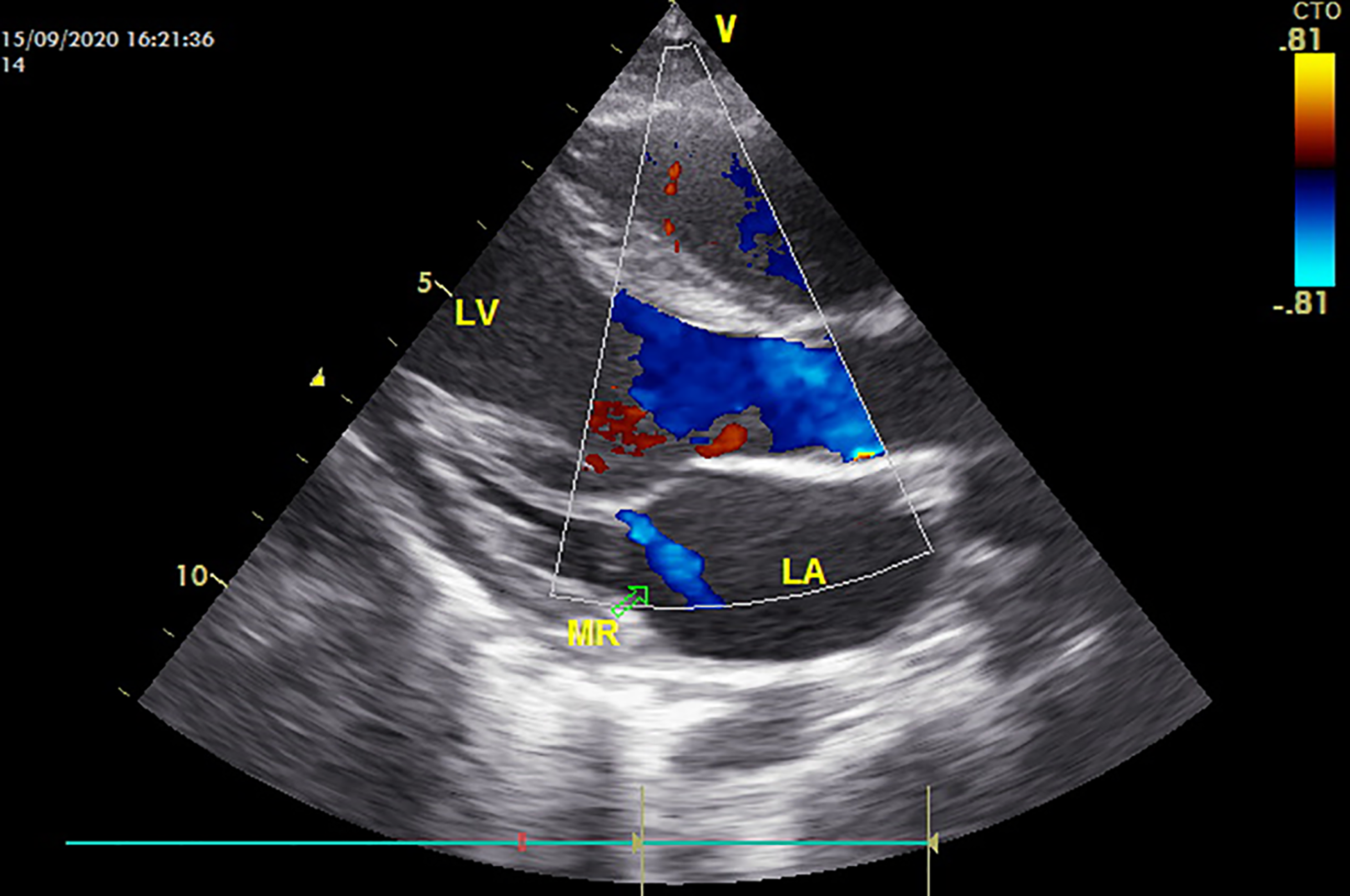
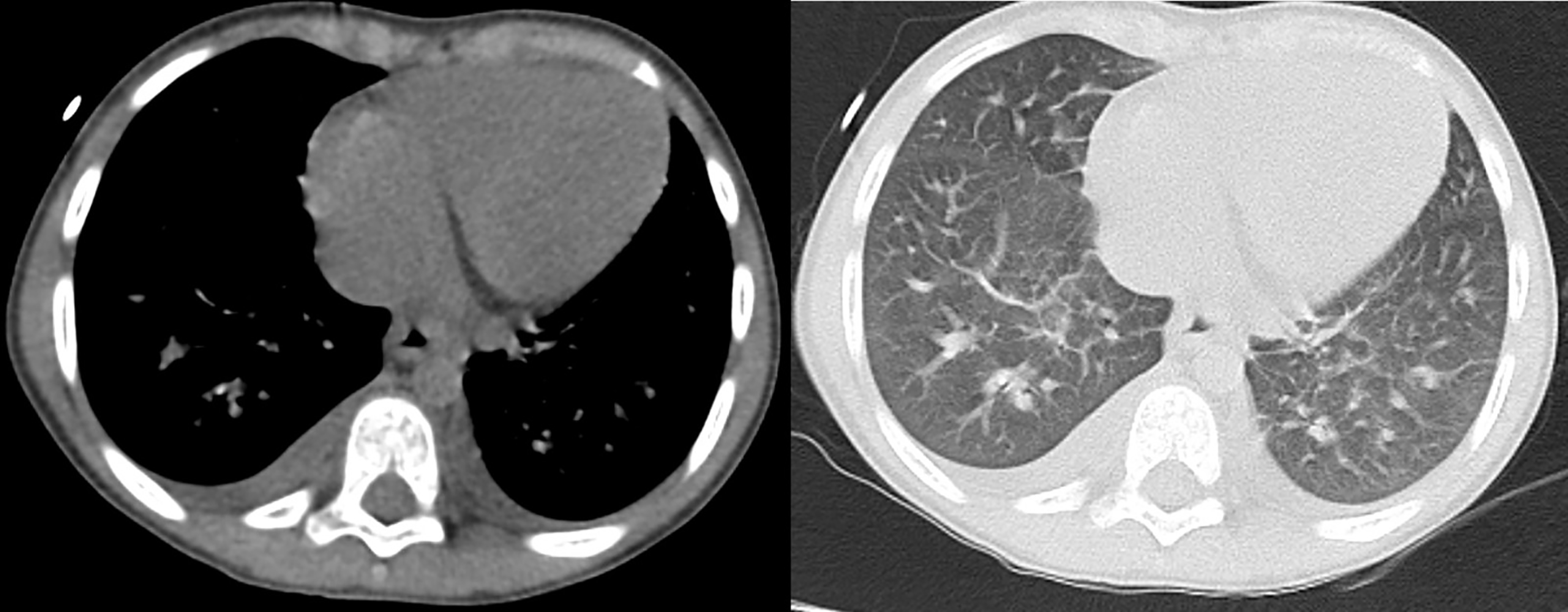
Seven hours later, the patient’s condition started to deteriorate. The patient had tachypnoeic respiration and moderate hypotension (80/35 mm Hg) developed. Thus, the Department of Pediatric Cardiology was consulted. Transthoracic echocardiography demonstrated a mild/moderate systolic dysfunction of the left ventricular (Ejection fraction 52%, SF 25%), and a first-degree mitral insufficiency was detected (Figure 3). Additionally, a notable increase in echogenicity of the coroner’s vessels was observed. A thorax CT scan was requested due to the patient’s worsening respiratory status. Diffuse ground-glass opacity was seen in association with interlobular septal thickening or peri-bronchovascular interstitial thickening. These findings were compatible with pulmonary oedema (Figure 4). Since all the rapid strep tests, viral serology, and COVID-19 PCR results were negative, our team re-examined the patient’s history. This time, the family informed us that their close family members had contracted COVID-19, but the family had never shown the clinical manifestation of COVID-19. The patient’s COVID-19 (SARS-CoV-2) IgG (10.27) was positive. However, the patient’s COVID-19 (SARS-CoV-2) IgM (0.557) was negative. The patient is diagnosed with multisystem inflammatory syndrome in children associated with cardiogenic shock. Milrinone and noradrenaline were promptly started for haemodynamic support.
TTE showing a first-degree of mitral insufficiency and systolic dysfunction.

Interlobular septal thickening and ground-glass opacities consistent with pulmonary oedema were observed in the lung parenchyma. Additionally, a distinct bilateral pleural effusion, more prominent on the right side, was noted. The cardiac silhouette shows mild enlargement.

Additionally, intravenous immunoglobulin (IVIG) (2 g/kg over 10 hours of infusion), methylprednisolone 20 mg twice daily, favipiravir 600 mg twice daily, and anakinra 50 mg twice daily SC were started. On the second day of hospitalisation, the patient’s condition was ameliorated. On the 10th day of hospitalisation, the patient was discharged from the hospital. Elevated D-dimer levels, troponin, ferritin, and a low platelet level, a low albumin level, and lymphopenia were signs of multisystem inflammatory syndrome in children, and COVID-19 PCR was negative. However, the COVID-19 IgG test was positive in our case. Persistent fever, mucosal findings, echogenicity of coroner’s vessels, hypotension, and laboratory findings (transthoracic echocardiography, thorax tomography, and baryochemical marker) led us to suspect that the clinical presentation could be cardiogenic shock with impaired cardiac function, a subtype of multisystem inflammatory syndrome in children associated with COVID-19.
Discussion
Cardiogenic shock with impaired cardiac function is a subtype of multisystem inflammatory syndrome in children. There have been reported cases of cardiovascular involvement, including inflammation of the heart muscle (myocarditis). Heart failure is prevalent, especially as a form of left ventricular insufficiency. Shock can occur when myocardial involvement has either ventricular or valve dysfunction. Respiratory symptoms are not an important feature. If a respiratory symptom is present in a patient, it is often associated with shock and serves as a premonition of heart failure. Some paediatric patients exhibit features of a cytokine storm characterised by elevated serum interleukin-6 levels and an immediate need for inotropic support to maintain normal cardiac function. Dilatation, which is one of the coronary artery abnormalities, is frequent. Some children can develop coronary artery aneurysms. 7–Reference Rowley9
It remains unclear to what extent its pathophysiology is like that of other paediatric inflammatory syndromes with similar clinical traits. Immunological activation and dysregulation of similar inflammatory pathways are the primary reasons behind clinical overlaps with syndromes of different causes, such as Kawasaki disease, toxic shock syndrome, or macrophage activation syndrome. The vital research issues regarding the underlying molecular threats that lead to the disorder following exposure to COVID-19 include identifying any possible genetic susceptibility factors, associations with specific viral variants, or molecular patterns that could trigger the autoimmune response. Reference Galeotti and Bayry10
In the literature, the first reported cases of the relationship between acute COVID-19 and skin involvement were reported from China, Turkey, and Italy. The cutaneous findings in the patients included an erythematous rash, chickenpox-like vesicles, and widespread urticaria. Visconti et al. investigated the diagnostic value of various cutaneous manifestations associated with SARS-CoV-2 infection. They highlighted that skin findings, such as erythematous rashes and vesicular eruptions, can serve as important clinical clues for COVID-19 diagnosis. Their study emphasised that recognising these dermatologic signs may facilitate early identification of infected patients, especially when respiratory symptoms are absent or minimal. Reference Visconti, Bataille and Rossi11 Baykal Selçuk et al. reviewed dermatologic manifestations observed in children diagnosed with multisystem inflammatory syndrome during the COVID-19 pandemic, reporting a broad spectrum of skin findings, including polymorphous rashes and mucocutaneous lesions. They underscored the significance of these cutaneous symptoms in aiding the timely diagnosis and management of multisystem inflammatory syndrome in children, which often presents with systemic inflammation and multiorgan involvement. (B) All of these skin findings were located in the lower extremities and trunk. Reference Baykal Selçuk, Ferhatosmanoğlu, Gayretli Aydın, Aksu Arıca and Yeşilbaş12
Another essential skin manifestation was that itching was minimal, and all lesions healed within four days. Reference Bursal Duramaz, Yozgat, Yozgat and Turel13–Reference Riphagen, Gomez, Gonzalez-Martinez, Wilkinson and Theocharis15 Skin manifestation of Kawasaki-like illness was a subtype of multisystem inflammatory syndrome in children, which was not the same as the skin rashes of patients with COVID-19. Although Kawasaki disease skin rash was polymorphous, varying from macular to maculopapular or morbilliform, it was never vesicular. It began on the trunk and spread over the extremities in a few days. Reference Yozgat, Uzuner and Bursal Duramaz16 Our case was cardiogenic shock with the subtype of multisystem inflammatory syndrome in children associated with COVID-19 in a 7-year-old boy. Unlike the skin rash formation in Kawasaki-like illness, cutis marmorata was present, indicating circulatory disorders in the skin, especially on the extremities.
There are differences between typical Kawasaki disease (KD) and multisystem inflammatory syndrome in children. First, KD has neutrophilic myocarditis, but multisystem inflammatory syndrome in children has lymphocytic myocarditis. Second, KD has high platelet counts, while multisystem inflammatory syndrome in children has low platelet counts. Third, IL-6 and ferritin are elevated in multisystem inflammatory syndrome in children but not in KD. Fourthly, lymphopenia is not seen in multisystem inflammatory syndrome in children, unlike in KD. Fifth, Lower albumin levels in multisystem inflammatory syndrome in children compared to KD. Last, KD often presents with normal troponin levels, but multisystem inflammatory syndrome in children is associated with elevated troponin levels. Reference Haella5,Reference Recalcati6,Reference Guan, Ni and Hu14,Reference Riphagen, Gomez, Gonzalez-Martinez, Wilkinson and Theocharis15
The first reported cases of multisystem inflammatory syndrome in children were initially described in the literature as a hyperinflammatory syndrome. Rihagen et al. reported an intriguing case of an asymptomatic child with COVID-19 who also had a hyperinflammatory syndrome with multiorgan involvement, which had a remarkably similar presentation to Kawasaki disease shock syndrome. Reference Riphagen, Gomez, Gonzalez-Martinez, Wilkinson and Theocharis15 Licciardi et al. reported two children who had profuse diarrhoea, persistent fever, and mucocutaneous involvement. Blood tests for these two patients revealed notably increased markers of inflammation, as well as lymphopenia and thrombocytopenia. Both patients had a high titre of IgG and IgM antibodies against the SARS-CoV-2 virus; however, the nasal swab for SARS-CoV-2 PCR testing was negative. Both patients responded exceptionally well to the IVIG treatment. Reference Licciardi, Pruccoli and Denina17 Similarly, our case had high inflammation markers, lymphopenia, thrombocytopenia, and a high titre of IgG against the SARS-CoV-2 virus during the first admission into the emergency department. Also, our case had an excellent response to IVIG treatment. Deza Leon MP et al. reported a unique case of a previously healthy 6-year-old girl who was initially admitted to the hospital with atypical Kawasaki disease symptoms. The patient later developed cardiogenic shock; thus, she was hospitalised in the extracorporeal membrane oxygenation (ECMO) unit for six days. The patient’s COVID-19 PCR test result from the nasopharyngeal swab was positive. The authors reported this case as COVID-19 COVID-19-associated paediatric multisystem inflammatory syndrome with cardiogenic shock. Reference Deza Leon, Redzepi and McGrath18 Our case was like Deza Leon’s case. Unfortunately, our case developed a rapid cardiogenic shock. Anti-inflammatory treatments, including IVIG, steroids, anakinra, cardiac milrinone, and adrenaline, were administered for support, and the patient showed improvement without the need for ECMO.
Bertoncelli et al. published a review article on cases with COVID-19, primarily presenting Kawasaki-like disease and various types of multisystem inflammatory syndrome in children. This review article highlights the importance of maintaining constant vigilance for additional clinical problems, such as myocardial injury and cardiovascular issues, to prevent misdiagnosing severe clinical entities. Reference Bertoncelli, Guidarini and Della Greca19 In our case, both cardiac dysfunction, cardiac involvement, and coronary changes developed. While there have been reported cases of skin findings associated with Kawasaki-like disease subtype multisystem inflammatory syndrome in children, to the best of our knowledge, this will be the first case reported of skin manifestation in cardiogenic shock exacerbated by a high cytokine storm. Cutis marmorata is characterised by a red-purple discolouration, similar to striae, resulting from circulatory disorders in the skin. Even though the skin findings in our case were similar to those in patients with Kawasaki-like illness and multisystem inflammatory syndrome in children, the most crucial difference was the presence of cutis marmorata, a result of the skin’s circulatory disorders.
The Royal College of Pediatrics and Child Health of the United Kingdom issued a guideline for physicians on how to manage this condition. 2 This guideline includes elevated markers of inflammation and coronary artery diseases, such as high levels of CRP and erythrocyte sedimentation rate, troponin, and triglycerides, and is also accompanied by hypoalbuminemia and lymphopenia. Multisystem inflammatory syndrome in children typically develops one month after the initial exposure to acute COVID-19 in patients. The antibody tests were positive in cases with multisystem inflammatory syndrome in children, but the results of COVID-19 PCR tests were negative in most cases. Similarly, our COVID-19 PCR test result was negative. The clinical presentation was a Kawasaki-like illness, and unfortunately, it turned into a severe clinical case with cardiogenic shock and impaired cardiac function.
Conclusion
Nowadays, due to the ongoing COVID-19 pandemic, if cutis marmorata is observed on the extremities in paediatric patients with both tachypnoeic breathing and Kawasaki-like illness-like findings, cardiogenic shock with impaired cardiac function associated with multisystem inflammatory syndrome in children should be suspected. Additionally, paediatric patients’ conditions can rapidly deteriorate due to a hyper-inflammatory state or cytokine storm. Most of these paediatric patients develop shock and heart failure. Furthermore, most of these cases require hospitalisation in the paediatric ICU.
Authors contributions
All authors participated in creating content for the manuscript, editing, and provided final approval for submission. No undisclosed authors contributed to the manuscript.
Financial support
This research received no specific grant from any funding agency or commercial or not-for-profit sectors.
Competing interests
The author(s) declare no potential conflicts of interest with respect to the research, authorship, and/or publication of this article.
Declaration of patient consent
The authors certify that they have obtained all appropriate patient consent forms. In the form the patient(s) has/have given his/her/their consent for his/her/their images and other clinical information to be reported in the journal. The patients understand that their names and initials will not be published, and due efforts will be made to conceal their identity, but anonymity cannot be guaranteed.