Holt–Oram syndrome is a rare, autosomal dominantly inherited syndrome characterised by upper limb anomalies and congenital cardiac defects.Reference Huang 1 It is the prototype of a group of disorders known as heart-hand syndromes. It is caused by a mutation in the TBX5 gene on chromosome 12q24.1. Atrial septal defect is the most commonly described cardiac malformation in patients with this syndrome.Reference Huang 1 Rarely, it can be associated with complex congenital heart defects. We report a unique case of Holt–Oram syndrome associated with aortopulmonary window.
Case report
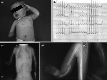
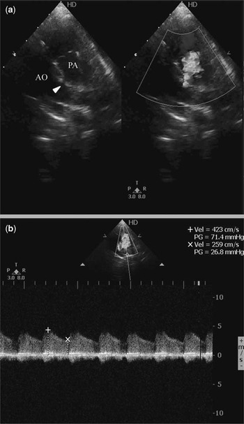
A 4-month-old male infant presented with hurried breathing and recurrent cough of 2 months’ duration. He was born at term to a 24-year-old primigravida with no significant antenatal history. On examination, the baby was active, weighed 6 kg, and had a heart rate of 138/minute and blood pressure of 90/60 mmHg in the left upper limb. Systemic oxygen saturation was 98% at ambient room air. Musculoskeletal examination revealed shortened mid segment of the right upper limb with flexion deformity of the elbow, radial deviation of the hand, and absent right thumb (Fig 1a). On auscultation, he had a continuous murmur in the left upper sternal border. Electrocardiogram showed sinus rhythm with normal axis and biventricular hypertrophy (Fig 1b). Chest X-ray revealed situs solitus, cardiomegaly, and pulmonary plethora (Fig 1c). X-ray of the right upper limb showed hypoplastic ulna with absence of the radius, first metacarpal and phalangeal bones (Fig 1d). There were no abnormalities in the other limbs (Fig 1e). Two-dimensional transthoracic echocardiogram revealed dilated left atrium and ventricle (Supplementary Video 1) with a 2 mm defect in the aortopulmonary septum (Fig 2a, arrowhead, Supplementary Video 2). Colour Doppler showed left-to-right shunt across the defect in both systole and diastole (Fig 2a, Supplementary Video 2). Continuous wave Doppler revealed both systolic and diastolic gradients across the defect with systolic gradient of 71 mmHg (Fig 2b). There was an additional small atrial septal defect with left-to-right shunt. Red blood cell, white blood cell, and platelet counts were normal. Other blood investigations were within normal limits. The infant’s father also had a similar defect involving both the upper limbs. However, he was not diagnosed with any heart defects. A diagnosis of Holt–Oram syndrome with atrial septal defect and aortopulmonary window was established. Genetic testing was not done because molecular genetic testing of TBX5 gene is currently not available in India. However, the unique features that differentiated this syndrome from other heart-hand syndromes were radial aplasia, absent thumb, and no associated haematological abnormalities. He was treated with diuretics followed by device closure of the defect using Amplatzer duct occluder II device (waist-3 mm, length-4 mm) (St. Jude Medical, Minnesota, United States of America). He was advised consultation with an orthopedic surgeon for corrective surgery of skeletal abnormalities. The parents were counselled regarding the possibility of recurrence of the defect in subsequent offspring and advised foetal echocardiographic evaluation of future pregnancies.
( a ) A shortened mid segment of the right upper limb and absent thumb. ( b ) The electrocardiogram showing normal axis and biventricular hypertrophy. ( c ) Cardiomegaly with pulmonary plethora. ( d ) X-ray of right upper limb showing absent radius, first metacarpal and phalyngeal bones. ( e ) Normal X-ray of the left upper limb.

( a ) Two-dimensional echocardiogram with colour Doppler showing aortopulmonary window with continuous left-to-right shunt. ( b ) Continuous wave Doppler showing significant gradient across the aortopulmonary window in both phases of the cardiac cycle.

Discussion
Holt and Oram first described this syndrome in 1960 when they reported on a family with atrial septal defects and congenital anomalies of the thumbs.Reference Holt and Oram 2 It is a rare autosomal dominant condition involving TBX5 gene in the long arm of chromosome 12 (12q24.1).Reference Basson, Cowley and Solomon 3 Approximately 40% of the cases are due to new mutations.Reference Huang 1 It is the prototype of heart-hand syndromes that comprises various upper limb abnormalities and congenital heart defects. The prevalence of this disorder is estimated to be 1 per 100,000 total births with a wide ethnic and geographic distribution.Reference Basson, Cowley and Solomon 3
The upper limb anomalies involve structures derived from the embryonic radial ray, typically radial, carpal, and thenar bones.Reference Basson, Cowley and Solomon 3 The abnormalities are often asymmetric, with the left side being more commonly affected.Reference Huang 1 Rarely, unilateral involvement is noted.Reference Aqrabawi, Sweitty and Quwar 4 The thumb is the most commonly affected structure and can be triphalangeal, hypoplastic, or completely absent.Reference Huang 1 Other abnormalities range from clinodactyly, brachydactyly, carpal bone dysmorphism, shortness of ulna and shortness of humerus to aplasia of radius (as in our case), and phocomelia.Reference Alizad and Seward 5
More than 85% of the individuals with the Holt–Oram syndrome have cardiac malformations.Reference Huang 1 , Reference Lin and Perloff 6 They are the most important determinants of morbidity and mortality in this condition. The most common anomalies are atrial septal defects of the secundum type and ventricular septal defects.Reference Huang 1 Rarely, it can be associated with complex congenital heart defects and conduction disturbances. Bruneau et alReference Bruneau, Logan and Davis 7 summarised the defects in 240 patients. Among these patients, 58% had an atrial septal defect and 28% had a ventricular septal defect. Less common anomalies included conduction defects, truncus arteriosus, mitral valve defects, patent ductus arteriosus, and tetralogy of Fallot. Other rare associations of the Holt–Oram syndrome include endocardial cushion defects, total anomalous pulmonary venous connection,Reference Sletten and Pierpont 8 and double-outlet right ventricle.Reference Awni 9
Aortopulmonary window is an uncommon malformation consisting of a communication, usually non-restrictive, between the adjacent walls of the ascending aorta and the pulmonary trunk. It is due to the maldevelopment of the truncal and aortopulmonary septum. Association of Holt–Oram syndrome with aortopulmonary window is extremely rare, and till date only one case report has been documented in the literature.Reference Ulrich, Schrickel and Dorn 10
Conclusions
Aortopulmonary window is a rare finding in patients with the Holt–Oram syndrome. The present case is the second report of this association. Cardiac defects are an important determinant of survival in this condition. Early diagnosis and treatment will improve the prognosis in this group of patients.
Acknowledgment
The authors are thankful to Dr Milan Bharadwaj for helping in preparing the images and videos.
Financial Support
This research received no specific grant from any funding agency, commercial or not-for-profit sectors.
Conflicts of Interest
None.