Abnormalities in the origin, course, and distribution of coronary arteries in the setting of normal atrioventricular and ventriculo-arterial connections are rare congenital anomalies that are found in 0.16–1.3% of patients undergoing diagnostic angiographyReference Akpinar, Sayin and Karabag 1 and in 0.6% of subjects in autopsy series.Reference Yuksel, Meric and Soylu 2 Although most of these anomalies are inherently benign, some are associated with myocardial ischaemia, angina, acute coronary events, arrhythmias, and sudden cardiac death.Reference Angelini 3 – Reference Cheitlin, De Castro and McAllister 5 Even benign variants are important in clinical practice as they pose a diagnostic and therapeutic challenge during diagnostic coronary angiography.
The predominant significance of congenital coronary artery anomalies in clinical practice is the potential of some of these anomalies to cause intermittent myocardial ischaemia, ischaemia-related arrhythmias, and sudden death. An abnormal origin, take-off angle, or a course between the great arteries are considered the major causes for myocardial ischaemia in congenital coronary anomalies.Reference Erdogan, Buyuklu and Aktoz 6 Beyond scientific reasoning, however, only little objective evidence is present to support the notion that these anomalies can cause reversible myocardial ischaemia or symptoms attributable to stable or unstable coronary disease. Moreover, previous observational studies have reported that atherosclerotic coronary artery disease is also prevalent in patients with coronary artery anomalies, and some have even suggested an increase in premature atherosclerotic disease due to abnormal intracoronary haemodynamics induced by coronary anomalies. As coronary artery disease is the most frequent cause of myocardial ischaemia and stable or unstable coronary events, it is therefore possible that coronary anomalies can be bystander lesions that do not influence clinical presentation or outcomes.
In this present retrospective study, we aimed to investigate clinical and angiographic characteristics of patients with coronary artery anomalies in the normally connected heart diagnosed at a tertiary referral centre and to compare these findings with an age- and sex-matched control group that included patients who underwent coronary angiography because of suspected atherosclerotic coronary artery disease. The presence and frequency of significant atherosclerosis in anomalous vessels were studied in a secondary analysis to investigate the possible association of an anomalous coronary artery with premature atherosclerosis.
Methods
Study design
Coronary angiograms performed between 2011 and 2015 were retrospectively reviewed for the presence of a coronary anomaly in at least one major epicardial coronary artery. A total of 219 patients diagnosed with coronary anomalies were found from 36,893 coronary angiography reports. Angiography records of these 219 patients were retrieved from an electronic medical database and reviewed by two experienced interventionalists (Y.Ç. and M.B.K.). The anomaly was considered present if both investigators agreed about the presence of the anomaly and its specific subset. A third investigator (T.S.G.) was invited if the initial reviewers had disagreements in a particular case. The following were accepted as coronary variants and not included in the final analysis: separate conal branch from the right coronary artery, “high take-off” right coronary artery or left main stem from the aorta, and separate left anterior descending artery and left circumflex artery from the aorta, with absent left main stem. In addition, coronary anomalies not involving changes in the origin, course, or distribution of major epicardial coronary arteries, such as coronary fistulae, those associated with other major CHD, or coronary abnormalities not hereditary in origin, such as coronary aneurysms, were excluded. After applying the aforementioned exclusion criteria, 108 patients were excluded from the study, and the final analysis included 111 patients with a definite diagnosis of coronary artery anomaly. A flow chart describing the selection of subjects is shown in Figure 1.
Flow diagram of subject recruitment. RCA=right coronary artery; CCA = congenital coronary anomaly.

In order to compare clinical and angiographic characteristics of individuals with coronary anomalies with subjects with normal coronary arteries, 110 age- and sex-matched patients who had previously undergone coronary angiography for symptomatic coronary artery disease, including patients with acute coronary events, were randomly selected to serve as a control group. Coronary angiograms of these patients were reviewed using the method defined previously for coronary anomalies.
Concomitant cardiac disease was defined as structural or functional cardiac disease that could provide an alternative explanation for the symptoms of patients, such as heart failure, severe valvulopathy, or ventricular arrhythmias. Patients with concomitant cardiac disease were included in the analysis if they had a symptom compatible with coronary artery disease. Patients who were asymptomatic at the time of coronary angiography were excluded.
Demographic and clinical data of the study population, including age, sex, symptoms, diabetes, hypertension, hypercholesterolaemia, and cigarette smoking status, were recorded.
Analysis of angiographic data
For all subjects within the study group, the anomaly was categorised into 10 major groups according to the origin, course, or distribution of the involved major epicardial coronary artery. In addition to the specific anomaly, patients were further divided into two major groups according to the Angelini classification.Reference Angelini 3 Primary analysis of these data included demographics, clinical presentation, presence of co-morbidities, and angiographic features of the patients.
All normal and anomalous major epicardial vessels were further evaluated for the presence of significant coronary atherosclerosis on the basis of available coronary angiograms. A vessel was said to have significant atherosclerosis if >50% diameter stenosis was present in at least two projections, as confirmed by the majority of investigators. Additional manifestations of atherosclerosis, such as positive re-modelling and coronary ectasia, were not included in the analysis. Patients with anomalies intrinsic to coronary anatomy, such as those with dual left, dual, or absent left circumflex artery or dual right coronary artery, were excluded from this secondary analysis, as these patients could not be classified using standard coronary nomenclature. The location of significant coronary artery lesions was classified as described earlier.Reference Scanlon, Faxon and Audet 7 After excluding these patients, 98 were included in this secondary analysis. Patients were grouped according to the type of anomalous epicardial vessel, and the presence of significant atherosclerotic coronary artery disease in these anomalous vessels was compared with the same non-anomalous artery of control patients.
Statistical methods
Continuous variables are expressed as mean±standard deviation, whereas categorical variables are given as percentages. Descriptive data, including the frequency of each anomaly, are provided for patients in the coronary artery anomaly group. For categorical data including symptoms, mode of presentation, and the presence of significant coronary artery disease in a major epicardial artery, the χ2-test and Fisher’s exact test were used as appropriate to compare groups. For continuous variables, Student’s t-test was used to compare coronary artery anomaly and control groups. A p-value <0.05 was accepted as significant if the test was within the 95% confidence interval. A second analysis with similar statistical methods was performed after exclusion of patients with concomitant cardiac disease.
Results
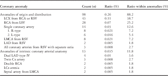
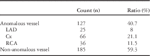
Among 36,893 coronary angiograms, 111 patients (0.30%) with coronary anomalies were found. The mean age was 57.3±11.3 (with a range from 23 to 80). There were 45 (40.6%) females and 66 (59.4%) males. Data regarding the frequency of each coronary anomaly are given in Table 1. Anomalous aortic origin of the left circumflex from the right sinus of Valsalva or from the right coronary artery was found as the most prevalent anomaly (43 out of 111 patients, 38.7%). The second most common anomaly was anomalous right coronary artery originating from the left sinus of Valsalva, present in 28 patients (25.2%). Single coronary artery (14 out of 111 patients, 12.6%) and left coronary artery arising from the right sinus of Valsalva (eight out of 111 patients, 7.2%) were other frequent anomalies in our study population. Table 2 provides details regarding the frequency of anomalous vessels in the study group.
Classification of coronary anomalies according to angiographic characteristics.

Cx=circumflex; LAD=left anterior descending artery; LCx=left circumflex; LMCA=left main coronary artery; LSV=left sinus of Valsalva; RCA=right coronary artery; RSV=right sinus of Valsalva
Distribution of anomalous vessels in 107 coronary anomaly patients.

Cx=circumflex; LAD=left anterior descending artery; RCA=right coronary artery
Two patients with a septal artery from the left main coronary artery and two patients with atretic left circumflex artery were excluded
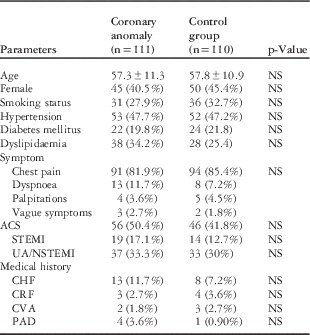
Baseline patient characteristics, as well as clinical presentation for the study and control groups, are provided in Table 3. In the coronary anomaly cohort, the predominant symptom was chest pain and was observed in 81.9% of patients. The frequency of chest pain as a symptom was similar to patients who underwent coronary angiography for suspected coronary artery disease (p=0.48, odds ratio: 0.77, 95% confidence interval=0.38–1.6). There were no statistically significant differences regarding the frequency of other symptoms. Presentation with an acute coronary event was more frequent in the study group (50% versus 42%), but this finding did not have statistical significance (p=0.2, odds ratio: 1.41, 95% confidence interval=0.83–2.40). Among patients with stable symptoms at presentation, 16 out of 55 patients within the coronary arterial anomaly group (29%) and 32 out of 64 patients (50%) in the control group had significant coronary artery disease on angiography, with a statistically significant difference between groups (p=0.02, odds ratio: 0.41, 95% confidence interval=0.19–0.87).
Demographic and clinical characteristics of patients with a congenital coronary anomaly and control patients.

ACS=acute coronary syndrome; CHF=chronic heart failure; CRF=chronic renal failure; CVA=cerebrovascular accident; NS=not significant; PAD=peripheral arterial disease; STEMI=ST segment elevation myocardial infarction; UA/NSTEMI=unstable angina/non-ST elevation myocardial infarction
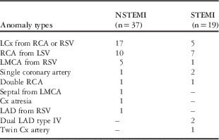
The diagnosis at admission was acute coronary syndrome in 56 (50.4%) out of 111 patients. Of these 56 patients, 37 (66.0%) had a diagnosis of unstable angina/non-ST segment elevation myocardial infarction, and the remaining 19 (34.0%) patients had a diagnosis of ST elevation myocardial infarction. In the study group, no significant coronary artery disease was found in 16 (43.2%) out of 37 unstable angina/non-ST elevation myocardial infarction patients, whereas only two out of 19 (10.5%) patients diagnosed with ST elevation myocardial infarction did not have significant coronary disease. In the control group, 11 out of 33 patients with unstable angina/non-ST elevation myocardial infarction (33%) and 1 out of 18 patients with ST elevation myocardial infarction (5.5%) did not have significant coronary artery disease at angiography. For acute coronary events, there was no significant difference between the study group and the control group regarding the frequency of significant coronary disease at angiography (p=0.28 for unstable angina/non-ST elevation myocardial infarction and p=0.74 for ST elevation myocardial infarction). Table 4 shows the distribution of patients admitted with acute coronary syndrome according to the coronary anomaly types.
Distribution of acute coronary syndrome patients according to the coronary anomaly types.

Cx, circumflex; LAD, left anterior descending artery; LCx, left circumflex; LMCA, left main coronary artery; LSV, left sinus of Valsalva; RCA, right coronary artery; RSV, right sinus of Valsalva; STEMI=ST segment elevation myocardial infarction; NSTEMI=non-ST elevation myocardial infarction
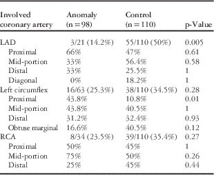
A smaller number of patients within the study group had atherosclerotic coronary artery disease compared with control patients, but the statistical significance was marginal (p=0.05, odds ratio: 0.69, 95% confidence interval=0.34–1.001). When anomalous vessels were compared with their counterparts in the control group, a significantly lower number of patients within the coronary anomaly group had significant atherosclerotic coronary artery disease in the left anterior descending artery (p=0.005). Among patients with an anomalous left circumflex or right coronary anomaly, the odds ratios for having significant coronary artery disease were similar to control patients without a coronary artery anomaly (Table 5). When present, atherosclerotic coronary artery disease was more frequently confined to proximal segments of the anomalous circumflex artery in patients with coronary anomalies involving the left circumflex artery (Table 5). For other anomalies, no significant differences were noticed regarding the location of the coronary artery lesions.
Comparison of the frequency of significant atherosclerotic coronary artery disease in patients with anomalous coronary arteries and control patients.

LAD=left anterior descending artery; RCA=right coronary artery
Significant coronary artery disease is defined as >50% angiographic stenosis in two different projections in a major epicardial vessel. Patients with intrinsic coronary artery anomalies were excluded and 98 patients (118 anomalous vessels) were included
Analysis after exclusion of patients with concomitant cardiac disease
There were six patients in each group (0.054% in both groups, p=0.98) who had concomitant cardiac disease that may cause symptoms compatible with coronary artery disease. A repeat analysis after exclusion of these patients showed that patients with stable symptoms less frequently had significant coronary atherosclerosis in at least one major epicardial vessel compared with controls (31.3% in coronary anomaly group versus 51.6% in the control group, p=0.03), whereas this figure was similar for patients with an acute coronary event (70.4 versus 77.3%, p=0.44). Similar to the original cohort, there were also no significant differences between the two groups when patients with acute coronary events were analysed within the ST elevation myocardial infarction (89.5 versus 92.3%, p=0.79) or the unstable angina/non-ST elevation myocardial infarction (60.0 versus 71.0%, p=0.35) subgroups. Finally, patients with a left anterior descending coronary artery anomaly had significantly less atherosclerosis in the left anterior descending artery compared with controls (16.7 versus 51.0%, p=0.002), but there were no differences for anomalous left circumflex (28.0 versus 35.6%, p=0.29) or anomalous right coronary artery (25.0 versus 36.5%, p=0.21).
Discussion
The incidence of coronary anomalies differs considerably between studies and ranges from 0.16–1.3%.Reference Akpinar, Sayin and Karabag 1 The incidence of a major epicardial coronary artery anomaly was 0.3% in our retrospective study, which is within the range reported by previous studies.Reference Click, Holmes, Vlietstra, Kosinski and Kronmal 8 , Reference Chaitman, Lesptrance, Saltiel and Bourassa 9 A major reason for this wide range reported by previous studies is the absence of a universally accepted definition for “coronary anomaly”. Some studies categorised every deviation from normal anatomy as a coronary artery anomaly, such as absence of the left main coronary artery, whereas others accepted these anomalies as variations. Anomalous left circumflex artery arising from the right coronary artery or from the right sinus of Valsalva was the most common coronary anomaly reported in the majority of previous studies,Reference Yuksel, Meric and Soylu 2 , Reference Click, Holmes, Vlietstra, Kosinski and Kronmal 8 , Reference Sivri, Aktoz, Yalta, Ozcelik and Altun 10 – Reference Sohrabi, Habibzadeh and Abbasov 12 whereas the remaining studies found that the right coronary artery arising from the left sinus of Valsalva was the most frequent anomaly.Reference Suryanarayana, Kollampare and Bin Riaz 13 – Reference Garg, Tewari, Kapoor, Gupta and Sinha 15 Our findings were comparable with the literature, with the most frequent anomaly being an anomalous left circumflex artery arising from the right sinus of Valsalva or from the right coronary artery, and right coronary artery arising from the left sinus of Valsalva was the second most common anomaly. Consistent with the previous literature, the majority of our patients were classified into Angelini Group A, which includes anomalies of origin and course.Reference Angelini 3
More than 80% of coronary anomalies are benign and do not cause ischaemic symptoms.Reference Yamanaka and Hobbs 16 The remaining anomalies, however, are cited as a cause of myocardial ischaemia, acute coronary events, malignant arrhythmias, and sudden cardiac death, with the latter particularly observed during or immediately after physical activity.Reference Erdogan, Buyuklu and Aktoz 6 , Reference Salah, de Voogt and Suat 17 The prognosis of a coronary anomaly is predominantly determined by its relation to the great arteries, as compression of a major epicardial vessel between the aorta and the pulmonary trunk is suggested as the major cause for myocardial ischaemia.Reference Yamanaka and Hobbs 16 Other mechanisms suggested to explain myocardial ischaemia included the following: acute angulation of the anomalous vessel at its proximal segment; kinking, torsion, or spasm of the vessel shaft; slit-like ostium of the coronary artery rendering the vessel vulnerable for acute closure; and intussusception of the anomalous artery and atherosclerotic coronary artery disease causing vessel obstruction.Reference Nath, Kennett and Politte 18 In our series, the most common symptom was chest pain, with or without anginal characteristics, and the incidence of chest pain was similar to age- and sex-matched patients who underwent angiography for suspected atherosclerotic coronary artery disease. This finding is in parallel with a previous substudy of the Coronary Artery Surgery Study, which reported that both coronary artery anomaly and control patients had angina – typical or atypical – as the most frequent symptom at admission, without any difference between the groups.Reference Click, Holmes, Vlietstra, Kosinski and Kronmal 8 In patients with stable symptoms, significant atherosclerotic coronary artery disease was less frequent in the coronary anomaly group, thus suggesting the presence of a coronary anomaly as a possible cause for chest pain; yet, a significant fraction of patients (29%) with a coronary anomaly and chest pain had concomitant obstructive coronary disease, suggesting that atherosclerosis is also common in this patient population and may cause myocardial ischaemia independent of the underlying coronary anomaly. Therefore, it is advisable to review all coronary segments for the presence of significant atherosclerotic coronary artery disease before concluding that the coronary anomaly per se is the cause of the symptoms.
In a proportion of patients with clinical, electrocardiographic, and laboratory features of myocardial infarction, no significant lesions were found on coronary angiograms. This can be observed in 1–12% of patients with a referring diagnosis of myocardial infarction,Reference Ammann, Marschall and Kraus 19 and could be as high as 30% in patients with unstable angina/non-ST elevation myocardial infarction.Reference Ong, Athanasiadis and Hill 20 Potential mechanisms for this phenomenon include coronary spasm, spontaneously lysed coronary thrombus, coagulation disturbances, endothelial dysfunction, and coronary artery anomalies.Reference Kardasz and De Caterina 21 An acute coronary syndrome was the initial diagnosis of admission in 17.2% of patients in a series of 238 patients with coronary artery anomalies.Reference Akpinar, Sayin and Karabag 1 In our study, the initial diagnosis was acute coronary syndrome in 50.4% of patients with a coronary anomaly. In the subset of patients with unstable angina/non-ST elevation myocardial infarction, 43.2% of patients in the coronary anomaly group had no significant atherosclerosis in any major epicardial vessel, which was not significantly different from the control group (33.3%, p=0.28). In both groups, patients with an ST elevation myocardial infarction had high rates of significant atherosclerotic stenosis in at least one major epicardial artery (94.4% in the control group and 89.4% in the anomaly group). Our findings question the importance of coronary artery anomalies as a cause of acute coronary events, especially in patients with ST segment elevation myocardial infarction, as the frequency of significant coronary artery disease did not differ between patients with a coronary anomaly and age- and sex-matched controls; yet, patients with unstable angina/non-ST elevation myocardial infarction and a coronary artery anomaly less frequently had significant atherosclerotic disease compared with control patients, although this finding did not reach statistical significance. Moreover, the culprit lesion causing the acute event – coronary anomaly or significant coronary artery disease – is unknown, and the symptoms might have been caused by the anomaly rather than the atherosclerotic obstruction in at least a proportion of patients. Therefore, these results should be interpreted with caution, and do not necessarily imply that all coronary artery anomalies are innocent bystander lesions not associated with an acute coronary event.
Congenital coronary anomalies were previously suggested as a cause for accelerated atherosclerosis in coronary arteries, due to haemodynamic effects caused by the anomalous vessel. Silverman et alReference Silverman, Bulkley and Hutchins 22 proposed that an abnormal angle at origin and a tortuous course accelerate atherosclerosis in anomalous arteries. Hutchins et alReference Hutchins, Miner and Boitnott 23 suggested that the unusual take-off angle and a more tortuous course of the proximal portion of the anomalous coronary artery predispose it to accelerated atherosclerosis. Liu et alReference Liu, Richardson and Taylor 24 have postulated that the junction point of the bound portion of the anomalous artery and the free portion as it wraps around the aorta is an area susceptible to lipid accumulation. Erdogan et alReference Erdogan, Buyuklu and Aktoz 6 hypothesised that the atherosclerotic process is accelerated before the take-off of an anomalous right coronary artery from the left anterior descending coronary artery. The literature is conflicting in this sense, as the results of some studies suggest an increase in atherosclerosis in patients with a coronary anomaly,Reference Click, Holmes, Vlietstra, Kosinski and Kronmal 8 , Reference Samarendra, Kumari, Hafeez, Vasavada and Sacchi 25 whereas findings from others did not find a similar relationship.Reference Eid, Itani, Al-Tannir, Sayegh and Samaha 11 , Reference Suryanarayana, Kollampare and Bin Riaz 13 , Reference Harikrishnan, Jacob and Tharakan 26 , Reference Page, Engel, Campbell and Thomas 27 This discrepancy is not surprising as the haemodynamic effect created by an anomaly could be substantially different from each other, even when two anomalies are grouped within the same subgroup. In this regard, abnormal branching patterns caused by a specific anomaly rather than the anomaly itself could be more important, as proximal points of branching were frequent sites for narrowing in normal coronaries.Reference Halon, Sapoznikov, Lewis and Gotsman 28 In the present report, we noted that patients with an anomalous left anterior descending artery had significantly less atherosclerotic lesions within the left anterior descending artery, whereas patients with an anomalous left circumflex or right coronary artery did not have an increased tendency for atherosclerosis in the respective arteries, as compared with controls. The sole study with a similar case–control design as the present study suggested an increase in the degree of stenosis in the left circumflex and a trend for lesser atherosclerosis in the left anterior descending coronary artery.Reference Click, Holmes, Vlietstra, Kosinski and Kronmal 8 The results of this aforementioned study and the present analysis are not directly comparable, as patients without significant stenosis were not included in the present study. In a separate analysis of 147 patients with coronary anomalies, with the majority of patients having a right coronary artery anomaly, anomalous arteries were compared with the remaining non-anomalous vessels of the same patient group. In the latter study, the prevalence of atherosclerotic disease was similar for anomalous coronary arteries and other non-anomalous vessels, suggesting that anomalous coronary arteries did not have a tendency for accelerated atherosclerosis.Reference Sohrabi, Habibzadeh and Abbasov 12 On the basis of the present results and previous literature, it is reasonable to suggest that coronary artery anomalies do not cause an increase in the frequency of significant atherosclerotic coronary artery disease. An increase in subclinical atherosclerotic lesions, however, remains a possibility that highlights the need for further research on the topic.
Notably, in nearly all the anomalies involving the left anterior descending coronary artery, the left anterior descending artery – or the left main coronary – took off directly from the right sinus of Valsalva and coursed anteriorly to reach the anterior segment of the left ventricle. Owing to this long course, the proximal part of the vessel lacked branching points, which can reduce the tendency for developing atherosclerosis. We have previously suggested a similar mechanism in a patient with type IV dual left anterior descending artery anomaly,Reference Çanga, Güvenç and İlhan 29 in whom all vessels except the long left anterior descending artery originating from the right sinus of Valsalva had extensive atherosclerosis. In contrast to the left anterior descending artery, anomalies involving the left circumflex or the right coronary artery were less uniform and had different origin points and multiple potential courses, thus creating branching points or zones compressed between the aorta and the pulmonary artery that could change local haemodynamics and create vulnerable zones for atherosclerosis. Our results show that the proximal segments of the left circumflex artery were more frequently involved with atherosclerotic disease in patients with an anomalous left circumflex artery, as compared with those with normal circumflex arteries (Table 5). A similar finding was also reported by Coronary Artery Surgery Study investigators, which was explained by the aforementioned postulate of Hutchins et al.Reference Click, Holmes, Vlietstra, Kosinski and Kronmal 8 , Reference Hutchins, Miner and Boitnott 23 This hypothesis, however, does not explain why this finding is limited to abnormal circumflex arteries, and not seen in anomalous the left anterior descending artery or right coronary arteries, or why the overall incidence of atherosclerotic lesions in anomalous circumflex arteries is lower than normal circumflex arteries. As stated earlier, the origin and course of the circumflex artery can be highly variable, which can render the proximal segment of the anomalous circumflex more vulnerable to atherosclerosis compared with other anomalous coronary arteries. It should be acknowledged that other mechanisms besides disturbed local haemodynamics could affect the frequency and distribution of atherosclerotic lesions in anomalous coronary vessels, which necessitates further studies on this subject.
Patients with severe concomitant cardiac disorders such as severe valvulopathy or heart failure may have symptoms that overlap with coronary artery disease, and the frequency of coronary artery disease may be different in these patient populations. Studies on patients who underwent coronary angiography before a cardiac operation for valvular disease have suggested a low prevalence of coronary artery disease, and the presence of coronary artery disease was rather confined to patients who were older and had aortic valve disease.Reference Sampaio, Jonke and Falcão 30 Ischaemic cardiomyopathy is the most common aetiology of congestive heart failure,Reference DM, Larson and Leip 31 and ~65% of patients with heart failure have demonstrable coronary artery disease.Reference Gheorghiade, Sopko and De Luca 32 In the present study, both groups had patients with a severe concomitant cardiac disease, although the number of patients were low (~5%) for both groups. After excluding patients with a severe concomitant cardiac disease, a repeat analysis showed that the main findings for the present analysis did not change. Whether these results could be extended to patients with severe concomitant cardiac disease remains unknown as the number of patients was too low to make an assumption.
Study limitations
The data were collected retrospectively from one centre, which severely limits the accuracy of the study. As the study design was retrospective and cross-sectional, long-term data on cardiovascular end points such as mortality, arrhythmias, repeat acute coronary events, repeat hospitalisations, etc. were not available. Data on myocardial ischaemia were not available for the majority of patients with coronary artery anomalies. Chest pain is a common symptom in the general population and does not necessarily confirm the presence of myocardial ischaemia. Coronary angiography is a luminographic method, which could not discriminate a normal vessel from an atherosclerotic vessel with extensive positive re-modelling. Neither CT coronary angiography nor invasive techniques were performed to determine subclinical atherosclerosis; therefore, the results presented in this study are valid only for angiographically significant coronary atherosclerosis. Although the results were not affected by the exclusion of patients with concomitant cardiac disease, some patients might have had undiagnosed cardiac or other conditions at the time of angiography, as echocardiography or other diagnostic modalities were not routinely used in all patients. Although a conventional classification scheme was used to define coronary segments in anomalous coronary arteries, this scheme was initially developed for normal coronary arteries, and could therefore give erroneous results when used for anomalous coronary vessels. Subgroup analyses as well as multivariate regression could not be performed because of the low number of patients within each subgroup. A longitudinal follow-up study, rather than a study with a case–control design, should provide more information on the susceptibility of anomalous coronary vessels to atherosclerosis. Only symptomatic patients with coronary anomalies were included in the present study, and the results should be interpreted within this context.
Conclusions
Congenital coronary anomalies in the otherwise normally structured heart are infrequent yet important entities that may lead to myocardial ischaemia without atherosclerotic coronary disease, misinterpretation of angiographic data, and difficulties in surgical or percutaneous interventions. Our findings indicate that patients with a coronary anomaly presenting with stable symptoms more frequently had non-obstructed coronary arteries at angiography, but nearly one-third of these patients still have significant coronary atherosclerosis. Approximately half of these patients were admitted to the hospital with unstable angina or myocardial infarction, but the frequency of significant atherosclerotic coronary disease did not differ from age- and sex-matched controls without a coronary anomaly. The frequency of significant coronary artery disease was found to be similar in anomalous left circumflex or right coronary arteries as compared with normal counterparts, whereas there was a tendency towards less atherosclerotic involvement in anomalous left anterior descending arteries. More studies on congenital coronary arteries, preferably with long-term follow-up data for cardiovascular end points, are needed to ascertain the relationship of coronary anomalies with coronary events and atherosclerosis.
Acknowledgements
None.
Financial Support
This research received no specific grant from any funding agency or from commercial or not-for-profit sectors.
Conflicts of Interest
None.
Ethical Standards
The authors assert that all procedures contributing to this work comply with the ethical standards of the Turkish national guidelines on human experimentation and with the Helsinki Declaration of 1975, as revised in 2008, and has been approved by the Haydarpasa Numune Hospital ethical committee.