INTRODUCTION
Microsporidia are a unique and diverse group of obligatory intracellular parasites (Keeling, Reference Keeling2009), now regarded as derived from fungi or fungi-like ancestors (Hirt et al. Reference Hirt, Logsdon, Healy, Dorey, Doolittle and Embley1999; Keeling and Fast, Reference Keeling and Fast2002). Microsporidia have been reported to infect almost all invertebrate phyla as well as most major vertebrate groups. Their ubiquitous presence and significant adaptations to a parasitic life, such as the reduction of mitochondria to vestigial organelles called mitosomes (Williams et al. Reference Williams, Hirt, Lucocq and Embley2002), support the argument that the origin of parasitism in this group is ancient (Vossbrinck et al. Reference Vossbrinck, Maddox, Friedman, Debrunner-Vossbrinck and Woese1987). Amongst Daphnia, an ecologically important genus of planktonic crustaceans inhabiting lakes and ponds, they constitute one of the most common groups of parasites (Green, Reference Green1974; Stirnadel and Ebert, Reference Stirnadel and Ebert1997; Ebert, Reference Ebert2005). Field studies on the occurrence of microsporidia in natural Daphnia communities have found that they usually first appear in late spring when host density is high and persist throughout summer and autumn, with prevalences in some cases rising up to 100% (Brambilla, Reference Brambilla1983; Bengtsson and Ebert, Reference Bengtsson and Ebert1998; Ebert et al. Reference Ebert, Hottinger and Pajunen2001; Wolinska et al. Reference Wolinska, Seda, Koerner, Smilauer and Petrusek2011a). Microsporidia infect a variety of Daphnia species across different habitats, ranging from tiny rock pools (Ebert et al. Reference Ebert, Hottinger and Pajunen2001) and temporary ponds (Decaestecker et al. Reference Decaestecker, Declerck, De Meester and Ebert2005), to permanent lakes (Wolinska et al. Reference Wolinska, Seda, Koerner, Smilauer and Petrusek2011a; Yan and Larsson, Reference Yan and Larsson1988).
Until now only microscopic techniques have been used to assess microsporidian infection patterns in natural Daphnia communities. Thus, the prevalence of microsporidia may have been largely underestimated, due to their minute size and intracellular lifestyle (Ebert, Reference Ebert2005). In particular, the early stages of infection are easily missed, as during this phase there are no visible signs of infection. Moreover, some microsporidian species are similar in appearance within the Daphnia host, making it difficult to discriminate species based on light microscopy (Stirnadel and Ebert, Reference Stirnadel and Ebert1997). Lastly, co-infections by different parasite species are common in Daphnia (Decaestecker et al. Reference Decaestecker, Declerck, De Meester and Ebert2005; Stirnadel and Ebert, Reference Stirnadel and Ebert1997; Wolinska et al. Reference Wolinska, Keller, Manca and Spaak2007). As a result of the underestimation of prevalences and/or misclassifications it is difficult to compare epidemic patterns across different habitats or between studies. For further detailed studies of this host-parasite system, it is thus crucial to obtain more powerful tools to detect and properly identify different microsporidian species. These methods should be applicable for screening a large sample of host individuals from a given population. Although the above-mentioned limitations would make microsporidia prime candidates for using a molecular approach for their identification, molecular techniques have scarcely been used to assess infection patterns in natural host populations (e.g. Hogg et al. Reference Hogg, Ironside, Sharpe, Hatcher, Smith and Dunn2002; McClymont et al. Reference McClymont, Dunn, Terry, Rollinson, Littlewood and Smith2005), and never in Daphnia.
The purpose of the present study was to describe microsporidian infection patterns across Daphnia communities inhabiting 8 different permanent water bodies, focusing on spatial and temporal variation. In addition, we wanted to determine the frequency of microsporidian co-infections within single hosts. Thus, we designed general and species-specific primers, amplifying a fragment of the microsporidian small subunit ribosomal DNA (hereafter referred to as SSU rDNA) and applied them to apparently infected Daphnia. In addition, some PCR products were sequenced to verify the specificity of the newly designed primers, as well as to confirm detected co-infections. As we detected some lineages that had never been sequenced before, we performed phylogenetic analyses to reveal their relationship to other microsporidian parasites of Daphnia.
MATERIALS AND METHODS
Origin of specimens and symptoms of infection
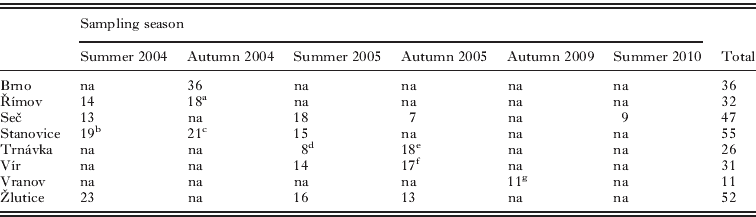
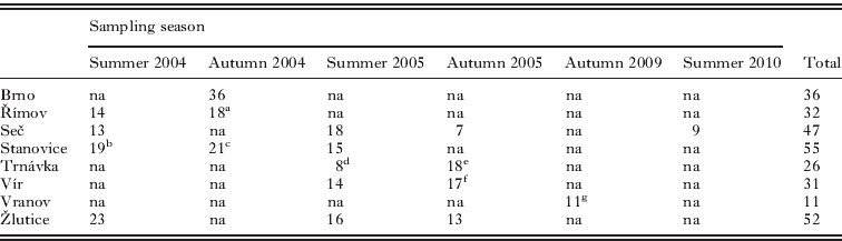
The infected Daphnia were selected from ethanol-preserved zooplankton samples originating from 8 man-made reservoirs in the Czech Republic. All sampled reservoirs have a similar canyon-shaped morphology, and were created between years 1934 and 1982. Stanovice and Žlutice (21 km apart) are located in the west of the country, Seč and Trnávka (47 km apart) in the centre, Brno and Vír (40 km apart) in the east, and Římov and Vranov (97 km apart) in the south (see Fig. 1). The Daphnia populations of these reservoirs consist mainly of common members from the European D. longispina complex (Petrusek et al. Reference Petrusek, Hobaek, Nilssen, Skage, Černý, Brede and Schwenk2008a): D. cucullata, D. galeata and D. longispina, as well as their interspecific hybrids (Seda et al. Reference Seda, Petrusek, Machacek and Smilauer2007b; Petrusek et al. Reference Petrusek, Seda, Macháček, Ruthová and Šmilauer2008b). The samples were collected in summer (July) and autumn (late September to October) of the years 2004 and 2005 (Table 1). Additionally, we analysed 2 more samples: from autumn 2009 (Vranov) and summer 2010 (Seč). The samples were collected from the entire water column, by hauling a plankton net (mesh size 170 μm) at 3 sampling stations along the reservoir's horizontal axis: near the river inflow, in the middle of the reservoir, and near the dam (see Table 1).
Location of reservoirs in the Czech Republic and taxon composition of microsporidian communities infecting the Daphnia longispina complex (samples from different dates are pooled). The ‘other’ group consists of 4 rare taxa (Gurleya vavrai, Mic4, Mic5 and Mic6). Total number of screened microsporidia is given next to the respective pie chart.

Number of analysed hosts infected with microsporidia, per reservoir and sampling season
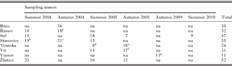
a–g Some or all analysed Daphnia individuals originated from inflow stations of the respective reservoirs (a: 18/18 individuals, b: 12/19, c: 21/21, d: 3/8, e: 15/18, f: 15/17), or from a middle station (g: 11/11).
na, Not analysed (due to low prevalence in 2004–2005, and due to unavailability of samples in 2009–2010).
The ethanol-preserved samples (96% ethanol) were screened for Daphnia with visible signs of microsporidian infections, using a stereomicroscope at X 50 magnification. Under the microscope, microsporidian infections appear as a fuzzy and opaque mass within the body cavity of their host (Fig. 2; see also Vossbrinck et al. Reference Vossbrinck, Andreadis, Vávra and Becnel2004; Refardt et al. Reference Refardt, Decaestecker, Johnson and Vávra2008). In an attempt to link the visible signs of infection to a particular parasite species or clade, infected Daphnia were photographed and assigned into categories based on the shape of the parasite mass. After molecular identification of parasites (as described below), their appearance was evaluated against parasite identity. To allow for the specificity of the assignment, only Daphnia infected with a single parasite species were taken into consideration for this comparison.
Picture of Daphnia infected with Berwaldia schaefernai, with (left; arrow) and without (right) spore mass proliferating into the host's head.

DNA extraction and primer test
Genomic DNA was isolated by overnight incubation of entire, single infected Daphnia with 100 μg/ml proteinase K (Merck KgaA, Darmstadt, Germany) in proteinase K buffer (10 mm Tris (pH 8·0), 100 mm NaCl, 25 mm EDTA (pH 8·0) and 0·1% SDS) at 55 °C. After heat inactivation of proteinase K for 12 min at 95°C, chitinous debris was removed and the DNA was precipitated from the supernatant using isopropanol. The DNA was then dissolved in 20–50 μl of sterile water (see Wolinska et al. Reference Wolinska, Giessler and Koerner2009). One general and four species-specific primer pairs (see Table 2) were designed, all amplifying a segment from the SSU rDNA of 4 microsporidia taxa identified in a previous study (Wolinska et al. Reference Wolinska, Giessler and Koerner2009), namely Berwaldia schaefernai, and unidentified lineages provisionally labelled as Mic1, Mic2 and Mic3. Primer specificity was evaluated using DNA isolated from several hosts infected by any of the 4 microsporidian taxa, obtained by Wolinska et al. (Reference Wolinska, Giessler and Koerner2009), see Supplementary Table 1, online version only. In addition, negative controls were included: DNA extracted from laboratory cultivated (i) uninfected Daphnia; (ii) Daphnia infected with the protozoan Caullerya mesnili, and (iii) Daphnia infected with the parasitic yeast Metschnikowia sp. These 2 microparasites frequently infect natural communities of the D. longispina complex (Wolinska et al. Reference Wolinska, Keller, Manca and Spaak2007, Reference Wolinska, Seda, Koerner, Smilauer and Petrusek2011a). Detection sensitivity of the primers in the case of co-infections was tested on artificially mixed parasite DNA, in various ratios (as obtained from Wolinska et al. Reference Wolinska, Giessler and Koerner2009) as well as by using parasite DNA isolated from naturally co-infected Daphnia (only available for B. schaefernai and Mic3 infections, from Wolinska et al. Reference Wolinska, Giessler and Koerner2009).
Description of primers used to amplify partial SSU rDNA regions from microsporidian parasite taxa
a The PCR cycle was identical for all primers: initial denaturation of 3 min at 95°C, followed by 35 cycles of 95°C (30 sec), primer specific annealing temperature Tm (30 sec) and 72°C (30 sec) with a final extension of 7 min at 72°C.
b Product length of the general primers varied depending on parasite taxa; 455 bp for B. schaefernai and Mic1, 451 bp for Mic2a and b variant likewise, 461 bp for Mic3a and 494 bp for Mic3b (variants of Mic2 and Mic3 were not considered separately, for further information on a and b variants, see Wolinska et al. Reference Wolinska, Giessler and Koerner2009). Gurleya vavrai had a product length of 456 bp. The bands of the newly sequenced taxa, Mic4, Mic5 and Mic6, were of the following length: 461 bp, 438 bp and 466 bp.
Application of primers on field samples
First, the general primers were used to verify microsporidian infections in Daphnia previously selected by microscopic examination (Table 1). Amplicon size in PCR varied between 438 and 494 bp, depending on the parasite taxon (Table 2). Then, species-specific primers were applied to those samples for which PCR products of the aforementioned size range were detected. PCRs were performed in a 20 μl reaction volume, including 2 μl of genomic DNA, DreamTaq buffer (resulting in a 2 mm concentration of MgCl2), 0·25 mm deoxynucleoside triphosphates (both from Fermentas, St. Leon-Rot, Germany) and 0·5 μ m of each primer pair (Metabion, Martinsried, Germany). Different amounts of DreamTaq polymerase were used: 0·1 U/μl for the general primer, 0·0625 U/μl for Berwaldia and Mic1 primers and 0·125 U/μl for Mic2 and Mic3 primers. Furthermore, for Mic2 and Mic3 primers a final MgCl2 concentration of 4 mm was used and 10% DMSO were added to the PCR reactions to improve PCR efficiency. Primer sequences, annealing temperatures, and PCR cycle settings are given in Table 2.
Sequencing
There were 2 main objectives for sequencing: (i) to verify whether the appropriately-sized bands obtained with the general primers, but not with any of the specific primers, represent microsporidian infections and, if so, to identify the parasite (attempted for all 59 such cases), (ii) to check the reliability of identification by species-specific primers (Table 2; in total 44 PCR products were sequenced, comprising representatives of all 4 focal microsporidian species and 27·7% of all detected co-infections). Before sequencing, PCR amplicons obtained with the general primers were cut from the gel and eluted using the QIAquick gel extraction kit (Qiagen, Hilden, Germany). The amplicons obtained with the species-specific primers were purified (using exonuclease I and shrimp alkaline phosphatase, both from Fermentas, St. Leon-Rot, Germany) and directly sequenced. The sequencing reactions were carried out using BigDye v1.1 sequencing mix and an ABI 3730 capillary sequencer (Applied Biosystems, Foster City, CA). The resulting chromatograms were carefully inspected for occasional polymorphic sites (i.e. those with overlapping or secondary peaks) indicating potential intragenomic (Wolinska et al. Reference Wolinska, Giessler and Koerner2009, Reference Wolinska, Spaak, Koerner, Petrusek, Seda and Giessler2011b) or intraspecific (in case of co-infection by different parasite strains) variation in the respective microsporidian taxon. Nucleotide composition of such polymorphic sites was expressed by the respective IUPAC codes. Representative sequences of newly characterized lineages (Mic4 to Mic6) have been deposited in GenBank (Accession numbers: JQ012755, JQ012756, JQ012757).
Data analysis
Spatial and temporal patterns in microsporidian communities
Differences in microsporidian taxon composition across and within the lakes (i.e. between different years or seasons) were determined with a G test of independence (Sokal and Rohlf, Reference Sokal and Rohlf1995). A sequential Bonferroni correction (Rice, Reference Rice1989) was used to assess significance in case of multiple tests performed within the same lake. In addition, to evaluate similarities in parasite community composition among different host populations, the Euclidean distance between each pair of lakes was computed from the presence/absence data as well as from the frequency data on microsporidian taxa composition (taking into consideration only the microsporidian taxa B. schaefernai, Mic1, Mic2 and Mic3, as the additionally recorded taxa occurred at low frequencies only). The correlation between pairwise geographical distances and Euclidean distances of community composition was evaluated using a Mantel test (5000 permutations) as incorporated in Past v2.07 (Hammer et al. Reference Hammer, Harper and Ryan2001).
Molecular identification and phylogeny reconstruction
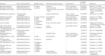
Obtained sequences (one representing each lineage) were compared with those deposited in the NCBI nucleotide database. First, we used similarity searches (BLAST) to check for the presence of corresponding sequences, and calculated the sequence divergence (using Kimura 2-parameter distance) of lineages that did not have any close match (<1% divergence) in GenBank from their most similar counterparts. Then we constructed a tree in order to reveal the relationships of these lineages. We included in the analysis sequences of related microsporidians known to infect Daphnia, as well as closely related microsporidian lineages infecting other hosts for each clade (Table 3). The selection of taxa was based on similarity searches and previously published microsporidia phylogenies (Refardt et al. Reference Refardt, Canning, Mathis, Cheney, Lafranchi-Tristem and Ebert2002, Reference Refardt, Decaestecker, Johnson and Vávra2008; Slothouber Galbreath et al. Reference Slothouber Galbreath, Smith, Terry, Becnel and Dunn2003; Franzen et al. Reference Franzen, Fischer, Schroeder, Schölmerich and Schneuwly2005; Vossbrinck and Debrunner-Vossbrinck Reference Vossbrinck and Debrunner-Vossbrinck2005; Wolinska et al. Reference Wolinska, Giessler and Koerner2009). A sequence of the basal fungus Basidiobolus ranarum was used as an outgroup. Sequences were aligned by MUSCLE (Edgar, Reference Edgar2004) incorporated in MEGA5 (Tamura et al. Reference Tamura, Peterson, Peterson, Stecher, Nei and Kumar2011). The poorly aligned, non-conserved and highly divergent regions were eliminated by Gblocks 0.91b (Castresana, Reference Castresana2000) using less-stringent settings, resulting in 402 bp long alignment. The relationship among taxa was inferred by the maximum likelihood method computed in PAUP 4.10b (Swofford, Reference Swofford2000) under the TIM3+I+G model selected by Akaike information criterion in jModeltest 0.1.1 (Posada, Reference Posada2008). Branch support in the resulting tree was estimated by nonparametric bootstrapping with 200 pseudoreplicates. Furthermore, we performed a Bayesian inference of phylogeny in MrBayes 3.1.2 (Ronquist and Huelsenbeck, Reference Ronquist and Huelsenbeck2003) with equivalent settings (6 substitution types, gamma-distributed rate variation with invariant sites) run for 100 000 generations, with 2 parallel runs of 4 chains, tree samples every 100 generations, and first 250 trees eliminated as the burn-in.
List of microsporidia and outgroup sequences used for the SSU rDNA phylogenetic analysis

a Microsporidia parasites found in terrestrial habitats or terrestrial life phase of the respective hosts.
RESULTS
Visible symptoms of microsporidia infections
In an attempt to link external signs of infection to a particular parasite species or clade, 238 microphotographs were analysed to classify visible symptoms of infections, but only 2 categories could be identified independently by 2 researchers (J.W. and S.W.): infections (i) with or (ii) without a spore mass proliferating into the host's head (Fig. 2). In 96·2% of cases, the assignment of symptoms into 2 categories matched between the investigators. All Daphnia that were assigned to the category (i) by either researcher (28 and 36 individuals, respectively) were infected with a single microsporidian taxon, Berwaldia schaefernai. However, Daphnia infected with B. schaefernai were also frequently classified into the category (ii), i.e. with no spore mass proliferating within the host's head (108 and 100 individuals, respectively; together with the 7 other microsporidian species uncovered by the genetic analysis).
Primer specificity
The general primers designed to amplify different microsporidian species (amplicon size of 438–494 bp; Table 2) also amplified Daphnia DNA with a PCR product of approximately 800 bp, and occasionally DNA of other microorganisms, some of which resulted in bands similar in size to those of microsporidia. We attempted to sequence all PCR products of the expected size, whose identity could not be revealed by species-specific primers (see Materials and Methods).The sequencing was successful for 41 of 59 such cases. In 34 cases, the sequenced bands represented microsporidia different from B. schaefernai, Mic1, Mic2 and Mic3, while in 7 cases they originated from other organisms (e.g. another Daphnia parasite, a bacterium Spirobacillus cienkowskii, as revealed by 100% match to sequences available in GenBank). In contrast, all species-specific primers were shown to amplify exclusively the respective microsporidian taxon and to perform reliably for co-infections as well.
Identification of microsporidia taxa
Across 290 Daphnia hosts that were identified as carriers of microsporidian parasites (based on the results obtained with species-specific primers and/or sequencing), 339 microsporidian infections were detected comprising 8 different taxa (see Supplementary Table 2; online version only). Apart from B. schaefernai previously studied in detail (Wolinska et al. Reference Wolinska, Spaak, Koerner, Petrusek, Seda and Giessler2011b), we could identify 1 additional lineage, Gurleya vavrai, to the species level. Although the analysed SSU rDNA fragment matched sequences assigned to 2 species: G. daphniae (with a complete match) and G. vavrai (differing by a single point mutation i.e. 99·8% match), we identified the microsporidium detected by us as the latter species because of the host taxon (Daphnia longispina complex); G. daphniae was described from D. pulex (Friedrich et al. Reference Friedrich, Winder, Schaffler and Reinthaler1996). Furthermore, the results of Refardt et al. (Reference Refardt, Canning, Mathis, Cheney, Lafranchi-Tristem and Ebert2002) suggest that the two names might actually be synonymous (i.e., referring to the same species), in which case G. vavrai would take precedence.
Then, we detected taxa provisionally labelled Mic1, Mic2 and Mic3 in a previous study on Daphnia microparasites (Wolinska et al. Reference Wolinska, Giessler and Koerner2009). Three remaining lineages, labelled Mic4, Mic5 and Mic6, did not have an exact match in the NCBI sequence databases. The sequence of rarely found Mic5 (only 2 detections in Seč reservoir), however, was closely related to Glugoides intestinalis, from which it diverged by 1·3% (Kimura 2-parameter distance) and 2 single-nucleotide indels (not considered in divergence calculations).
Mic4 was related to 2 Daphnia parasites, Ordospora colligata and previously recorded Mic3, diverging from both to a similar extent (8·6 and 9·2%, respectively). Despite originating from 2 different reservoirs (Brno and Trnávka), the 5 available sequences of Mic4 were almost uniform (a single nucleotide position was polymorphic in 1 isolate but fixed in all 4 others).
Similarly, 5 sequences of Mic6 from 2 reservoirs (Seč and Trnávka) showed little intraspecific variation (1 fixed point mutation in a single isolate, and 3 other positions with occasional polymorphisms). The sequence best-matching to Mic6 was relatively divergent (on average 12·9%) and belonged to Fibrillanosema crangonycis, a parasite of amphipods (Slothouber Galbreath et al. Reference Slothouber Galbreath, Smith, Terry, Becnel and Dunn2003).
The relatedness of these taxa to other selected microsporidians infecting Daphnia with available sequence data was assessed using maximum likelihood and Bayesian inference methods; both resulting in trees with identical topology (Fig. 3). Three major clades were identified. Clade I contained a number of relatively closely related lineages infecting Daphnia (including Berwaldia schaefernai, Gurleya vavrai, and Mic1) among which also the mosquito parasite Senoma globulifera was clustered. The 2 other clades consisted of more divergent lineages. Clade II was further divided into 3 well-supported subclades, including species known to infect Daphnia gut epithelium (Ordospora colligata, Glugoides intestinalis) and 4 microsporidian lineages detected in the reservoirs (Mic2 to Mic5). Finally, a divergent Clade III included the newly sequenced microsporidian Mic6 together with Fibrillanosema crangonycis.
Relationship of the selected microsporidian taxa (estimated by the maximum likelihood method based on partial SSU rDNA sequences), including 8 microsporidians detected in the present study (highlighted in bold). For species infecting hosts other than Daphnia, the host taxa are given in parentheses. The numbers at selected branches indicate bootstrap values for the maximum likelihood, and posterior probability of the Bayesian inference. Branch lengths are based on the expected numbers of nucleotide substitutions per site.

Spatial and temporal patterns in microsporidian communities
Microsporidian taxon composition differed significantly across Daphnia communities originating from the 8 reservoirs (G=306; d.f.=28; P<0·001), with either B. schaefernai (5 reservoirs), Mic1 (2 reservoirs) or Mic3 (1 reservoir) being the dominant taxon (Fig. 1). The total number of microsporidian taxa detected in the different reservoirs ranged from 2 to 7 (from 1 to 5 in samples collected on the same date; Supplementary Table 2, online version only). In general, B. schaefernai was the most abundant taxon (56·6% of all detected infections) with Brno being the only reservoir in which this species was not detected. In the Seč and Vír reservoirs, all 4 common taxa (i.e. B. schaefernai, Mic1, Mic2 and Mic3) coexisted. Among the rare taxa, Mic4 was detected 5 times (in Brno and Trnávka), as was Mic6 (in Seč and Trnávka), whereas Mic5 was detected only twice (in Seč), and G. vavrai was detected only once (in Seč).
There were seasonal but not interannual changes in taxon composition (none of the 3 summer-to-summer comparisons, whereas 3 of 6 summer-to-autumn comparisons resulted in significant differences: Fig. 4). Specifically, we observed either changes in parasite taxon proportions (in Stanovice) or a complete replacement of particular taxa (in Římov and Seč). A Mantel test revealed a significant relationship between geographical distance of the reservoirs and microsporidian taxon composition, when calculated for presence/absence (R2=0·13, P=0·027) but not for frequency data (R2<0·01, P=0·414). This contrasting result was due to differences in relative proportions of the same taxa in different reservoirs. For example, Stanovice and Žlutice, being only 21 km apart, were inhabited exclusively by the same 2 microsporidian taxa (B. schaefernai and Mic1); however, these occurred at different frequencies (Fig. 1).
Taxon composition of microsporidian communities infecting the Daphnia longispina complex across 8 reservoirs in the Czech Republic and across sampling seasons. Other sampling seasons were not taken into consideration due to low prevalence values. Brno and Vranov reservoirs are not shown as the samples were collected in a single season. The ‘other’ group consists of 4 rare taxa (Mic4, Mic5, Mic6 and Gurleya vavrai). Sample size is given at the top of each bar. A, Autumn; S, Summer (*P<0·05; **P<0·01; ***P<0·001; ns, not significant).

Co-infection pattern within host individuals
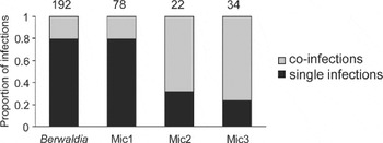
In total, 47 out of 290 analysed Daphnia (i.e. 16·2%) were infected with more than 1 microsporidian taxon; 45 double-infections and 2 triple-infections were detected. Berwaldia schaefernai and Mic1 mostly occurred in single infections, whereas Mic2 and Mic3 mostly occurred in co-infections (Fig. 5). Co-infections rarely happened between closely related taxa; we detected only 8 cases of B. schaefernai–Mic1 co-infections (despite these being the 2 most commonly detected taxa), and a single case of Mic2–Mic3 co-infection.
Proportions of each parasite occurring in single or co-infections. Sample size is given at the top of each bar.

DISCUSSION
Identification of microsporidia taxa
In the present study, using sequences of the SSU rDNA gene, we detected 8 taxa of microsporidian parasites across 8 investigated communities of the Daphnia longispina hybrid complex. The patterns of sequence similarity summarized in Fig. 3 were consistent with previously published phylogenies for microsporidia (Refardt et al. Reference Refardt, Canning, Mathis, Cheney, Lafranchi-Tristem and Ebert2002; Slothouber Galbreath et al. Reference Slothouber Galbreath, Smith, Terry, Becnel and Dunn2003; Franzen et al. Reference Franzen, Fischer, Schroeder, Schölmerich and Schneuwly2005;Vossbrinck and Debrunner-Vossbrinck, Reference Vossbrinck and Debrunner-Vossbrinck2005; Wolinska et al. Reference Wolinska, Giessler and Koerner2009). In addition, our study discovered at least 2 microsporidian taxa, which had not been genetically analysed before (Mic4, Mic6, and possibly Mic5; see below).
The most common parasites were Berwaldia schaefernai and the unidentified microsporidium Mic1. The Mic1 lineage is closely related to B. schaefernai, Larssonia obtusa, and Gurleya vavrai (Wolinska et al. Reference Wolinska, Giessler and Koerner2009), all shown to negatively impact host fecundity (Vávra and Larsson, Reference Vávra and Larsson1994; Bengtsson and Ebert, Reference Bengtsson and Ebert1998). These species probably have indirect life cycles, as their maintenance in the laboratory has failed repeatedly (J.W., personal observation and Vávra, Reference Vávra1964), and because they are closely related to the family Amblyosporidae (Refardt et al. Reference Refardt, Canning, Mathis, Cheney, Lafranchi-Tristem and Ebert2002), a group of microsporidia which infect mosquitoes and use copepods as intermediate hosts (Sweeney et al. Reference Sweeney, Hazard and Graham1985; Vossbrinck et al. Reference Vossbrinck, Andreadis, Vávra and Becnel2004). The previously observed low genetic differentiation between B. schaefernai infecting the different Daphnia populations (Wolinska et al. Reference Wolinska, Spaak, Koerner, Petrusek, Seda and Giessler2011b) also supports the hypothesis that this parasite is transmitted through a mobile vector, probably insects. However, another well-studied species belonging to this clade, Binucleata daphniae, does have a direct life cycle (Refardt et al. Reference Refardt, Decaestecker, Johnson and Vávra2008).
The lineages Mic2 to Mic5 detected in the present study belong to a relatively diverse group (clade II in Fig. 3) containing taxa infecting Daphnia gut epithelium (Ordospora colligata, Glugoides intestinalis, and Microsporidium sp. DP-1-19 possibly conspecific with Mic2). However, as all investigated taxa appeared in our study as clear fuzzy and opaque mass within the body cavity of Daphnia, it seems that multiple host tissues are infected within the Daphnia by these parasites (Mic2 to Mic5). The identity of Mic5 requires further analyses – although the sequence data clearly indicate that it is closely related to Glugoides intestinalis, the latter has been reported from other host species (D. magna and D. pulex) and with different infection symptoms (unlike Mic5, G. intestinalis infections seem to be limited to Daphnia gut epithelium; Larsson et al. Reference Larsson, Ebert, Vávra and Voronin1996). Mic5 might thus be either a sister species to G. intestinalis or it is conspecific but its infection progresses differently in an alternative host (D. longispina complex).
Spatial and temporal patterns in microsporidian communities
The taxon composition of microsporidian communities differed among the 8 studied reservoirs, with the lakes in closer geographical proximity hosting more similar assemblages of microsporidian taxa. Aggregated distributions are likely to be caused by dispersal between neighbouring habitats (e.g. Bengtsson and Ebert, Reference Bengtsson and Ebert1998). We suppose that at least some of the observed parasites have good dispersal capabilities via airborne vectors, as discussed above for B. schaefernai and its relatives (see also Wolinska et al. Reference Wolinska, Spaak, Koerner, Petrusek, Seda and Giessler2011b). Brno is the only reservoir in which we did not detect B. schaefernai infections; however, comparisons of limnological and hydrological parameters, as well as Daphnia host community structure, did not reveal any major differences between Brno and the other reservoirs (see Seda et al. Reference Seda, Petrusek, Machacek and Smilauer2007b). Thus, the reasons for the variation in parasite community structure remain uncertain.
Within reservoirs, the parasite community composition was similar between consecutive summers. Thus, microsporidians were able to overcome periods of low host density (i.e. in winter; Wolinska et al. Reference Wolinska, Keller, Bittner, Lass and Spaak2004). This could be facilitated by forming resistant spores deposited in lake sediments (Ebert, Reference Ebert1995), by surviving unfavourable periods in alternate hosts (Becnel and Andreadis, Reference Becnel, Andreadis, Wittner and Weiss1999) or within the host's diapausing stages themselves (Canning et al. Reference Canning, Barker, Page and Nicholas1985). In contrast, parasite composition changed within years, from summer to autumn. Seasonal changes in microsporidia abundance is a commonly observed phenomenon in Daphnia (Green, Reference Green1974; Brambilla, Reference Brambilla1983; Lass and Ebert, Reference Lass and Ebert2006), and density-dependent transmission (associated with host life cycle), environmental factors (especially temperature) and the ability to survive outside the host have been argued to shape the observed patterns (Ebert, Reference Ebert1995; Lass and Ebert, Reference Lass and Ebert2006). However, with some microsporidian parasites being host species-specific (Solter and Maddox, Reference Solter and Maddox1998; Stirnadel and Ebert, Reference Stirnadel and Ebert1997) or even genotype-specific (Little and Ebert, Reference Little and Ebert1999), seasonal changes in parasite community structure may also be related to changes in taxon and/or clonal composition of the host population. Such seasonal changes in D. longispina communities have been frequently observed (Keller et al. Reference Keller, Wolinska, Tellenbach and Spaak2007; Seda et al. Reference Seda, Kolarova, Petrusek and Machacek2007a; Spaak, Reference Spaak1996), including in the reservoirs studied here (A.P. and J.S., unpublished data). Conversely, no major interannual changes in host community structure were detected between the consecutive summers of 2004 and 2005; host distribution patterns were relatively stable in the 8 studied Czech reservoirs (Petrusek et al. Reference Petrusek, Seda, Macháček, Ruthová and Šmilauer2008b). These findings support the idea of an interdependency of host and parasite occurrence. Nonetheless, further studies are required to test this hypothesis. Specifically, by simultaneously following the changes in host community structure (e.g. by using microsatellite markers, Yin et al. Reference Yin, Wolinska and Gießler2010) and parasite community structure (using the species-specific primers developed here), one would be able to test for the dependence of specific parasite taxa on certain hosts.
Co-infection patterns within host individuals
Co-infection of a host with multiple parasite species is not the exception but rather a rule in nature (Lipsitch and Moxon, Reference Lipsitch and Moxon1997; Petney and Andrews, Reference Petney and Andrews1998). The 2 pairs of related microsporidian species (i.e. B. schaefernai with Mic1, and Mic2 with Mic3) showed similar proportions of single and co-infections, respectively. Here, relatedness may result in similar abilities to invade and seize host resources. Moreover, closely related parasites were rarely found in co-infections with one another. With the immune response being pathogen-specific (Kurtz and Armitage, Reference Kurtz and Armitage2006), relatedness could prove a disadvantage to a parasite invading an already infected host; such a parasite may encounter an already activated immune system. This mechanism may result in strong priority effects (e.g. de Roode et al. Reference de Roode, Helinski, Anwar and Read2005) and apparent competition between related species. However, experimental infection studies (similar to those of Ben-Ami et al. Reference Ben-Ami, Rigaud and Ebert2011 and Lohr et al. Reference Lohr, Yin and Wolinska2010) are required to reveal the true levels of competition between parasites.
Conclusions
Despite the existence of some visible morphological characteristics, mainly B. schaefernai showing spore mass proliferation within the host's head, discrimination between the 8 microsporidian taxa is not possible using the traditional screening method of light microscopy. By applying a molecular approach, we were able to overcome not only the problem of unambiguous identification but also to detect otherwise unseen multiple infections. What still remains to be tested is the sensitivity of the applied methods and hence their applicability to detect infections at early stages. Nevertheless, when seeking to investigate large host samples for microsporidian infection patterns, to gain more detailed insights into the diversity of parasites or to compare findings among studies and investigators, molecular methods should be considered in future parasite surveys of Daphnia infections.
ACKNOWLEDGEMENT
We thank Rita Jaenichen and Sabine Radetzki for assistance with the molecular work, and Jennifer Lohr, Mingbo Yin, Volker Witte and anonymous referees who provided valuable comments that helped to improve the manuscript.
FINANCIAL SUPPORT
This work was supported in part by the German Research Foundation (WO 1587 ⁄ 2–1, SPP 1399), the Grant Agency of the Academy of Sciences of the Czech Republic (IAA600960901), the STRESSFLEA project of the European Science Foundation EUROCORES Programme EuroEEFG (funded through the Czech Science Foundation project EEF/10/E022), and by departmental research money provided by Wilfried Gabriel.