Introduction
Fascioliasis in domestic ruminants caused by Fasciola gigantica inflicts an annual economic loss of around US$3 billion to the livestock sector in Asia and Africa (Spithill et al., Reference Spithill, Smooker and Copeman1999) and is an emerging human health problem in several countries (Mas-Coma et al., Reference Mas-Coma, Bargues and Valero2005; Moghadami & Mardani, Reference Moghadami and Mardani2008). Fasciola gigantica prevalence in some ruminants ranges from 80 to 100% in several countries of West Africa and Asia (Sharma et al., Reference Sharma, Dhar and Raina1989; Sanyal, Reference Sanyal2001; Keyyu et al., Reference Keyyu, Kassuku, Msalilwa, Monrad and Kyvsgaard2006). The infection is treated by regular use of anthelmintics but, due to the development of anthelminthic resistance and the cost of treatment involved, vaccination would be preferred as the long-term control measure. Several Fasciola proteins under evaluation as potential vaccine candidates are glutathione-S-transferase, cathepsin-L proteases, leucine aminopeptidase, saposin-like proteins and fatty acid binding proteins (Wijffels et al., Reference Wijffels, Salvatore, Dosen, Waddington, Wilson, Thompson, Campbell, Sexton, Wicker, Bowen, Friedel and Spithill1994; Dalton et al., Reference Dalton, McGonigle, Rolph and Andrews1996; Morrison et al., Reference Morrison, Colin, Sexton, Bowen, Wicker, Friedel and Spithill1996; Tendler et al., Reference Tendler, Brito, Vilar, Serra-Freire, Diogo, Almeida, Delbem, Silva, Savino, Garratt, Katz and Simpson1996; Estuningsih et al., Reference Estuningsih, Smooker, Wiedosari, Widjajanti, Vaiano, Partoutomo and Spithill1997).
Fatty acid binding protein (FABP) is a prominent vaccine candidate against fascioliasis and schistosomiasis (Tendler et al., Reference Tendler, Brito, Vilar, Serra-Freire, Diogo, Almeida, Delbem, Silva, Savino, Garratt, Katz and Simpson1996; Estuningsih et al., Reference Estuningsih, Smooker, Wiedosari, Widjajanti, Vaiano, Partoutomo and Spithill1997; Ramajo et al., Reference Ramajo, Oleaga, Casanueva, Hillyer and Muro2001; Nambi et al., Reference Nambi, Yadav, Raina, Sriveny and Saini2005). Trematode parasites lack a de novo pathway of lipid metabolism, utilizing host fatty acids and sterols for their lipid metabolism and parasite FABPs as transport molecules for host fatty acids (Meyer et al., Reference Meyer, Meyer and Bueding1970; Ockner, Reference Ockner1990; Pankao et al., Reference Pankao, Sirisriro, Grams, Vichasri-Grams, Meepool, Kangwanrangsan, Wanichanon, Ardseungneon, Viyanant, Upatham and Sobhon2006). FABPs, the cytosolic proteins, are concentrated in the parenchymal cell type 2 of Fasciola, with the tegumental tissue consuming a major supply of fatty acids from these parenchymal cells for the maintenance of its integrity. Thus, FABP molecules are targeted to attack the parasite tegumental membrane integrity, creating an imbalance in the homeostatic mechanism of the parasite and thereby aiding in its removal from the host (Pankao et al., Reference Pankao, Sirisriro, Grams, Vichasri-Grams, Meepool, Kangwanrangsan, Wanichanon, Ardseungneon, Viyanant, Upatham and Sobhon2006).
Nucleic acid vaccination induces both humoral and cellular immune responses of the host against infectious agents (Zhang et al., Reference Zhang, Taylor, Johansen and Bickle2002; Garzon et al., Reference Garzon, Berraondo, Crettaz, Ochoa, Vera, Lasarte, Vales, Van Rooijen, Ruiz, Prieto, Zulueta and Gonzalez-Aseguinolaza2005; Kennedy et al., Reference Kennedy, Spithill, Tennent, Wood and Piedrafita2006). However, DNA vaccination efficacy is low and a number of strategies have been developed for adjuvanting plasmid DNA for enhanced DNA delivery, increased magnitude or duration of gene expression, protecting DNA against DNases and endocytic degradation or facilitating uptake of the DNA into cells (Boussif et al., Reference Boussif, Lezoualch, Zanta, Mergny, Scherman, Demeneix and Behr1995; Hattori et al., Reference Hattori, Kawakami, Nakamura, Yamashita and Hashida2006). The success of nucleic acid-based vaccination depends upon the delivery vehicle's ability to selectively and efficiently deliver nucleic acid to the target cells with minimal toxicity. Polyethylenimine (PEI), a cationic polymer with varying molecular sizes and structures, is widely used as a gene delivery vehicle for its versatility and high transfection efficiency (Boussif et al., Reference Boussif, Lezoualch, Zanta, Mergny, Scherman, Demeneix and Behr1995; Wiseman et al., Reference Wiseman, Goddard, McLelland and Colledge2003; Akinc et al., Reference Akinc, Thomas, Klibanov and Langer2005; Bonnet et al., Reference Bonnet, Erbacher and Bolcato-Bellemin2008). In particular, the recent generation of linear PEI is considered a more efficient in vivo delivery vehicle than the first generation of branched PEI (Bonnet et al., Reference Bonnet, Erbacher and Bolcato-Bellemin2008). But, so far, use of cationic polymers for gene delivery has been restricted to viral and bacterial DNA targeting and in gene therapy strategies. No earlier reports on the use of a cationic polymer in DNA vaccination against parasites such as Fasciola or Schistosoma are available, apart from a preliminary study to evaluate an immune response to F. gigantica FABP when delivered in plasmid DNA with branched PEI in mice and rabbits (Raina et al., Reference Raina, Tripathy, Sriveny, Gupta, Samanta, Gupta, Singh, Tewari, Banerjee, Sanjay Kumar and Yadav2009). DNA vaccination trials performed so far with Fasciola antigens have shown effectiveness of DNA vaccines in evoking potent immune responses (Smooker et al., Reference Smooker, Steeper, Drew, Strugnell and Spithill1999, Reference Smooker, Kennedy, Steeper, Christopoulos and Spithill2001; Wedrychowicz et al., Reference Wedrychowicz, Lamparska, Kesik, Kotomski, Mieszczanek, Jedlina-Panasiuk and Plucienniczak2003; Espino et al., Reference Espino, Osuna, Gil and Hillyer2005; Kennedy et al., Reference Kennedy, Spithill, Tennent, Wood and Piedrafita2006). The present communication reports on the delivery of the plasmid DNA encoding F. gigantica FABP in conjunction with mannosylated linear polyethylenimine to the peritoneal cells of mice, to study the antibody and cytokine responses evoked to this DNA in these laboratory animals.
Materials and methods
Experimental animals
Thirty, 6-week-old, Swiss albino mice were obtained from the Laboratory Animal Resource Section, Indian Veterinary Research Institute (IVRI), Izatnagar and reared in the Small Animal House of the Division of Parasitology, IVRI, Izatnagar. All animal studies were conducted in accordance with the institute Animal Ethics Committee guidelines on the use of experimental animals.
Cloning of F. gigantica fatty acid binding protein gene
Fasciola gigantica fatty acid binding protein gene (399 bp) was cloned in a mammalian expression vector pVR1020 with a cytomegalovirus early promoter/enhancer sequence and tissue plasminogen activator signal sequence (Vical Inc., San Diego, California, USA). The gene was amplified by polymerase chain reaction, using primers with a BamHI restriction site in each primer (forward primer: 5′-A CCG GAT CCG CTG ACT TTG TGG GTT CG-3′; reverse primer: 5′-A CCG GAT CCT CAC GCT TTG AGC AGA GT-3′) and cloned immediately downstream and in frame with the signal sequence of the expression vector, for the secretion of the expressed protein. Plasmid DNA was propagated in Escherichia coli strain DH5α and endotoxin-free plasmid DNA was obtained by using Endofree Plasmid Maxi kit (Qiagen, GmbH, Germany).
Polyethylenimine complexation of the plasmid DNA
In vivo-jet polyethylenimine, a 22 kDa linear, mannosylated cationic polymer (Poly plus-transfection, France) was used for the gene delivery in mice peritoneal cells. In vivo-jet PEI-mannose was provided as a ready-to-use solution at 150 mm nitrogen concentration. The complexation of the PEI with DNA was done at N/P ratio 5, using the formula:

(1 μg of DNA contains 3 nmoles of anionic phosphate).
The appropriate quantity of PEI was mixed, dropwise, with the DNA, in a 5% final concentration of glucose, vortexed and incubated for 15 min at room temperature (RT) before immunizing the animals.
DNA immunization of mice
Group I mice received 50 μg naked DNA and Group II 50 μg DNA complexed with PEI-mannose per mouse, respectively. Group III served as non-immunized control mice. Groups IV and V of mice received mannosylated PEI and 5% glucose, respectively, as the control groups for PEI-mannose-DNA and naked-DNA groups. Each group comprised ten mice. Immunization of the mice was done via the intraperitoneal route and each mouse was administered DNA at a 3-week interval before a final booster was given, with either recombinant fatty acid binding protein or plasmid DNA. Mice in each group (Group I and Group II) were divided into two subgroups of five mice after three DNA immunizations. One of the two subgroups of the DNA-PEI-mannose group was boosted with recombinant FABP (60 μg protein/mouse in Freund's incomplete adjuvant, intraperitoneally) and the other subgroup with DNA-PEI-mannose complex. Likewise, of the two subgroups of the naked-DNA group, one was boosted with recombinant protein and the other received a DNA booster. Blood from the immunized mice was sampled from the tail vein at 2-week intervals, pre- and post-immunization, for determination of the antibody response.
Enzyme-linked immunosorbent assay
The 96-well flat-bottomed polystyrene microtitre plates (Greiner, Germany) were coated with 2 μg/ml recombinant FABP in carbonate–bicarbonate buffer, pH 9.6, at 4°C overnight. The plates were washed (3 × ) with phosphate-buffered saline, pH 7.2, containing 0.05% Tween-20 (PBS-T) for 15 min. A 300 μl volume of 3% skimmed milk in phosphate-buffered saline (PBS) was then dispensed into each well and incubated for 2 h at 37°C to block the free sites, followed by PBS-T washing. Serum samples were diluted 1:50 and 1:100, respectively in 1% skimmed milk–PBS and incubated with antigen-coated wells for 60 min at 37°C. Following three washes with PBS-T, mouse secondary antibody–horseradish peroxidase (HRP) conjugated (diluted in 1% skimmed milk–PBS, 1:5000 to 1:10,000) was incubated per well at 37°C for 1 h. The plates were washed with PBS-T, and orthophenylene diamine (OPD) substrate in citrate-phosphate buffer, pH 5.0, was dispensed into each well. The reaction was stopped with 50 μl of 3N HCl and absorbance read at 490 nm on an ELISA reader (Bio-Rad Laboratories, California, USA). The volumes of antigen, test serum and secondary antibody used in the assay were each 100 μl/well.
Mouse immunoglobulin isotype response to DNA immunization was detected by using goat anti-mouse IgG, IgG1, IgG2a and IgG2b antibodies coupled to HRP (Santa Cruz Biotechnology, Santa Cruz, California, USA) at appropriate dilution.
Cytokine analysis by flow cytometry
Splenocyte culture
All the experimental mice were sacrificed 6 days after the last booster. The spleen from each mouse was retrieved under sterile conditions in a laminar flow hood, flushed with PBS to release splenocytes and the cell suspension was treated with 0.83% sterile ammonium chloride for 5 min to lyse red blood cells. Splenocytes were washed with RPMI-1640 twice. Cell viability by trypan blue test and cell density by Neubauer's haemocytometer were determined and cells finally suspended in RPMI-1640 containing 10% fetal calf serum. Approximately 2 × 10⁶ cells/ml were dispensed in each well of a 24-well culture plate in duplicate for all the three groups of mice. One of each duplicate well was stimulated with 10 μg recombinant FABP. The culture plate was incubated at 37°C in a CO2 incubator for 48 h. The cells were pelleted at 1600 rpm for 15 min at 4°C and the culture supernatant stored at − 20°C until analysis.
Flow cytometry
BD Mouse Th1/Th2 Cytometry Bead Array Kit (BD Bio-Sciences, Singapore) was used to measure the concentration of interleukin (IL)-2, IL-4, IL-5, interferon-γ (IFN-γ) and tumour necrosis factor (TNF) cytokines. The procedure was carried out as per the manufacturer's instructions. Briefly, a standard curve for each cytokine was prepared with defined set of concentrations from 20 to 5000 pg/ml. Fifty microlitres of the mixed capture beads were added to the assay tubes. Fifty microlitres of the cytokine standard dilutions and test sample were added to the control assay tubes and test assay tubes, respectively. Fifty microlitres of the Mouse Th1/Th2 PE-detection reagent was added to the tubes and incubated for 2 h at room temperature in the dark. One millilitre of wash buffer was added to the assay tubes and centrifuged at 1500 rpm for 5 min. A 400 μl volume of wash buffer was used to resuspend the bead pellet and the samples were analysed on a flow cytometer (BD FACS Calibur, BD Bio-Sciences).
Statistical analysis
Statistical analysis was by one-way ANOVA using SPSS 10.0 software (SPSS Inc., Chicago, Illinois, USA).
Results
Humoral response
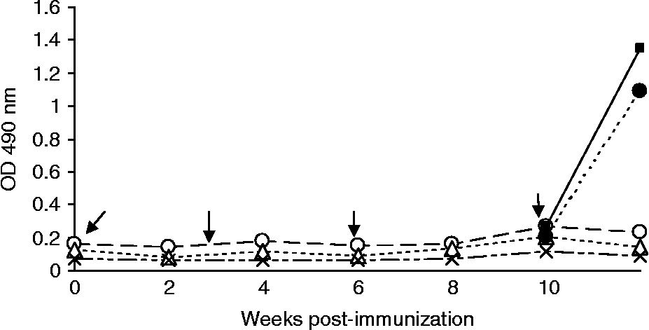
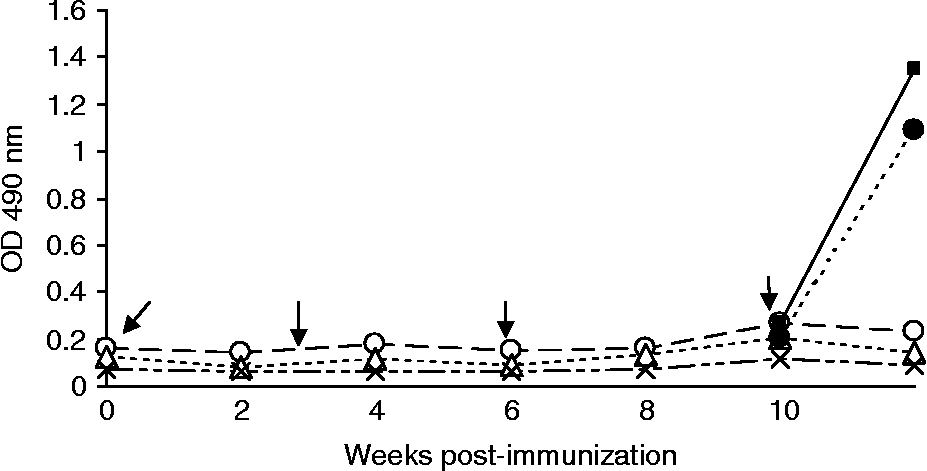
Antibody titre (IgG) was probed in mice receiving naked DNA (Group I), DNA complexed to PEI-mannose (Group II) and other control groups (Groups III, IV and V) at 2-week intervals post-immunization. The naked-DNA group of mice (Group I) did not show a significant increase in the IgG titre (P>0.05) for three DNA immunizations and the same results were obtained in Group II mice receiving mannosylated PEI–DNA (fig. 1). However, in each subgroup of Group I and Group II mice boosted with recombinant FABP, the IgG antibody titre increased steeply (to OD490 1.0–1.4) (fig. 1). This IgG antibody response was statistically significant (P < 0.05) when compared with the response elicited by the mice of subgroups boosted with DNA (naked or PEI-mannose complexed), where there was no perceptible increase in the antibody titre. However, the difference in the IgG antibody response between PEI-mannose DNA complexed and naked-DNA groups post-protein boost was statistically insignificant (P>0.05).
Mean IgG antibody responses in mice immunized with naked DNA and PEI-mannose DNA, respectively. Each subgroup of naked DNA and PEI-mannose DNA was protein boosted. Arrows indicate immunization at 3-week intervals. ○, PEI-DNA-immunized group; △, naked-DNA-immunized group; × , non-immunized group; ■, PEI-DNA protein-boosted subgroup; ●, naked-DNA protein-boosted subgroup.
Isotype responses of the IgG1, IgG2a and IgG2b of these mice to DNA immunization did not show any significant (P>0.05) increase in both the PEI-mannose complexed or naked-DNA groups, when probed after the first and second boosters, respectively (data not shown). No antibody isotype response was probed after protein boosting of these mice.
Cytokine response
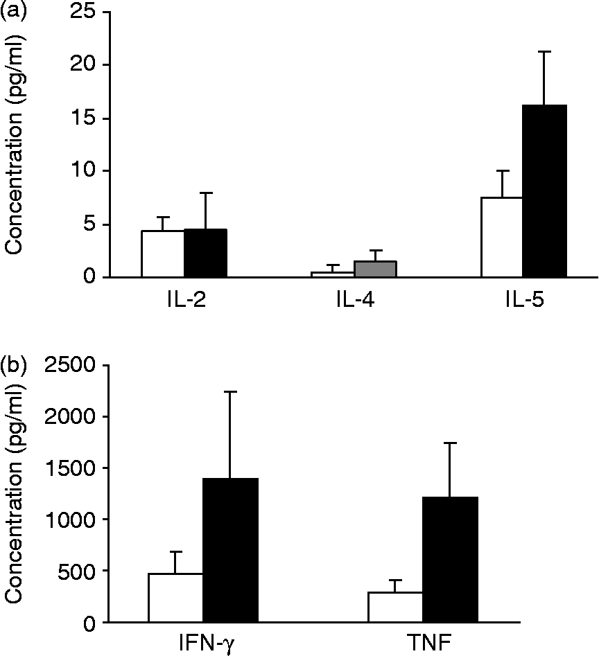
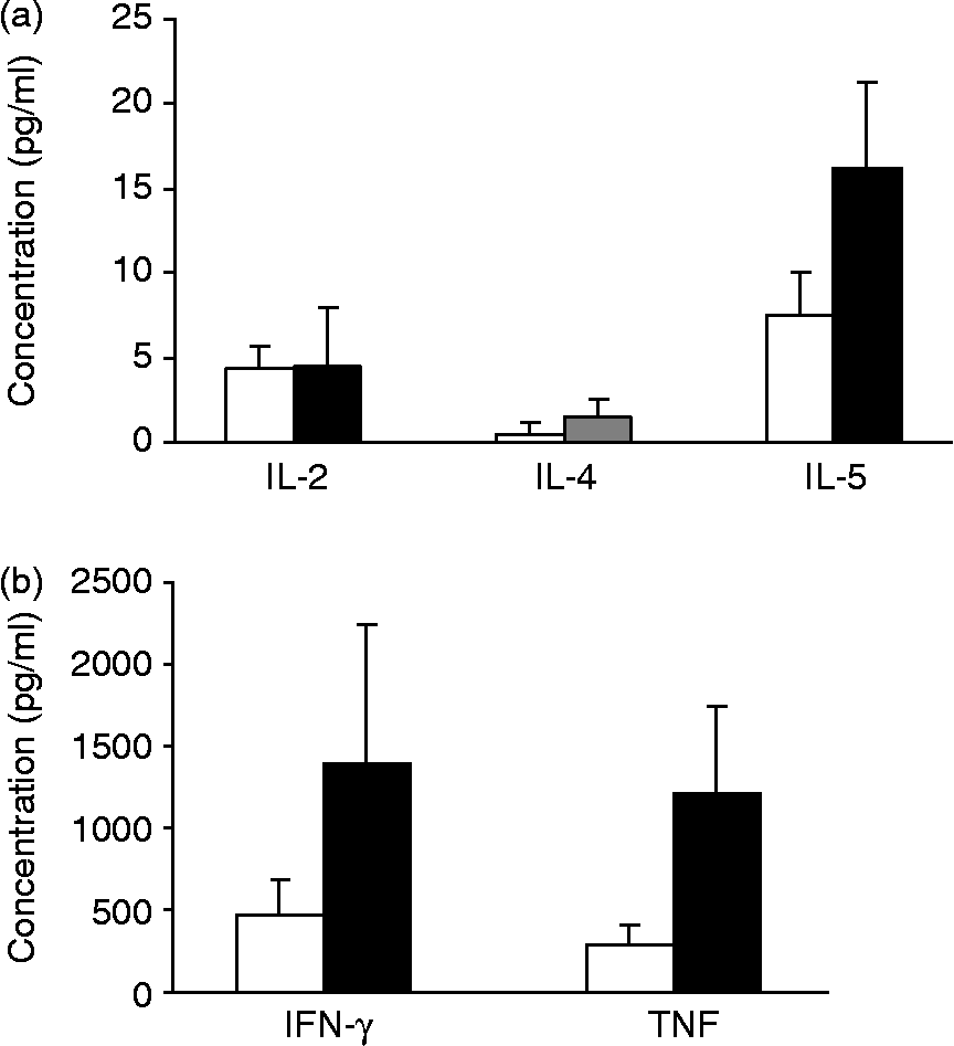
Concentrations of IL-2, IL-4, IL-5, IFN-γ and TNF were measured in the splenocyte culture supernatant after in vitro stimulation with recombinant FABP. Re-stimulation of the splenocyte culture of PEI-mannose and naked-DNA immunized mice with recombinant FABP antigen showed that the production of cytokines was antigen specific due to the priming of immune cells with DNA encoding FABP (fig. 2).
Mean concentrations of cytokines IL-2, IL-4, IL-5, IFN-γ and TNF in in vitro FABP-antigen-stimulated versus un-stimulated splenocytes of the PEI-mannose-DNA immunized group (n = 5; P < 0.05). Cytokines were probed after the final boost with DNA. Black bars, FABP-antigen-stimulated group; white bars, FABP-antigen-unstimulated group.
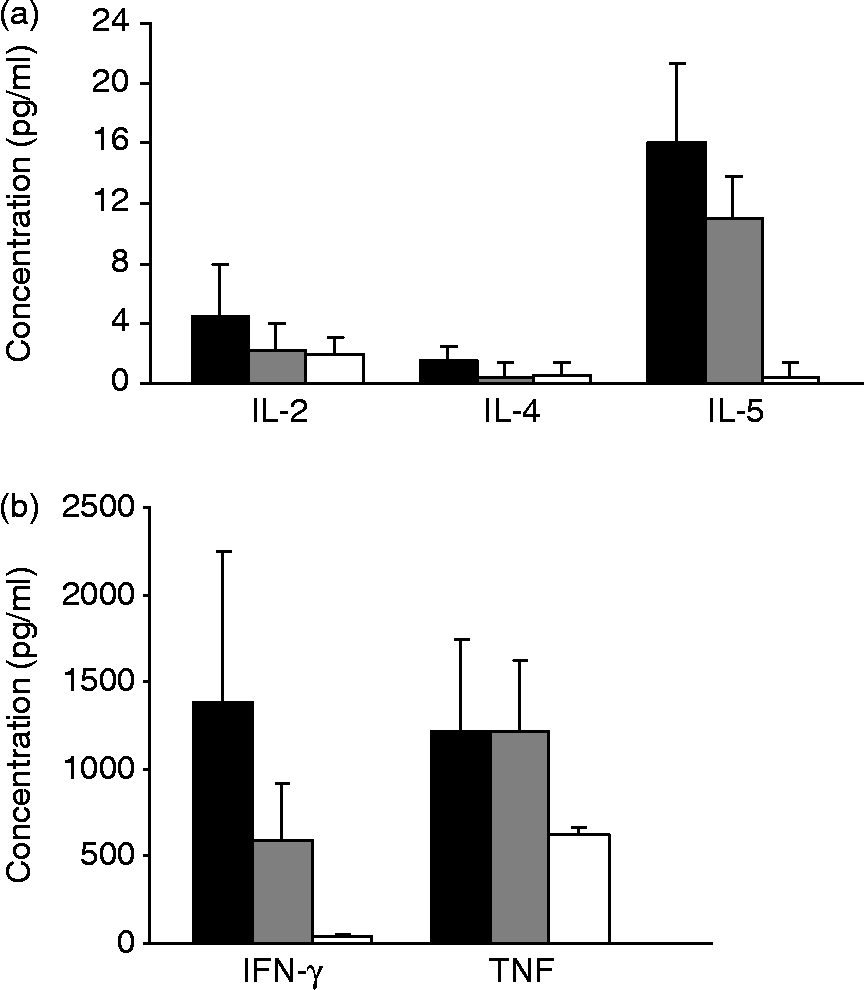
The IL-2 concentration in the splenocyte culture supernatant stimulated with recombinant FABP was slightly higher for the PEI-mannose group (4.2 pg/ml) than naked-DNA group (2.6 pg/ml) but the difference was statistically insignificant (P>0.05) (fig. 3a).
Mean cytokine concentrations of IL-2, IL-4, IL-5, IFN-γ and TNF in PEI-mannose-DNA, naked-DNA and non-immunized control groups (n = 5; P < 0.05) on in vitro antigenic stimulation of splenocytes with recombinant FABP after the final boost with DNA. Black bars, PEI-DNA-immunized group; grey bars, naked-DNA-immunized group; white bars, non-immunized group.
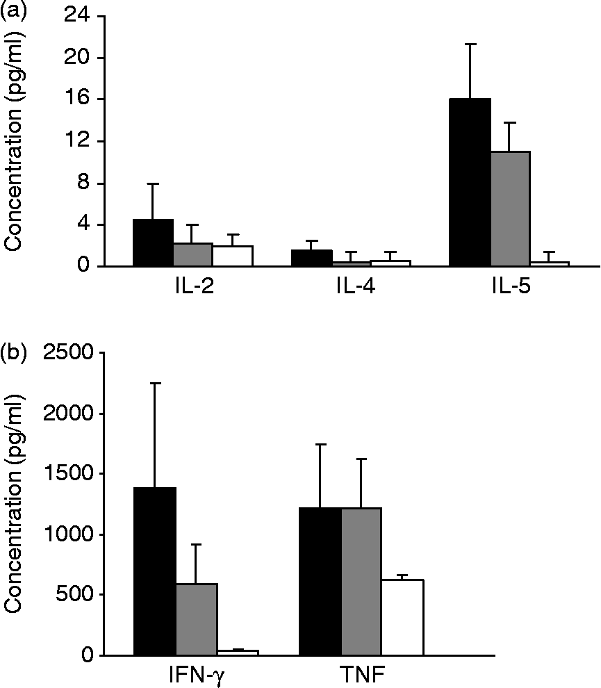
IL-4 concentration in the in vitro stimulated splenocyte culture in both the immunized groups was at a low level of 1.5–2.0 pg/ml (fig. 3a). However, there was a significant difference in the IL-5 concentration between the immunized and the un-immunized control groups (P < 0.05), with concentrations of IL-5 of 15.4, 11.5 and 1.3 pg/ml in PEI-DNA, naked-DNA and un-immunized groups, respectively (fig. 3a).
Out of the five cytokines screened for their expression in response to DNA immunization, only pro-inflammatory cytokines, i.e. IFN-γ and TNF, showed a significantly higher expression. There was a significant overexpression of IFN-γ (1387 pg/ml) on antigenic stimulation of PEI-mannose-DNA group, 2.4-fold higher concentration than the naked-DNA group (594 pg/ml) (fig. 3b). This difference was statistically significant (P < 0.05) between the two immunized groups. The TNF concentration on in vitro antigenic stimulation was 1208 and 1217 pg/ml, respectively, in the naked-DNA and PEI-mannose-DNA groups (fig. 3b), with an insignificant difference between the two immunized groups (P>0.05).
The two subgroups, one each of the PEI-mannose-DNA and naked-DNA group, that received final boosting (a fourth injection) with the recombinant FABP were simultaneously probed for the production of the above five cytokines. The concentration of each of the five cytokines in these protein-boosted subgroups was significantly lower compared to the DNA-boosted subgroups (P < 0.05) (data not given).
Discussion
Immunological control of Fasciola infection by vaccination seems a realistic possibility, although at present there is no vaccine available against this parasite. Most of the work on DNA vaccination in helminthic infections has been conducted on Schistosoma, where certain DNA vaccine formulations did partially protect mice against S. japonicum and S. mansoni, and also some reports of no protection in mice with DNA vaccine formulations based on paramyosin, Sj22, Sj62, Sm26–28 glutathione-S-transferase (GST) are available (Waine et al., Reference Waine, Yang, Scott, McManus and Kalinna1997; Zhang et al., Reference Zhang, Taylor, Johansen and Bickle2002; Dai et al., Reference Dai, Wang, Yu, Xu, Peng, He, Liu, Zhou and Liu2007). DNA vaccine trials with Schistosoma antigens conducted in large animals such as sheep, goats, buffaloes and pigs have shown encouraging results (Shi et al., Reference Shi, Zhang, Ye, Lin, Cai, Shen, Bickle and Taylor2002; Zhu et al., Reference Zhu, Ren, Da'dara, Harn, Xu, Si, Yu, Liang, Ye, Yin, He, Xu, Cao and Hua2004; Tang et al., Reference Tang, Zhou, Chen, Luo, Wang, Chen, Huang, Zeng and Yi2007) but, compared to Schistosoma, little work on exploiting the DNA vaccine potential of Fasciola antigens has been done so far.
Strategic targeting of a DNA vaccine to the immune cells has been shown to enhance the immune response (Lisziewicz et al., Reference Lisziewicz, Trocio, Whitman, Varga, Xu, Bakare, Erbacher, Fox, Woodward, Markham, Arya, Behr and Lori2005; Hattori et al., Reference Hattori, Kawakami, Nakamura, Yamashita and Hashida2006; Kawakami et al., Reference Kawakami, Ito, Charoensit, Yamashita and Hashida2006, Reference Kawakami, Higuchi and Hashida2008; Kennedy et al., Reference Kennedy, Spithill, Tennent, Wood and Piedrafita2006) and the first report on such targeted delivery of a DNA vaccine against F. hepatica was F. hepatica pro-cathepsin B antigen, delivered to the immune cells in sheep by combining the use of CpG motifs with CTLA-4 antigen, which elicited both antibody and cellular responses (Kennedy et al., Reference Kennedy, Spithill, Tennent, Wood and Piedrafita2006). In the present study, naked DNA or mannosylated linear polyethylenimine-DNA complex delivered intraperitoneally to the antigen-presenting cells of mice, i.e. macrophages and dendritic cells, did not evoke a significant humoral immune response with respect to IgG and its isotypes IgG1, IgG2a and IgG2b. These results are contradictory to several of the DNA vaccination protocols for Fasciola spp. Smooker et al. (Reference Smooker, Kennedy, Steeper, Christopoulos and Spithill2001) reported that DNA encoding F. gigantica FABP induced an IgG1 dominant response with significant IgE, IgG2a and IgG2b responses (mixed Th1/Th2 response), while immunization with F. hepatica cathepsin-L5 DNA induced a Th2 response in mice. Mixed Th1/Th2 type of immune responses to F. hepatica saposin-like protein-2 plasmid DNA in BALB/c mice, with the production of IgG1, IgG2, IgE and high levels of IL-10 and IFN-γ, were observed by Espino et al. (Reference Espino, Osuna, Gil and Hillyer2005). Clonorchis sinensis FABP-encoding plasmid DNA induced IgG2 antibodies predominantly, with production of IFN-γ but no IL-4 in Sprague–Dawley rats, indicating a typical Th1-dominated immune response with this protein (Lee et al., Reference Lee, Kim, Sohn, Lee and Yong2006). In contrast, Waine et al. (Reference Waine, Yang, Scott, McManus and Kalinna1997) reported no antibody response in mice immunized with cDNA constructs coding for S. japonicum 26 and 28 kDa GST, FABP, calreticulin, glyceraldehyde-3-phosphate dehydrogenase and 22.6 kDa membrane-associated antigen. In all the above vaccination protocols, no gene delivery vehicle was used and DNA was administered through the intramuscular route. The present DNA administration was done via the intraperitoneal route in mice, which appears to have influenced the outcome of the antibody and overall responses in the naked DNA and polymer-adjuvanted groups, as the route of immunization is known to influence the outcome of the immune response. In our earlier intramuscular immunization study on Swiss albino mice with the same naked recombinant DNA construct, a significant production of antibodies was evoked (Raina et al., Reference Raina, Tripathy, Sriveny, Gupta, Samanta, Gupta, Singh, Tewari, Banerjee, Sanjay Kumar and Yadav2009) and the same construct failed to evoke antibody production when delivered intraperitoneally in this strain of mice. However, protein boosting of the DNA-primed mice showed a steep increase in the IgG antibody titre, indicating that antibody-producing immune cells were pre-sensitized by DNA immunization. Therefore, it is speculated that antibody response in these DNA-primed mice could be boosted on challenge infection with F. gigantica.
The cell-mediated immune response probed by measuring IFN-γ, IL-2, IL-4, IL-5 and TNF cytokines showed that PEI-mannose-DNA group caused significant up-regulation of two pro-inflammatory cytokines IFN-γ and TNF on in vitro splenocyte stimulation, while IL-2 and IL-4 did not show a significant level of expression. Interleukin-5 showed a twofold increase on in vitro antigenic stimulation in both the immunized groups, although its expression was very low compared to IFN-γ and TNF. This indicates that DNA immunization evoked a predominant Th1-type response, though a Th2 response in lieu of IL-5 expression was also elicited.
Polyethylenimine, a cationic polymer, is known to be an efficient carrier of DNA, the transfection efficiency of which relies on extensive endosome buffering with its consequent swelling and rupture, thus providing the DNA/PEI particles an escape mechanism in the cell (Boussif et al., Reference Boussif, Lezoualch, Zanta, Mergny, Scherman, Demeneix and Behr1995). Several reports on DNA vaccination using PEI as a versatile adjuvant are available (Boussif et al., Reference Boussif, Lezoualch, Zanta, Mergny, Scherman, Demeneix and Behr1995; Oh et al., Reference Oh, Park, Kang, Ko, Kim and Kim2003; Garzon et al., Reference Garzon, Berraondo, Crettaz, Ochoa, Vera, Lasarte, Vales, Van Rooijen, Ruiz, Prieto, Zulueta and Gonzalez-Aseguinolaza2005), with systemically delivered PEI-DNA complexes resulting in the transduction of lung, spleen, liver, heart, brain and kidney, causing no histopathological changes in these organs (Oh et al., Reference Oh, Kim, Yoon, Kim, Yang and Kim2001). The use of PEIs with different molecular weights, structures and chemical modifications, and different nitrogen/phosphate ratios, has a bearing on the immune response and cytotoxicity (Wightman et al., Reference Wightman, Kircheis, Rossler, Carotta, Ruzicka, Kursa and Wagner2001; Kunath et al., Reference Kunath, Harpe, Fischer, Petersen, Bickel, Voigt and Kissel2003; Garzon et al., Reference Garzon, Berraondo, Crettaz, Ochoa, Vera, Lasarte, Vales, Van Rooijen, Ruiz, Prieto, Zulueta and Gonzalez-Aseguinolaza2005). In the present experiment, DNA at 50 μg concentration and polyethylenimine (linear, 22 kDa) at a single N/P ratio of 5 were used, and other DNA concentrations with different N/P ratios were not worked out. The correct formulation of the N/P ratio allows for control of DNA condensation and size of the resulting complexes which, in turn, will affect not only interactions with the cell membrane and release from the endosome but also intracellular trafficking and possibly nuclear entry, dissociation of complexes and gene expression. Analysing and improving each of these steps may allow us to achieve the ultimate goal of highly sustained, regulatable gene expression in targeted tissue.
Generally, helminth infections are manifested by suppression of Th1 function and induction of T cells which express cytokines characteristic of the Th2 subset (Clery et al., Reference Clery, Torgerson and Mulcahy1996; Brady et al., Reference Brady, O'Neill, Dalton and Mills1999; O'Neill et al., Reference O'Neill, Brady, Callanan, Mulcahy, Joyce, Mills and Dalton2000). Studies in cattle, sheep and buffaloes have demonstrated that the T-cell response is polarized towards a Th2 response with Fasciola infection (Moreau et al., Reference Moreau, Chauvin and Boulard1998; Waldvogel et al., Reference Waldvogel, Lepage, Zakher, Reichel, Eicher and Heussler2004; Molina, Reference Molina2005). Whether this selective induction of Th1/Th2 response results in the elimination of the helminth or the chronicity of the infection, depends on the host–parasite model (Else et al., Reference Else, Finkelman and Grencis1994). Therefore, understanding the basic immune responses in each host–parasite system and identification of lymphocyte subsets responding to Fasciola infection and the cytokines they produce are important in characterizing the protective and non-protective immune effector mechanisms in operation against this parasite.
Acknowledgements
The authors express their thanks to the Director, Indian Veterinary Research Institute, Izatnagar, for providing the necessary facilities to conduct this work.