

Introduction
The deep evolutionary record of hominins in Africa, together with the continent’s diversity of ecological regions and climatic zones, suggest that a long history of interaction between humans and parasites is to be found on this landmass. Indeed, when the first evidence of parasitic diseases in ancient human populations was identified in 1910, it was in archaeological material from the African continent (Ruffer Reference Ruffer1910).
Helminths are parasitic worms (flukes, tapeworms and roundworms) that commonly infect humans and various animal species. Many are intestinal parasites, spread through faecal-oral transmission or the consumption of undercooked meats, and infection by such species may be identified archaeologically through the presence of eggs in fossilised excrement (coprolites), in mummified tissue or in sediment sampled from the abdominal region of burials. One of the earliest incidences of roundworm infection—byascarides (Ascaris lumbricoides) and whipworm (Trichuris trichiura)—was identified in a human coprolite from excavations in Kruger Cave in South Africa, dating to 10 000–7000 BC (Evans et al. Reference Evans1996). Yet, despite this early date, research on parasitic diseases in ancient populations in the African continent has received less attention than other world regions (Stehr & Ponce Reference Stehr and Ponce2023; Mitchell Reference Mitchell2024), and much of the available information has been obtained through the study of mummies and mummified remains originating from the territory of Egypt (Mitchell Reference Mitchell2024). For example, the earliest evidence of tapeworm (Taenia sp.) infection in Africa derives from the 3200-year-old mummy of an adolescent who lived in the final century of the New Kingdom (c. 1550–1077 BC) (Hart et al. Reference Hart1977). Eggs of Taenia sp. tapeworms and Ascaris sp. roundworms were also found in the contents of canopic jars from the mastaba (tomb) of Akhethetep in Saqqara, dating to the Twenty-fifth Dynasty (715–656 BC) (Harter et al. Reference Harter2003). Taenia sp. eggs were also discovered during excavations at the ed-Deir cemetery in the Kharga Oasis (Le Bailly et al. Reference Le Bailly2010). Mummified remains may also preserve adult helminths, as seen in intestinal samples taken from two mummies from Saqqara, dating to 400–300 BC, which preserved Dibothriocephalus sp. tapeworms, transmitted through the consumption of raw fish (Harter et al. Reference Harter2003).
Palaeoparasitological evidence for the Nubian region (the Nile Valley between the First and Fourth cataracts) is scarcer. Research on intestinal parasites, specifically, seems limited to Sai Island, located between the Second and Third Nile Cataracts, where eggs of Taenia sp. and Diphyllobothrium sp. tapeworms were found in faeces from the pelvic area of two naturally mummified bodies dated to between 700 and 300 BC (Harter et al. Reference Harter2003). This article adds to the available literature by determining the parasitological spectrum of sediment sampled from the surface of the sacrum in 26 burials, as well as estimating the prevalence of intestinal infection and reconstructing the diet and sanitary conditions of the medieval population buried in the Southern Necropolis of the Deraheib site in Sudan.
Archaeological context
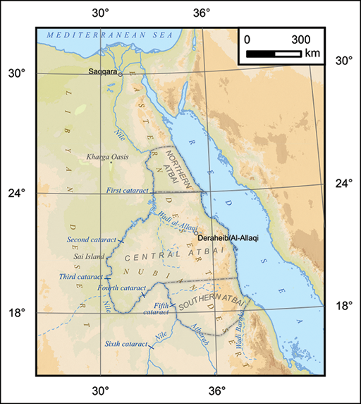
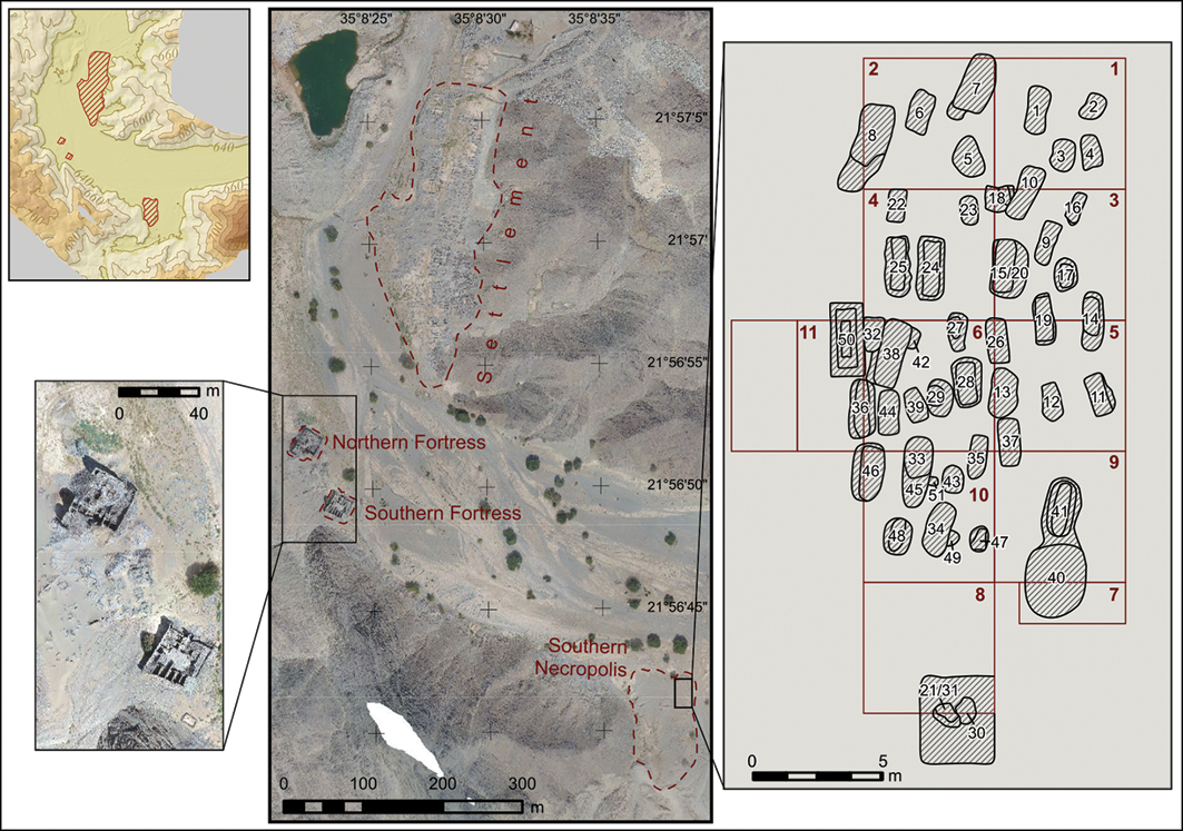
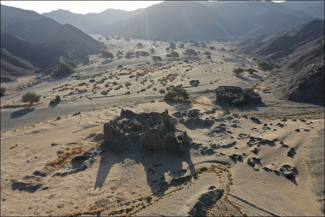
The medieval (ninth–eleventh centuries AD) cemetery at Deraheib (Republic of Sudan) has been excavated since 2017 by the Nubian Archaeological and Anthropological Expedition of the Lomonosov Moscow State University Research Institute and Museum of Anthropology under an excavation licence issued by the Sudanese National Corporation for Antiquities & Museums (Krol et al. Reference Krol2022: 100–124; 2023: 132–49) (Figure 1). The archaeological site of Deraheib can be identified with the historic town of al-Allaqi, often mentioned in medieval Arabic written sources. It includes a settlement area, consisting of two rows of residential buildings with a Friday Mosque at the centre (Krol Reference Krol and Matveev2024), that spreads along the eastern bank of a wadi (dry riverbed), the residence of a local ruler designated as the Northern Fortress (Krol & Tolmacheva Reference Krol and Tolmacheva2024), a fortified caravanserai known as the Southern Fortress, and the Southern Necropolis (Figures 2 & 3).
Map showing the location of Deraheib and geographical features mentioned in the text (figure by K. Kunavin & A. Krol).

Figure 1 Long description
The map displays the location of Deraheib and various geographical features in Egypt. It includes the Nile River, the Mediterranean Sea, and several oases such as the Kharga Oasis. The map also highlights different regions like Northern Atbai, Central Atbai, and Southern Atbai. Key locations such as Saqqara and the positions of various cataracts along the Nile River are marked. The map uses color shading to differentiate between land and water features, with blue representing water bodies and various shades indicating different geographical regions.
Main features of the Deraheib site (figure by K. Kunavin, E. Semenov, O. Kalinina & A. Krol).

Figure 2 Long description
The map of the Deraheib site includes a detailed layout of the area with various labeled sections. It highlights the Northern Fortress, Southern Fortress, and Southern Necropolis, each marked with dashed red lines. The map also features a grid with numbered sections, indicating specific areas of interest. A smaller inset map shows a zoomed-in view of the fortresses, providing more detailed structures within those areas. The map includes coordinates and scale bars for reference, helping to understand the spatial relationships and dimensions of the site.
The northern (left) and southern (right) fortresses with a view towards the Southern Necropolis between the mountains to the top right (photograph by Kiril S. Samurski).

Figure 3 Long description
The image captures the northern fortress on the left and the southern fortress on the right, with a view towards the Southern Necropolis situated between the mountains in the top right corner. The landscape is arid, featuring scattered vegetation and rocky terrain. The ruins of the fortresses are prominently displayed, with the northern fortress appearing more intact than the southern one. The scene is bathed in sunlight, casting long shadows and highlighting the rugged beauty of the desert environment.
Excavations have focused on Building 2 and Building 3 (the Mosque) in the settlement, the south-western and north-western walls of the Northern Fortress, and the Southern Necropolis. These confirm that the city of al-Allaqi was occupied between the ninth and twelfth centuries AD, dates corroborated by written sources (Krol Reference Krol and Zaytzev2020: 203–17). During this period, the city marked the centre of the gold-bearing region of Wadi al-Allaqi, which included the bed of the dry river together with its numerous tributaries. One of the routes used by caravans to cross the Nubian Desert also passed through Deraheib and pilgrims travelling for the Hajj would also use this route (Krol Reference Krol2018).
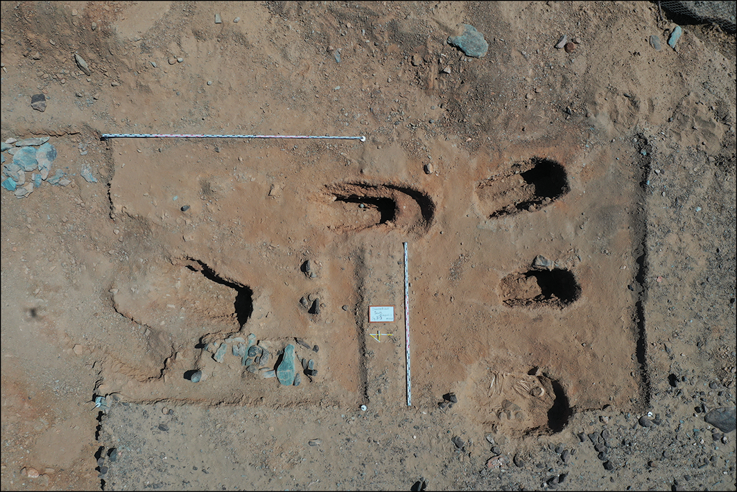
Since 2018, the Nubian Expedition has been conducting research in the Southern Necropolis (Figure 4). So far, 50 burials have been excavated, 44 of which relate to the Islamic period (ninth–eleventh centuries AD) of the site (Fedorchuk et al. Reference Fedorchuk2024). The remains of fabric discovered in some graves demonstrate that most of the bodies were wrapped in shrouds. Fragments of these shrouds and grape seeds (Vitis vinifera) recovered from grave 6 were selected for radiocarbon determinations (see online supplementary material (OSM)). Resultant dates fall into the period between the second half of the ninth and the beginning of the eleventh centuries (Krol et al. Reference Krol2019: 140–41).
Southern Necropolis. View of the area excavated in the fourth season, February 2022. Vertical scale is 3m, horizontal scale is 5m (photograph by Kiril S. Samurski).

Figure 4 Long description
The image presents an aerial view of the excavated area at the Southern Necropolis during the fourth season in February 2022. The excavation site is marked with a grid layout, with vertical and horizontal scales of 3 meters and 5 meters, respectively. Several pits of varying sizes and shapes are visible, some containing artifacts such as pottery shards and other small objects. The ground appears to be a mix of reddish-brown soil with patches of lighter and darker areas, indicating different layers of excavation. A measuring stick is placed vertically in the center of the image for scale, and a small sign with an arrow points to a specific feature within the grid. The overall scene suggests an archaeological excavation in progress, with careful documentation of the site's features.
Materials and methods
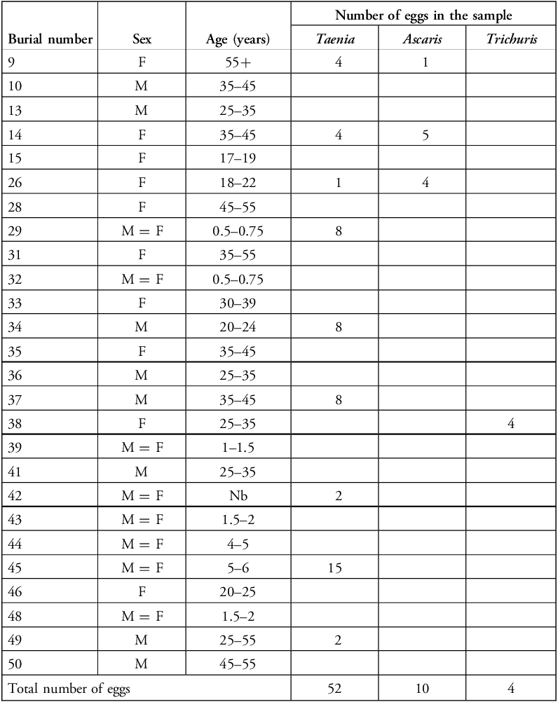
Sediment samples were taken directly from the surface of the sacrum from 26 individuals (see Table 1) while clearing the burials in the Southern Necropolis. Control samples were also taken from the filling of the grave under the skull of each individual.
Summary information for burials included in analysis.

Table 1 Long description
The table presents data on 26 individuals, detailing their burial number, sex, age, and the number of eggs found for Taenia, Ascaris, and Trichuris. The table has 26 rows and 6 columns, with headers for Burial number, Sex, Age (years), and the number of eggs for Taenia, Ascaris, and Trichuris. Notable entries include burial number 29, with 8 Taenia eggs, and burial number 45, with 15 Ascaris eggs. The total number of eggs for Taenia is 52, for Ascaris is 10, and for Trichuris is 4.
The samples, each measuring 50g, were rehydrated in 0.5 per cent disodium phosphate solution for two to three days under laboratory conditions (Callen & Cameron Reference Callen and Cameron1960). The coarse particles were separated on a 0.5mm mesh sieve, and the fine fraction was precipitated by centrifugation and washed several times with distilled water until the supernatant was clear. After the final centrifugation, the remaining pellet of material was flooded with 10 per cent hydrochloric acid for 30 minutes to promote decalcification and then washed twice, centrifuged with distilled water and finally fractionated using a set of laboratory sieves with cells from 200 to 20μm. All fractions less than 200μm were washed off the sieves into separate Petri dishes and viewed under a Motic AE 31 inverted microscope to identify helminth eggs, which were then examined in detail, photographed and measured under a Zeiss Axio Imager Z1 microscope with an AxioCam HRc camera using AxioVision software v.4.8.
Osteological assessment of each individual, including age estimation and sex determination, was conducted in accordance with standard methods (Alekseev & Debets Reference Alekseev and Debets1964; Alekseev Reference Alekseev1966). The age of adult individuals was determined based on characteristics of both the skull and the postcranial skeleton; as many of these age-estimation methods have limited accuracy, wide age brackets of 10 years were assigned (Todd Reference Todd1920; McKern & Stewart Reference McKern and Stewart1957; Alekseev & Debets Reference Alekseev and Debets1964; Ubelaker Reference Ubelaker1989), the stage of epiphyseal fusion and the length of long bone diaphyses (Ubelaker Reference Ubelaker1989). Individuals were grouped by age and biological sex for analysis. Biological sex was not determined for subadults.
Parasite eggs were identified and categorised taxonomically in accordance with the methods of Ash and Orihel (Reference Ash and Orihel2007), Gaevskaya (Reference Gaevskaya2015) and the Center for Disease Control (2023). The length and width of each egg were measured and grouped by species to provide metrics on absolute difference (the range from minimum measurement to maximum measurement) and arithmetic mean (M) with standard deviation (SD) as descriptive statistics for egg measurement traits. Calculations of numerical parameters and graphing were performed using the Microsoft Excel (2019 version). The software Quantitative Parasitology v.3.0 was used to calculate the prevalence (Pr, %) of parasitosis in the population, including confidence intervals, to compare prevalence data between biological sex and age groups, and to verify the presence/absence of statistically significant differences between the groups (Reiczigel & Rózsa Reference Reiczigel and Rózsa2005; Reiczigel et al. Reference Reiczigel2020).
Results
Eggs of intestinal parasites were detected in 10 of the sediment samples taken from the surface of the sacrum (Table 1). No helminth eggs were identified in the control samples.
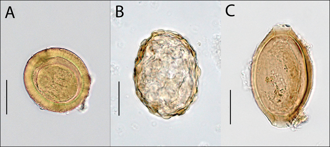
Eggs of tapeworms of the Taenia genus were identifiable in nine samples. These are characterised by their spherical shape and thick, brown shell with radial striations (Figure 5A). Oncospheres with embryonic hooks were detected inside some of the Taenia eggs with good preservation. Light brown, oval-shaped eggs with thick shells, knobby surfaces and amorphous content that correspond morphologically to the Ascaris genus of roundworms were identified in three samples (Figure 5B). Eggs with an elongated barrel shape characteristic of roundworms of the Trichuris genus were found in one of the samples (Figure 5C).
Parasite eggs that indicate the possible presence of Taenia saginata (A), Ascaris lumbricoides (B) and Trichuris trichiura (C). Scale bars = 20μm (figure by authors).

Figure 5 Long description
The image consists of three microscopic views of parasite eggs. Image A shows a round egg with a double outer shell, indicating the possible presence of Taenia saginata. Image B displays an oval-shaped egg with a rough outer shell, suggesting Ascaris lumbricoides. Image C presents an elongated egg with a smooth outer shell, indicative of Trichuris trichiura. Each image includes a scale bar measuring 20 micrometers. The eggs are identified and categorized taxonomically following specific methods and measured for length and width to provide descriptive statistics on egg measurement traits.
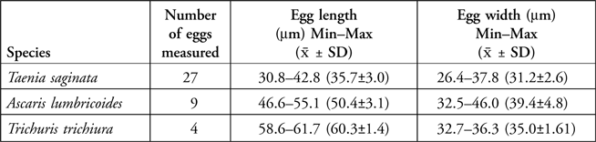
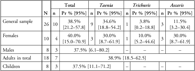
The total number of eggs identified and the descriptive statistics for each species are shown in Table 2. The overall prevalence of helminth infection in the sampled population of the necropolis is 38.5 per cent (95% confidence interval: 21.2–57.8%), with Taenia predominating (Table 3). This prevalence does not differ significantly between adults and subadults (F = 1.000; p≥0.05), nor were any significant differences apparent between male and female cohorts when considering both the total number of positive samples (F = 1.000; p≥0.05) and the breakdown of samples by parasite species (F = 1.000; p≥0.05 for Taenia helminths, F = 0.0915; p≥0.05 for soil-transmitted helminths: Ascaris and Trichuris helminths).
Summary information for identified parasite eggs.

Table 2 Long description
The table presents summary information for identified parasite eggs, detailing three species: Taenia saginata, Ascaris lumbricoides, and Trichuris trichiura. It includes the number of eggs measured for each species, along with the minimum and maximum egg length and width in micrometers, and the mean with standard deviation. Taenia saginata has 27 eggs measured, with lengths ranging from 30.8 to 42.8 micrometers and widths from 26.4 to 37.8 micrometers. Ascaris lumbricoides has 9 eggs measured, with lengths ranging from 46.6 to 55.1 micrometers and widths from 32.5 to 46.0 micrometers. Trichuris trichiura has 4 eggs measured, with lengths ranging from 58.6 to 61.7 micrometers and widths from 32.7 to 36.3 micrometers.
Prevalence (Pr %) and confidence intervals (95%) of infection by intestinal parasites in the necropolis population.

Table 3 Long description
The table presents data on the prevalence and confidence intervals of infection by intestinal parasites in a necropolis population. It includes columns for the total number of eggs identified, the number of positive samples, prevalence percentage, and 95% confidence intervals for each species of parasite. The table is divided into rows for the general sample, females, males, adults in total, and children. The general sample shows a 38.5% prevalence of helminth infection, with Taenia being the most predominant. Females have a 40% prevalence, while males have a 37.5% prevalence. Adults in total have a 38.9% prevalence, and children have a 37.5% prevalence. The table also includes data for Trichuris and Ascaris infections, with varying prevalence rates across different groups.
N = the number of people surveyed; n = the number of people affected by parasitosis.
Discussion
Three species of tapeworm of the Taenia genus infect modern human populations and all three have been reported in Africa (Ale et al. Reference Ale2014; Braae et al. Reference Braae2015; Dermauw et al. Reference Dermauw2018; Hendrickx et al. Reference Hendrickx2019; Saratsis et al. Reference Saratsis2019; Eichenberger et al. Reference Eichenberger2020). Infection by beef tapeworm (Taenia saginata), medically referred to as taeniasis, has the greatest reported incidence across the African continent (Dermauw et al. Reference Dermauw2018; Hendrickx et al. Reference Hendrickx2019; Saratsis et al. Reference Saratsis2019; Eichenberger et al. Reference Eichenberger2020). Infection with pork tapeworm (Taenia solium) (cysticercosis) is slightly lower but is likely to be underestimated due to insufficient laboratory diagnosis (Braae et al. Reference Braae2015). Most incidents of human infection with pork tapeworm are reported in West, Central and South Africa, with a few isolated cases in North Africa. This is primarily due to the religious taboo against farming and eating pigs in predominantly Muslim areas (Saratsis et al. Reference Saratsis2019). Asian tapeworm (Taenia asiatica), which also infects pigs, has not been recorded in Africa (Ale et al. Reference Ale2014).
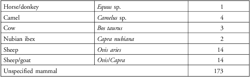
Written accounts and archaeological data indicate that the population occupying al-Allaqi practiced Islam. It is therefore probable that the tapeworm eggs discovered during our research derive from beef tapeworm. This suggestion is supported by the absence of pig bones in the zooarchaeological material from the site, which includes cattle remains instead (Table 4). The Taenia sp. eggs identified in the samples have a similar morphology to eggs from other species of the Taeniidae family (Echinococcus, Hydatigera), but these are obligate parasites of predatory mammals, and only species of the genus Taenia infect humans.
Faunal remains from Deraheib.

Table 4 Long description
A table listing faunal remains from Deraheib, detailing species and their respective counts. The table has seven rows and three columns. The first column lists the species: Horse/donkey, Camel, Cow, Nubian ibex, Sheep, Sheep/goat, and Unspecified mammal. The second column provides the scientific names: Equus sp., Camelus sp., Bos taurus, Capra nubiana, Ovis aries, Ovis/Capra, and Unspecified mammal. The third column shows the counts: 1, 4, 3, 2, 14, 14, and 173 respectively. The table highlights the prevalence of unspecified mammals with a count of 173, followed by sheep and sheep/goat with counts of 14 each.
Roundworms of the genus Trichuris are numerous worldwide, with the species Trichuris trichiura, Trichuris suis and Trichuris vulpis being of greatest modern epidemiological importance, affecting humans/primates, pigs and dogs, respectively (Bundy & Cooper Reference Bundy and Cooper1989; Callejón et al. Reference Callejón2015). Human whipworm (Trichuris trichiura) is a species-specific helminth that is most pathogenic to humans and is responsible for the majority of human trichuriasis cases reported worldwide (Bethony et al. Reference Bethony2006; Areekul et al. Reference Areekul2010). Morphologically, the eggs of pork whipworm (Trichuris suis) are almost indistinguishable from human whipworm eggs, but their contribution to human morbidity (disease load) is minimal due to the transient presence of the parasite in the human intestine, which lasts no more than a few weeks (Bundy & Cooper Reference Bundy and Cooper1989; Cutillas et al. Reference Cutillas2009). The main hosts of Trichuris vulpis are dogs and humans are rarely affected by this helminth (Singh et al. Reference Singh1993; Márquez-Navarro et al. Reference Márquez-Navarro2012). While the morphology of eggs from each species is similar, Trichuris vulpis eggs are larger, exceeding the variability of the much smaller Trichuris trichiura and Trichuris suis eggs (Bundy & Cooper Reference Bundy and Cooper1989; Singh et al. Reference Singh1993; Márquez-Navarro et al. Reference Márquez-Navarro2012; Phosuk et al. Reference Phosuk2018). Thus, consideration of epidemiology, morphology and archaeological context (burials corresponding to Islamic practices and a dearth of pig bones in the zooarchaeological assemblage), we can conclude with a high degree of certainty that the Trichuris sp. eggs discovered at Deraheib derive from human whipworm.
Two species of roundworms of the Ascaris genus—human (Ascaris lumbricoides) and pork (Ascaris suum) ascarids—pose the greatest risk of infection in humans. Eggs of these helminths cannot be distinguished morphologically due to the high genetic similarity of the two species and cases of cross-infection of hosts are documented (Zhou et al. Reference Zhou2020). Some researchers even question whether there are two distinct species (Leles et al. Reference Leles2012). Thus, we cannot determine the species affiliation of the Ascaris eggs from the Deraheib samples based on morphometric characteristics. However, application of the same reasoning used above allows us to suggest that the eggs likely derive from the human ascarid.
Reconstruction of diet
Data pertaining to the diet of the population inhabiting medieval al-Allaqi is scarce. The discovery of Taenia saginata eggs and the implied high prevalence of taeniasis in the medieval population of Deraheib indicate that beef was probably part of the daily diet rather than an exotic product, like the coconuts and almonds discovered in the Northern Fortress (Krol & Tolmacheva Reference Krol and Tolmacheva2024: 15). However, the zooarchaeological assemblage recovered from middens near the north-western wall of the Northern Fortress is dominated by the bones of sheep/goats with only small numbers of other identified domestic (horse/donkey, camel, cow) and wild animals (Nubian ibex) (see Table 4).
Despite the small number, the presence of cow bones is significant and, taken together, the zooarchaeological and palaeoparasitological data could indicate that nomads of the northern part of the Nubian desert may have bred cattle in the ninth–eleventh centuries, which would only have been possible if environmental conditions were more humid than today. Wetter conditions are also described in contemporaneous written sources. For example, al-Mas’udi, writing in about the tenth century, notes that the inhabitants of al-Allaqi made use of rainwater and sourced water from a spring that flowed through the middle of the city (Barbier de Meynard & Pavet de Courteille Reference Barbier de Meynard and Pavet de Courteille1864: 50). In an eleventh-century account, Al-Bakrī mentions that the city of al-Allaqi was rich in pomegranates, producing these in abundance (Vantini Reference Vantini1975: 244), and the grape seeds discovered in the mid-tenth-century burial 6 of the Southern Necropolis may indicate that grapes were also grown locally. The cultivation of pomegranates and grapes would require large volumes of water, which could in turn suggest that the amount of rainfall in the Wadi al-Allaqi and Jebel Elba region was sufficient to support cattle.
At present, cattle breeding is not possible in this part of Sudan as there is an insufficient fodder base (Krzywinski Reference Krzywinski, Krzywinski and Pierce2001: 56–58); the nomadic Beja people, specifically the Bisharin group occupying the area of Deraheib, herd only camels, goats and sheep (Sandars Reference Sandars1933: 144–47; Weschenfelder Reference Weschenfelder, Barnard and Duistermaat2012). The Beja are mentioned on multiple occasions in medieval Arabic sources that describe events that took place in the Central Atbai during the ninth–eleventh centuries, when gold mining and caravan trade flourished in this region of the Nubian Desert (Zaborski Reference Zaborski1965). However, the sources mention only camels and goats, not cattle.
It is possible that the Beja inhabiting the Southern Atbai, a region that reportedly received a considerable amount of precipitation in the tenth century, may have brought cattle to the mines of al-Allaqi. In reference to the ‘clans’ and ‘tribes’ occupying this area, Arab geographer Ibn Hawqal (d. AD 986) mentions that “they are all cattle-breeders and all nomads” (Vantini Reference Vantini1975: 161). Beja people from the nomadic Hadendova group inhabiting Western Sudan traditionally bred cattle but repeated droughts in the 1980s and increased aridity have forced them, like many other pastoral groups in the Sahel zone, to abandon cattle herding and to rear camels, sheep and goats instead (Magid Reference Magid, Barnard and Wendrich2008: 444).
Regardless of the mechanisms through which cattle appeared in the upper reaches of al-Allaqi, the presence of the helminth eggs in the sediment samples indicate that the people buried in the Southern Necropolis consumed raw and/or insufficiently heat-treated cattle meat. Gastronomic habits may have contributed to the high prevalence of taeniasis in the medieval population of al-Allaqi, which affected at least 18.8 per cent of the population (see Table 3, confidence intervals). Early twentieth-century ethnographic accounts indicate that the nomads of the Eastern Desert of Egypt ate raw offal: “when an animal is killed, the Arabs often eat the liver and kidney raw, adding a little salt. Some Ababda of my acquaintance killed an ibex, and devoured all the intestines raw, squeezing the contents of the gall-bladder over them as sauce” (Murray Reference Murray1935: 89). A popular dish in modern Sudan is kamounia, often made with raw beef and beef liver. In neighbouring Ethiopia, taeniasis cases are often associated with local dishes such as kitfo, tibs and kurt, featuring raw beef as the main ingredient (Dermauw et al. Reference Dermauw2018).
Parasitic diseases and sanitary conditions
The prevalence of soil-transmitted helminthiases is not uniform across Africa. The highest prevalence of human whipworm infection is found in South Africa (9.58%), the lowest in North Africa (0.59%) (Behniafar et al. Reference Behniafar2024). The prevalence of human ascarid infection presents a similar situation: Sub-Saharan Africa demonstrates the highest prevalence of ascariasis (11.66%), while North and West Africa have the lowest (2.04%) (Holland et al. Reference Holland2022). Thus, the areas with the highest prevalence of soil-transmitted helminthiases are also those that have high average relative humidity, high average annual precipitation and high average annual temperature (Holland et al. Reference Holland2022).
The presence of Ascaris and Trichuris eggs in four samples indicates that infection by helminths that mature in soil (geohelminths) was not uncommon in the population buried in the Southern Necropolis. This, in turn, indicates the realisation of the conditions necessary for the completion of the geohelminth life cycle (high constant humidity, high temperature and shaded soil), allowing them to infect human hosts. Such conditions may have occurred in wells, where constant humidity may have favoured the maturation of human whipworms and ascarids. Additionally, the life cycle of geohelminths could be completed in buildings where water was constantly used for domestic needs and shade was perpetually available. All sampled females were found to have geohelminth infection, potentially providing a link with the domestic sphere, although these findings were not statistically significant. Nomads could also have brought infections into urban areas.
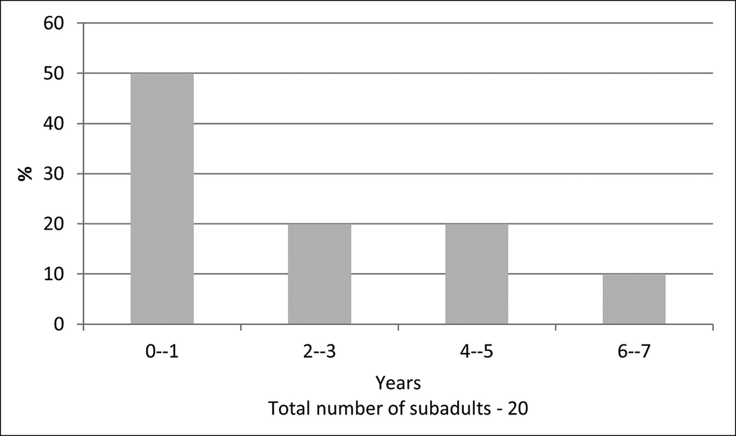
Cribra orbitalia, visible porosity of the bone of the upper orbit (above the eye), is often viewed as an indirect indication of poor sanitary conditions and a high-disease environment. It may develop in response to anaemia, the insufficiency of healthy red blood cells, which is brought about by a variety of external and internal causes, from nutrient deficiencies or congenital metabolic disorders to various parasitic infections (Wapler et al. Reference Wapler2004). Of the 43 individuals buried according to Islamic rites in the Southern Necropolis, cribra orbitalia was noted in 23 (54.5%). Most (17 individuals) were between the ages of six months and 6–7 years, with affected individuals making up 85 per cent of this age group. Six adults (26%) are affected. In combination with a consideration of age-distributed mortality rates (Figure 6), where the first year of life accounts for 50 per cent of mortality, the high prevalence of cribra orbitalia among subadults indicates that the inhabitants of al-Allaqi were affected by a suite of negative factors during infancy and early childhood. Not least among these, and contributing to the high first-year mortality, may have been parasitic infection.
Mortality profile of subadults buried in the Southern Necropolis (figure by authors).

Conclusion
The palaeoparasitological data presented here expand our understanding of the diet and sanitary conditions experienced by the medieval population buried in the al-Allaqi necropolis. A high prevalence of taeniasis is indicated through the identification of tapeworm eggs in multiple burials, likely reflecting infection by beef tapeworm (Taenia saginata) contracted through the consumption of insufficiently heat-treated meat from cattle. Infection by pork tapeworm is less likely in light of the zooarchaeological assemblage and Islamic taboos regarding the rearing and eating of pigs. The presence of human whipworm and ascarides eggs in the sediment samples may indicate generally poor sanitary and hygienic conditions in the associated settlement, potentially predominantly affecting females and subadults. These findings add to palaeoparasitological studies of the African continent, moving away from a focus on ancient Egyptian populations and demonstrating how parasitology can complement other research strands to build more informed reconstructions of various aspects of medieval life.
Acknowledgements
We acknowledge the core role of the National Corporation for Antiquities and Museums (NCAM) of the Sudan, for permitting the expedition campaigns, and for the ever-helpful participation of its staff. We are very grateful to Elena V. Bokova and Daniil V. Kambulov for financial support and friendly assistance.
Funding statement
This work was done with the support of Moscow State University Program of Development, Project No. 23-SCH02-22 (agreement number 004179) Scientific and Educational School of Lomonosov Moscow State University Preservation of world cultural and historical heritage ‘History, anthropology and ethnology of Northeast Africa in the context of environmental changes over the last 13 000 years (experience of interdisciplinary research)’.
Online supplementary material (OSM)
To view supplementary material for this article, please visit https://doi.org/10.15184/aqy.2026.10306 and select the supplementary materials tab.