Totally anomalous pulmonary venous return accounts for up to 3% of all cases of congenitally malformed hearts,Reference Friedman and Silverman1 and is typically classified into four subtypes according to the site of drainage of the pulmonary veins to the systemic venous circulation.Reference Darling, Rothurney and Craig2 These are the supracardiac, cardiac, infracardiac and mixed variant, with the supracardiac pattern being the most common, and mixed being the least. As afflicted patients commonly present with respiratory distress and cyanosis, it is a great mimic of pulmonary disease and differentiating between the two can be difficult. In this report, we describe a newborn infant who presented in the first hour of life with respiratory failure and cardiopulmonary collapse, referred to our centre for extracorporeal membrane oxygenation support.
Though various types of mixed drainage have been described,Reference Delisle, Ando and Calder3–Reference Kung, Gao, Wong, Sklansky, Uzunyan and Wood6 in our patient we discovered supracardiac totally anomalous return, but with a residual venous connection to the left atrium. This unusual anatomy confounded the diagnosis, and delayed consideration of surgery.
Case report
Baby EF was born at term, weighing 3.63 kilograms, after an uneventful pregnancy and labour. She deteriorated rapidly after birth, with severe cyanosis and bradycardia requiring intubation and ventilation. The chest X-ray showed a fine reticular pattern across both lung fields, and surfactant was given. She required very high ventilatory pressures, and was given trials of high frequency oscillatory ventilation and inhaled nitric oxide, with little improvement in oxygenation. Her best partial pressure of oxygen in arterial blood was only 2.8 kilopascals.
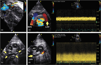
Cross-sectional and Doppler echocardiographic interrogation performed at the referring hospital demonstrated tricuspid and pulmonary regurgitation, but otherwise was interpreted to show a structurally normal heart. At this stage, she was referred to our centre for extracorporeal membrane oxygenation. Repeat echocardiography performed prior to cannulation for veno-arterial extracorporeal membrane oxygenation at our centre again showed a structurally normal heart. We noted that the pulmonary veins were difficult to visualize, but pulmonary venous connection with the left atrium was clearly seen, and proven by colour-flow mapping (Fig. 1a).
Cross sectional echocardiography with colour flow mapping in an apical four-chamber anatomical view (a), while on veno-arterial extracorporeal membrane oxygenation. Pulmonary venous flow draining into the left atrium (LA) was clearly visualised (arrow), albeit that no separated connections with individual pulmonary veins was seen. Right atrium (RA) and superior caval vein (SVC) drained via the venous cannula. RV=Right ventricle, LV=Left ventricle. Subsequent echocardiography in the suprasternal frontal view (b) demonstrates a pulmonary venous confluence (PVC). Arrows indicate all four pulmonary veins connected to the confluence. *=Pericardial effusion, AO=Aorta, PA=Pulmonary trunk. Colour flow mapping and pulsed Doppler technique from the suprasternal view (c) reveal the left vertical vein (LVV) with upward flow coded in red. Constant low velocity flow (0.15 m/s) Doppler signal taken from LVV indicates pulmonary flow distal to the obstruction. SVC=Superior caval vein. Colour flow mapping and pulsed Doppler technique from suprasternal view (d) show the narrow left vertical vein (LVV) coursing anterior to the left brachicephalic vein (arrow), with local obstruction at the level of left bronchus and left pulmonary artery. AO=Aorta, PA=Pulmonary trunk.

On the 6th day of extracorporeal membrane oxygenation, she developed severe hemorrhagic complications, with a large right-sided haemothorax leading to mediastinal shift, which caused malfunction of the circuit. Flows had to be reduced to maintain continuity of the circuit. At this stage, echocardiographic interrogation was repeated, and revealed an obstructed supracardiac totally anomalous pulmonary venous connection. All four pulmonary veins joined a confluence which then drained to the left brachiocephalic vein via a severely obstructed left vertical vein (Figs 1b–d). The vertical vein at the level of the left pulmonary artery and arterial duct did not exceed 1.5 millimetres in diameter. The maximal velocity of continuous flow through the vein was 1.5 metres per second. Reversed upward flow was seen on colour-flow mapping in both the left vertical vein and superior caval vein, with both draining into the venous cannula of the oxygenation circuit. Unfortunately, despite our best efforts, the neonate deteriorated further. Surgical correction of her cardiac lesion was not feasible in view of severe uncontrolled haemorrhage from the right hemithorax. After discussion between the medical team and family, intensive care support was withdrawn.
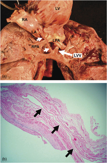
At post-mortem, the heart showed usual arrangement with concordant segmental connections. All four pulmonary veins came to a confluence behind the heart (Fig. 2a). From the left side of this confluence, a vein of 0.4 centimetres diameter, and 3.7 centimetres in length, ascended between the left pulmonary artery and the left main bronchus to insert into the brachiocephalic vein inferior to the junction of the left subclavian and jugular veins. This vertical vein was narrowed to a diameter of 0.15 centimetres diameter where it passed behind the pulmonary artery. Another small vein less than 0.1 centimetres in diameter, and 1 centimetre long, ascended from the junction of the venous confluence and the vertical vein and connected to the left atrium. The left atrium was diminutive. There was a small defect on its posterior aspect, which represented the point of insertion of the small vein from the pulmonary confluence. Histologically, a venous channel was seen in the left atrial wall running obliquely from the deficient epicardial surface to the lumen (Fig. 2b).
Panel (a) shows the dissected heart and lungs. The heart has been pulled forward and lifted upward to disclose the structures on its posterior aspect. The pulmonary veins come to a confluence, from the left side of which arises the vertical vein (LVV). From the right side of this vein, a small vessel (short arrows) runs to the left atrium (LA). RA=Right atrium, LV=Left ventricle, RPA=Right pulmonary artery, LPA=Left pulmonary artery. Histology (b) reveals a venous channel (arrows) running from the junction of the pulmonary venous confluence and left vertical vein to the left atrium.

Discussion
Our infant had anomalous connection of the pulmonary veins to a systemic venous site, as well as some, albeit inadequate, venous connection with the left atrium. This unusual combination confounded the initial echocardiographic findings, and her cardiac lesion only became apparent when the flows through the circuit providing extracorporeal membrane oxygenation were reduced.
In cases of obstructed pulmonary venous connection, early diagnosis combined with emergent surgery can be life-saving. With improved diagnostic tools and surgical skills, long-term survival rates have been quoted to be as high as 98%.Reference Bando, Turrentine and Ensing7 This is especially relevant, as outcomes are poor for neonates with congenitally malformed hearts who are referred for extracorporeal membrane oxygenation because of respiratory problems, with mortality rates quoted at 50%.Reference Brown, Miles and Sullivan8
Our unusual case demonstrates the need to be vigilant to the possibility of a cardiac lesion in patients with presumed severe neonatal “pulmonary disease” who are unresponsive to conventional therapy. Where doubt exists, echocardiography should be repeated, and all pulmonary veins should be visualized to exclude rare permutations of totally anomalous pulmonary venous connection.
Acknowledgement
The authors are grateful for, and would like to acknowledge the contribution of Dr Michael Ashworth in the provision of post mortem details.